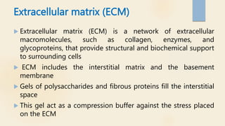
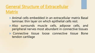
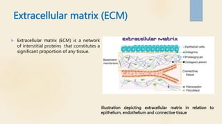
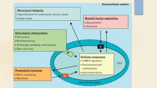
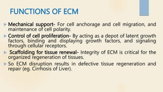
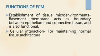
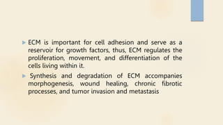
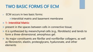
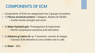
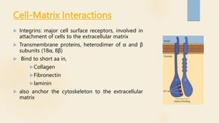
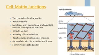
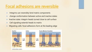
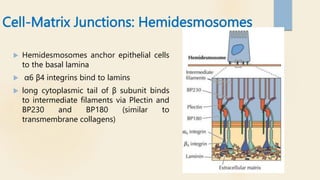
2. Extracellular matrix (ECM)
Extracellular matrix (ECM) is a network of extracellular
macromolecules, such as collagen, enzymes, and
glycoproteins, that provide structural and biochemical support
to surrounding cells
ECM includes the interstitial matrix and the basement
membrane
Gels of polysaccharides and fibrous proteins fill the interstitial
space
This gel act as a compression buffer against the stress placed
on the ECM
3. General Structure of Extracellular
Matrix
Animal cells embedded in an extracellular matrix Basal
laminae: thin layer on which epithelial cells rest.
Also surrounds muscle cells, adipose cells, and
peripheral nerves most abundant in connective tissues
Connective tissue loose connective tissue Bone
tendon cartilage
4. Extracellular matrix (ECM)
Extracellular matrix (ECM) is a network
of interstitial proteins that constitutes a
significant proportion of any tissue.
ilIustration depicting extracellular matrix in relation to
epithelium, endothelium and connective tissue
5.
6. FUNCTIONS OF ECM
Mechanical support- For cell anchorage and cell migration, and
maintenance of cell polarity.
Control of cell proliferation- By acting as a depot of latent growth
factors, binding and displaying growth factors, and signaling
through cellular receptors.
Scaffolding for tissue renewal- Integrity of ECM is critical for the
organized regeneration of tissues.
So ECM disruption results in defective tissue regeneration and
repair (eg. Cirrhosis of Liver).
7. FUNCTIONS OF ECM
Establishment of tissue microenvironments-
Basement membrane acts as boundary
between epithelium and connective tissue, and
is also functional.
Cellular interaction- For maintaining normal
tissue architecture.
8. ECM is important for cell adhesion and serve as a
reservoir for growth factors, thus, ECM regulates the
proliferation, movement, and differentiation of the
cells living within it.
Synthesis and degradation of ECM accompanies
morphogenesis, wound healing, chronic fibrotic
processes, and tumor invasion and metastasis
9. TWO BASIC FORMS OF ECM
ECM occurs in two basic forms:
interstitial matrix and basement membrane
1- Interstitial Matrix:
present in the spaces between cells in connective tissue;
it is synthesized by mesenchymal cells (e.g., fibroblasts) and tends to
form a three-dimensional, amorphous gel.
Its major constituents are fibrillar and nonfibrillar collagens, as well
as fibronectin, elastin, proteoglycans, hyaluronate, and other
elements
10. The basement membrane:
lies beneath the epithelium
It is synthesized by overlying epithelium and
underlying mesenchymal cells
Its major constituents are amorphous
nonfibrillar type IV collagen and laminin
11. COMPONENTS OF ECM
Components of ECM are categorized into 3 groups of proteins;
1) Fibrous structural proteins- Collagens, Elastins & Fibrillin.
Confer tensile strength and recoil.
2) Water-hydrated gels- Proteagylcans & Hyaluronan
Permit compressive resistance and lubrication.
3) Adhesive gl roteins & r y- F bronectin, Laminin & integan
Connect ECM elements to one another and to cells.
4) Water - 65%
12. Collagens
Collagens are fibrous structural proteins that confer tensile strength.
Collagens are composed of three separate polypeptide chains braided into a ropelike
triple helix.
About 30 collagen types have been identified
Some collagen types (e.g., types I, II, III, and V) form fibrils by virtue of lateral cross-
linking of the triple helices. These are called the fibrillarcollagens, and form a major
proportion of the connective tissue in healing wounds and particularly in scars
Other collagens are nonfibrillar and present in basement membrane (type IV)
The tensile strength of the fibrillar collagens derives from their cross-linking, which is the
result of covalent bonds.
This process is dependent on vitamin C; therefore, children with vitamin C
deficiencyhave skeletal deformities, bleed easily because of weak vascular wall basement
membrane, and heal poorly
13. Elastin
the ability of tissues to recoil and return to a baseline
structure after physical stress is conferred by elastic
tissue.
This is especially important in the walls of: vessels , the
uterus, skin, and ligaments.
14. Proteoglycans and Hyaluronan
Proteoglycans form highly hydrated compressible gels conferring resilience
and lubrication (such as in the cartilage in joints).
They consist of long polysaccharides called glycosaminoglycans (examples
are dermatan sulfate and heparan sulfate) linked to a protein backbone.
Hyaluronan a huge molecule composed of many disaccharide repeats
without a protein core, is also an important constituent of the ECM.
Because of its ability to bind water, it forms a viscous, gelatin-like matrix
15. Adhesive glycoproteins and adhesion
receptors
Adhesive glycoproteins and adhesion receptors are structurally diverse
molecules involved in cell-to-cell adhesion, the linkage between cells and
ECM, and binding between ECM components.
The adhesive glycoproteins include fibronectin (major component of the
interstitial ECM) and laminin (major constituent of basement membrane).
The adhesion receptors, also known as cell adhesion molecules (CAMs), are
grouped into four families: immunoglobulins, cadherins, selectins, and
integrins
16. Fibronectin
Fibronectin is a large disulfide-linked heterodimer
synthesized by a variety of cells, including fibroblasts, monocytes, and
endothelium.
Main adhesion protein of connective tissue
Fibronectins can bind to a wide spectrum of ECM components (e.g.,
collagen, fibrin, heparin, and proteoglycans) and can also attach to cell
integrins.
Fibronectin mRNA has two splice forms, which generate:
1- tissue fibronectin forms fibrillar aggregates at wound healing sites 2-
plasma fibronectin binds to fibrin to form the provisional blood clot of a
wound, which serves as a base for ECM deposition and re-epithelialization
17. Laminin
Laminin is the most abundant glycoprotein in
basement membrane.
Adhesion protein of basal laminae
connects cells to underlying ECM components
such as type IV collagen and heparan sulfate,
mediating attachment to basement membrane.
laminin can also modulate cell proliferation,
differentiation, and motility.
18. Cell-Matrix Interactions
Integrins: major cell surface receptors, involved in
attachment of cells to the extracellular matrix
Transmembrane proteins, heterodimer of α and β
subunits (18α, 8β)
Bind to short aa in,
Collagen
Fibronectin
laminin
also anchor the cytoskeleton to the extracellular
matrix
19. Cell-Matrix Junctions
Two types of cell-matrix junction
Focal adhesions:
bundles of actin filaments are anchored to β
subunits of integrins via α-actinin
Vinculin via talin
Assembly of focal adhesions
Focal complex: small group of integrins
RecruiteTalin, Vinculin, α-actinin and Formin
Formin initiates actin bundles
20. Focal adhesions are reversible
Integrins can reversibly bind matrix components
change conformation between active and inactive states
Inactive state: integrin heads turned close to cell surface
Cell signaling extends heads to matrix
Migrating cells: focal adhesions form at the leading edge
21. Cell-Matrix Junctions: Hemidesmosomes
Hemidesmosomes anchor epithelial cells
to the basal lamina
α6 β4 integrins bind to lamins
long cytoplasmic tail of β subunit binds
to intermediate filaments via Plectin and
BP230 and BP180 (similar to
transmembrane collagens)