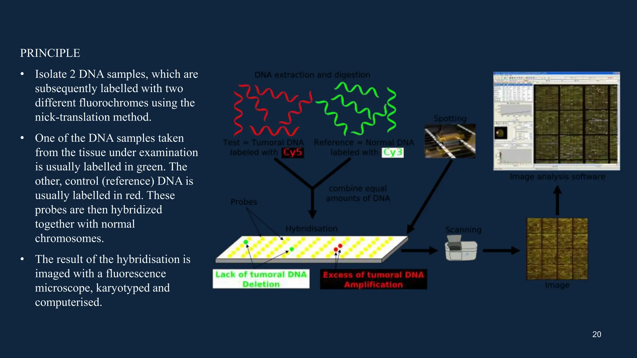
CYTOGENETIC MAP
INTRODUCTION
• Genomics is the study of entire genomes, including the complete set of genes , their nucleotide sequence,
organization and their interactions within a species and with other species.
• Gene mapping refers to the process of determining the location of genes on chromosomes. Today, the most
efficient approach for gene mapping involves sequencing a genome and then using computer programs to
analyze the sequence to identify the location of genes.
• A genetic map provides (also called a linkage map) shows the relative location of genetic markers (reflecting
sites of genomic variants) on a chromosome.
• A physical map is a representation of the physical distance, in nucleotides, between genes or genetic markers.
• Both genetic linkage maps and physical maps are required to build a complete picture of the genome.
• cytogenetic mapping is the process of identifying the physical location of genes and other genetic markers on
chromosomes. It combines techniques from cytogenetics, which focuses on the study of chromosomes, and
molecular genetics, which delves into the structure and function of genes.
• The mapping process typically involves staining chromosomes to produce unique banding patterns that help
distinguish different regions. These patterns act as a blueprint for determining the relative positions of genes.
2
WHAT IS CYTOGENETIC MAP
• Cytogenetic is the study of chromosomes and their role in heredity.
• The methods that scientists use to analyze chromosomes, chromosome abnormalities associated with disease,
the roles that chromosomes play in sex determination and changes in chromosomes during evolution
• Particularly important are visually distinct regions, called light and dark bands, which give each of the
chromosomes a unique appearance.
• A cytogenetic map is produced after staining metaphase chromosomes with a particular dye mixture and
visualizing the dark and light-coloured bands under microscope.
• Each chromosome pair stains with its own characteristic banding pattern. The bands correlate approximately
with the DNA sequence underlying it: AT-rich areas stain darkly, GC-rich areas lightly.
• Each band is numbered to help identify a particular region of a chromosome. This method of mapping a gene to
a particular band of the chromosome is called cytogenetic mapping.
3
Importance of cytogenetic mapping
• Cytogenetic maps depict the location and order of markers along chromosomes.
• Cytogenetic maps are important in genome research as they relate the genetic data and molecular sequences to
the morphological features of chromosomes.
• In evolutionary studies, it helps understand the rearrangements and structural variations that occur within and
between species. By comparing the chromosome maps of different organisms, scientists can trace evolutionary
relationships and identify genetic changes that contributed to speciation.
• In medical genetics, cytogenetic mapping plays a critical role in diagnosing genetic disorders.
• Chromosomal aberrations, such as deletions, duplications, inversions, or translocations, can lead to numerous
conditions. By mapping these abnormalities, identification of the exact regions involved and development of
targeted diagnostic tests or therapeutic interventions.
• Cancer research also benefits from cytogenetic mapping. By analyzing tumor cells, researchers can identify
chromosomal rearrangements and aberrations associated with different types of cancer
• Cytogenetic mapping is crucial for prenatal screening, enabling the early detection of chromosomal
abnormalities in fetuses.
4
TYPES
• IDIOGRAM MAPS
• LINKAGE MAPS
• PHYSICAL MAPS
• FISH MAPS
• COMAPARATIVE MAPS
5
IDIOGRAM MAPS
A set of chromosomes represent the genome and the genetic information of an organism. Each chromosome has
a unique shape, size and a set of genes. The number and physical structures of chromosomes reveal important
information regarding the organism. Karyotyping is a technique performed to examine the complete set of
chromosomes in a cell.
• A karyotype is a diagram that shows the chromosomal number and constitution in the cells’ nucleus. It
consists of a whole set of homologous chromosome pairs, arranged in decreasing series of their size.
• By analyzing the karyotype of the organism, it is possible to detect genetic disorders and other information
about the individual.
• Idiogram is a diagrammatic representation or a schematic diagram of a karyotype of a species. Idiogram
shows the chromosome maps indicating the locations of genes as bands.
• It is not an actual picture of total chromosomes of a cell. However, ideogram provides much information
about each chromosome. It provides locations of individual genes present in a chromosome.
6
Moreover, idiograms are useful in identifying various abnormalities associated with a range of chromosomal
disorders and determining the links between structural abnormalities and individual genes correlated with
various diseases or syndromes.
7
GENETIC MAPPING
• Genetic mapping, also known as linkage mapping, refers to the method used to create genetic maps to estimate
the positions of genes in chromosomes.
• Genetic mapping starts by using linkage analysis to study the frequency of recombination between genes.
• Recombination frequency indicates genetic linkage and helps to determine whether the genes are linked
together or not. A centimorgan (cM) is a unit that describes a recombination frequency of 1%.
• As the distance between two genes increases, the likelihood of recombination occurring also increases,
resulting in a higher recombination frequency between them.
• The recombination frequency is determined by using the number of recombinant individuals compared to the
total number of offspring. This frequency is expressed as a percentage.
8
PHYSICAL MAPPING
• Physical mapping involves determining the precise locations of DNA sequences on chromosomes. It uses base
pair as the unit of measurement.
• Physical maps provide a direct representation of the physical structure of a chromosome and the positions of
genes along its DNA sequence.
• Markers used in physical mapping include ESTs (Expressed Sequence Tags), STS (Sequence Tagged Site)
markers, and genome-wide DNA sequences
FLUORESCENCE IN SITU HYBRIDIZATION (FISH) MAPPING
• It is the most convincing technique for locating the specific DNA sequences, diagnosis of genetic diseases, gene
mapping, and identification of novel oncogenes or genetic aberrations contributing to various types of cancers.
• FISH involves annealing of DNA or RNA probes attached to a fluorescent reporter molecule with specific target
sequence of sample DNA, which can be followed under fluorescence microscopy.
• The technique has lately been expanded to enable screening of the whole genome simultaneously through
multicolor whole chromosome probe techniques such as multiplex FISH or spectral karyotyping or through an
array-based method using comparative genomic hybridization.
9
COMAPARATIVE MAPS
• Comparative mapping is a study how the genomes relate across species and genera and even families.
• Comparative mapping involves establishing the relative genomic positions of orthologous gene pairs in two
species
• The concept started with comparative mapping experiments using RFLP markers between two species that led to
the discovery of conserved linear orders of marker loci across related species.
10
CYTOGENETIC MAPPING METHODOLOGIES
Flowcytometry
• Flow Cytometry measures multiple characteristics of individual particles flowing in single file in a stream of fluid.
• Light scattering at different angles can distinguish differences in size and internal complexity,whereas light emitted from
fluorescently labelled antibodies can identify a wide array of cell surface and cytoplasmic antigens.
• This approach makes flow cytometry a powerful tool for detailed analysis of complex populations in a short period of time.
Principle
• Flow cytometry measures optical and fluorescence characteristics of single cells.
• Fluorescent dyes may bind or intercalate with different cellular components such as DNA or RNA
• When labelled cells are passed by a light source, the fluorescent molecules are excited to a higher energy state.
• Upon returning to their resting states, the fluorochromes emit light energy at higher wavelengths.
• The use of multiple fluorochromes, each with similar excitation wavelengths and different emission wavelengths allows several
cell properties to be measured simultaneously.
• Commonly used dyes include propidiumiodide , phycoerythrin, and fluorescein
11
Human/rodent somatic-cell hybrid mapping
12
Advantages of flow cytometry
This method is individual celloriented which renders it quite suitable for quantification of cells from a
heterogeneous cell sample.
It is quite a rapid method measurement being done at a rate of 1000 cells in a second.
Disadvantages of flow cytometry
1. Sensitivity: These are mostly designed to primarily analyse mammalian cells and so for bacteria, it does
not function that well.
2. Portability : Commercially available flow cytometers are not portable and hence not suitable for field
work.
3. Prior to use most of flow cytometers require adjustment.
4. Most of the flow cytometers are costly
13
APPLICATIONS
1. DNA Content Analysis :
The measurement of cellular DNA content by flow cytometry uses fluorescent dyes, such as propidium iodide, that
intercalate into the DNA helical structure . The fluorescent signal is directly proportional to the amount of DNA in
the nucleus and can identify gross gains or losses in DNA. Abnormal DNA content, also known as DNA content
aneuploidy”
, can be determined in a tumour cell population . Although conventional cytogenetic can detect smaller
DNA content differences, flow cytometry allows more rapid analysis of a larger number of cells.
2. Genome size Analysis:
Analytical information about the physical size of plant genomes and their state of replication is easily obtainable
from flow cytometry. Knowing the number of base pairs in a genome is valuable for studies of new species. Flow
cytometry provides a fast and accurate way to look at changes in genome size during evolution and differentiation.
Establishment of ploidy and aneuploidy changes during tissue culture, and examination of intra- and inter-specific
variation of DNA content can all be important in plant hybridization, breeding, and genetic manipulation programs.
3. Application also includes high speed sorting for construction of chromosome specific libraries that establish
feasibility for the Human Genome Program and the construction of chromosome specific fluorescent in situ
hybridization for research and clinical applications
14
FLORESCENCE IN-SITU HYBRIDIZATION TECHNIQUE
Fluorescence in situ hybridization is a method that allows for the detection of whole bacterial cells and analysis of
chromosomes via the labelling of the specific nucleic acids with fluorescently labelled oligonucleotide probes
First, short sequences of single-stranded DNA probes are prepared. These probes hybridize or bind to complementary
nucleic acids because they are labelled with fluorescent tags, allow the researchers to see the location of those
sequences of DNA .
The results are viewed , usually with a scanning laser microscope
15
Principle
a) The basic elements of FISH are a DNA probe and a target
sequence.
b) Before hybridization, the DNA probe is labeled by various
means, such as nick translation, random primed labeling, and
PCR. Two labeling strategies are commonly used: indirect
labeling (left panel) and direct labeling (right panel). For
indirect labeling, probes are labeled with modified nucleotides
that contain a hapten, whereas direct labeling uses nucleotides
that have been directly modified to contain a fluorophore.
c) The labeled probe and the target DNA are denatured.
d) Combining the denatured probe and target allows the
annealing of complementary DNA sequences.
e) If the probe has been labeled indirectly, an extra step is
required for visualization of the nonfluorescent hapten that
uses an enzymatic or immunological detection system.
Whereas FISH is faster with directly labeled probes, indirect
labeling offers the advantage of signal amplification by using
several layers of antibodies, and it might therefore produce a
signal that is brighter compared with background levels. 16
APPLICATIONS
• Detection of chromosomal aberrations: FISH and other in situ hybridization procedures are important in the
clinical diagnosis of various chromosomal abnormalities, including deletions, duplications, and translocations.
• Using FISH to analyse interphase chromosomes: Since the introduction of FISH, cytogeneticists have been able to
analyse interphase chromosomes as well as the metaphase chromosomes used in karyotypes
• Analysis of Somaclonal Variations: These variations in tissue culture are considered as novel source of genetic
variation for crop improvement.
17
COMPARATIVE GENOMIC HYBRIDIZATION
• One of the most significant developments for FISH in relation to genome-wide screening was the introduction of
comparative genomic hybridization (CGH) in 1992.
• This modification of quantitative two colour fluorescence in situ hybridization utilizes genomic DNA from the
sample under test to generate a map of DNA copy number changes in tumour genomes making it an ideal tool for
analysing chromosomal imbalances in archived tumour material and for examining possible correlations between
findings and tumour phenotypes
Advantages of comparative genomic hybridization
1. Visualisation of deletions and duplications in very small DNA segments
2. Searches of the whole genome without prior knowledge about the chromosomal aberration at hand
3. Analysis without the need for specific probes
4. The detection of the presence of amplified genes in cancer and maps their location
5. Unlike FISH, CGH is able to; Identify the chromosome with the aberration; identify the specific location from
which the extra material originated; FISH is only able to identify the chromosome with the particular aberration
18
Limitations of comparative genomic hybridization
1. For rearrangements that do not involve genomic imbalances, such as balanced chromosome translocations and
inversions, the use of CGH is limited.
2. Whole-genome copy number changes (ploidy changes) cannot be detected.
3. CGH provides no information about the structural arrangements of chromosome segments that are
involved in gains and losses.
19
PRINCIPLE
• Isolate 2 DNA samples, which are
subsequently labelled with two
different fluorochromes using the
nick-translation method.
• One of the DNA samples taken
from the tissue under examination
is usually labelled in green. The
other, control (reference) DNA is
usually labelled in red. These
probes are then hybridized
together with normal
chromosomes.
• The result of the hybridisation is
imaged with a fluorescence
microscope, karyotyped and
computerised.
20
CONCLUSION
The cytogenetic map provide valuable insight into
the chromosomal features and genetic variations
within a given genome. Understanding the precise
locations of a genes and structural changes on
chromosomes enhances the knowledge of genetic
disorders and contributes to advancements in
medical research and diagnosis.
As technology advances, cytogenetic mapping
provides high resolution techniques enhancing
ability to detect chromosomal changes .
From classic banding techniques to advanced
molecular techniques like FISH, CGH these
methods enable to visualize characteristics and
analyze chromosome at varying levels of resolution
21
REFEENCE
• Liehr, T. (Ed.). (2021). Cytogenomics. Academic Press.
• Shah, M., Varshney, P., Patel, P., Patel, D., & Meshram, D. (2012). Cytogenetic mapping techniques: An approach
to genome analysis. Research & Reviews in BioSciences, 7, 209-219.
• Primrose, S. B., & Twyman, R. (2006). Principles of gene manipulation and genomics. John Wiley & Sons.
• https://www.wur.nl/en/article/comparative-genomics.htm
• https://www.nature.com/scitable/topicpage/chromosome-mapping-idiograms-302/
• https://www.nature.com/scitable/content/principles-of-fluorescence-in-situ-hybridization-35120/
22

CYTOGENETIC MAP................ ppt.pptx

  • 1.
  • 2.
    INTRODUCTION • Genomics isthe study of entire genomes, including the complete set of genes , their nucleotide sequence, organization and their interactions within a species and with other species. • Gene mapping refers to the process of determining the location of genes on chromosomes. Today, the most efficient approach for gene mapping involves sequencing a genome and then using computer programs to analyze the sequence to identify the location of genes. • A genetic map provides (also called a linkage map) shows the relative location of genetic markers (reflecting sites of genomic variants) on a chromosome. • A physical map is a representation of the physical distance, in nucleotides, between genes or genetic markers. • Both genetic linkage maps and physical maps are required to build a complete picture of the genome. • cytogenetic mapping is the process of identifying the physical location of genes and other genetic markers on chromosomes. It combines techniques from cytogenetics, which focuses on the study of chromosomes, and molecular genetics, which delves into the structure and function of genes. • The mapping process typically involves staining chromosomes to produce unique banding patterns that help distinguish different regions. These patterns act as a blueprint for determining the relative positions of genes. 2
  • 3.
    WHAT IS CYTOGENETICMAP • Cytogenetic is the study of chromosomes and their role in heredity. • The methods that scientists use to analyze chromosomes, chromosome abnormalities associated with disease, the roles that chromosomes play in sex determination and changes in chromosomes during evolution • Particularly important are visually distinct regions, called light and dark bands, which give each of the chromosomes a unique appearance. • A cytogenetic map is produced after staining metaphase chromosomes with a particular dye mixture and visualizing the dark and light-coloured bands under microscope. • Each chromosome pair stains with its own characteristic banding pattern. The bands correlate approximately with the DNA sequence underlying it: AT-rich areas stain darkly, GC-rich areas lightly. • Each band is numbered to help identify a particular region of a chromosome. This method of mapping a gene to a particular band of the chromosome is called cytogenetic mapping. 3
  • 4.
    Importance of cytogeneticmapping • Cytogenetic maps depict the location and order of markers along chromosomes. • Cytogenetic maps are important in genome research as they relate the genetic data and molecular sequences to the morphological features of chromosomes. • In evolutionary studies, it helps understand the rearrangements and structural variations that occur within and between species. By comparing the chromosome maps of different organisms, scientists can trace evolutionary relationships and identify genetic changes that contributed to speciation. • In medical genetics, cytogenetic mapping plays a critical role in diagnosing genetic disorders. • Chromosomal aberrations, such as deletions, duplications, inversions, or translocations, can lead to numerous conditions. By mapping these abnormalities, identification of the exact regions involved and development of targeted diagnostic tests or therapeutic interventions. • Cancer research also benefits from cytogenetic mapping. By analyzing tumor cells, researchers can identify chromosomal rearrangements and aberrations associated with different types of cancer • Cytogenetic mapping is crucial for prenatal screening, enabling the early detection of chromosomal abnormalities in fetuses. 4
  • 5.
    TYPES • IDIOGRAM MAPS •LINKAGE MAPS • PHYSICAL MAPS • FISH MAPS • COMAPARATIVE MAPS 5
  • 6.
    IDIOGRAM MAPS A setof chromosomes represent the genome and the genetic information of an organism. Each chromosome has a unique shape, size and a set of genes. The number and physical structures of chromosomes reveal important information regarding the organism. Karyotyping is a technique performed to examine the complete set of chromosomes in a cell. • A karyotype is a diagram that shows the chromosomal number and constitution in the cells’ nucleus. It consists of a whole set of homologous chromosome pairs, arranged in decreasing series of their size. • By analyzing the karyotype of the organism, it is possible to detect genetic disorders and other information about the individual. • Idiogram is a diagrammatic representation or a schematic diagram of a karyotype of a species. Idiogram shows the chromosome maps indicating the locations of genes as bands. • It is not an actual picture of total chromosomes of a cell. However, ideogram provides much information about each chromosome. It provides locations of individual genes present in a chromosome. 6
  • 7.
    Moreover, idiograms areuseful in identifying various abnormalities associated with a range of chromosomal disorders and determining the links between structural abnormalities and individual genes correlated with various diseases or syndromes. 7
  • 8.
    GENETIC MAPPING • Geneticmapping, also known as linkage mapping, refers to the method used to create genetic maps to estimate the positions of genes in chromosomes. • Genetic mapping starts by using linkage analysis to study the frequency of recombination between genes. • Recombination frequency indicates genetic linkage and helps to determine whether the genes are linked together or not. A centimorgan (cM) is a unit that describes a recombination frequency of 1%. • As the distance between two genes increases, the likelihood of recombination occurring also increases, resulting in a higher recombination frequency between them. • The recombination frequency is determined by using the number of recombinant individuals compared to the total number of offspring. This frequency is expressed as a percentage. 8
  • 9.
    PHYSICAL MAPPING • Physicalmapping involves determining the precise locations of DNA sequences on chromosomes. It uses base pair as the unit of measurement. • Physical maps provide a direct representation of the physical structure of a chromosome and the positions of genes along its DNA sequence. • Markers used in physical mapping include ESTs (Expressed Sequence Tags), STS (Sequence Tagged Site) markers, and genome-wide DNA sequences FLUORESCENCE IN SITU HYBRIDIZATION (FISH) MAPPING • It is the most convincing technique for locating the specific DNA sequences, diagnosis of genetic diseases, gene mapping, and identification of novel oncogenes or genetic aberrations contributing to various types of cancers. • FISH involves annealing of DNA or RNA probes attached to a fluorescent reporter molecule with specific target sequence of sample DNA, which can be followed under fluorescence microscopy. • The technique has lately been expanded to enable screening of the whole genome simultaneously through multicolor whole chromosome probe techniques such as multiplex FISH or spectral karyotyping or through an array-based method using comparative genomic hybridization. 9
  • 10.
    COMAPARATIVE MAPS • Comparativemapping is a study how the genomes relate across species and genera and even families. • Comparative mapping involves establishing the relative genomic positions of orthologous gene pairs in two species • The concept started with comparative mapping experiments using RFLP markers between two species that led to the discovery of conserved linear orders of marker loci across related species. 10
  • 11.
    CYTOGENETIC MAPPING METHODOLOGIES Flowcytometry •Flow Cytometry measures multiple characteristics of individual particles flowing in single file in a stream of fluid. • Light scattering at different angles can distinguish differences in size and internal complexity,whereas light emitted from fluorescently labelled antibodies can identify a wide array of cell surface and cytoplasmic antigens. • This approach makes flow cytometry a powerful tool for detailed analysis of complex populations in a short period of time. Principle • Flow cytometry measures optical and fluorescence characteristics of single cells. • Fluorescent dyes may bind or intercalate with different cellular components such as DNA or RNA • When labelled cells are passed by a light source, the fluorescent molecules are excited to a higher energy state. • Upon returning to their resting states, the fluorochromes emit light energy at higher wavelengths. • The use of multiple fluorochromes, each with similar excitation wavelengths and different emission wavelengths allows several cell properties to be measured simultaneously. • Commonly used dyes include propidiumiodide , phycoerythrin, and fluorescein 11
  • 12.
  • 13.
    Advantages of flowcytometry This method is individual celloriented which renders it quite suitable for quantification of cells from a heterogeneous cell sample. It is quite a rapid method measurement being done at a rate of 1000 cells in a second. Disadvantages of flow cytometry 1. Sensitivity: These are mostly designed to primarily analyse mammalian cells and so for bacteria, it does not function that well. 2. Portability : Commercially available flow cytometers are not portable and hence not suitable for field work. 3. Prior to use most of flow cytometers require adjustment. 4. Most of the flow cytometers are costly 13
  • 14.
    APPLICATIONS 1. DNA ContentAnalysis : The measurement of cellular DNA content by flow cytometry uses fluorescent dyes, such as propidium iodide, that intercalate into the DNA helical structure . The fluorescent signal is directly proportional to the amount of DNA in the nucleus and can identify gross gains or losses in DNA. Abnormal DNA content, also known as DNA content aneuploidy” , can be determined in a tumour cell population . Although conventional cytogenetic can detect smaller DNA content differences, flow cytometry allows more rapid analysis of a larger number of cells. 2. Genome size Analysis: Analytical information about the physical size of plant genomes and their state of replication is easily obtainable from flow cytometry. Knowing the number of base pairs in a genome is valuable for studies of new species. Flow cytometry provides a fast and accurate way to look at changes in genome size during evolution and differentiation. Establishment of ploidy and aneuploidy changes during tissue culture, and examination of intra- and inter-specific variation of DNA content can all be important in plant hybridization, breeding, and genetic manipulation programs. 3. Application also includes high speed sorting for construction of chromosome specific libraries that establish feasibility for the Human Genome Program and the construction of chromosome specific fluorescent in situ hybridization for research and clinical applications 14
  • 15.
    FLORESCENCE IN-SITU HYBRIDIZATIONTECHNIQUE Fluorescence in situ hybridization is a method that allows for the detection of whole bacterial cells and analysis of chromosomes via the labelling of the specific nucleic acids with fluorescently labelled oligonucleotide probes First, short sequences of single-stranded DNA probes are prepared. These probes hybridize or bind to complementary nucleic acids because they are labelled with fluorescent tags, allow the researchers to see the location of those sequences of DNA . The results are viewed , usually with a scanning laser microscope 15
  • 16.
    Principle a) The basicelements of FISH are a DNA probe and a target sequence. b) Before hybridization, the DNA probe is labeled by various means, such as nick translation, random primed labeling, and PCR. Two labeling strategies are commonly used: indirect labeling (left panel) and direct labeling (right panel). For indirect labeling, probes are labeled with modified nucleotides that contain a hapten, whereas direct labeling uses nucleotides that have been directly modified to contain a fluorophore. c) The labeled probe and the target DNA are denatured. d) Combining the denatured probe and target allows the annealing of complementary DNA sequences. e) If the probe has been labeled indirectly, an extra step is required for visualization of the nonfluorescent hapten that uses an enzymatic or immunological detection system. Whereas FISH is faster with directly labeled probes, indirect labeling offers the advantage of signal amplification by using several layers of antibodies, and it might therefore produce a signal that is brighter compared with background levels. 16
  • 17.
    APPLICATIONS • Detection ofchromosomal aberrations: FISH and other in situ hybridization procedures are important in the clinical diagnosis of various chromosomal abnormalities, including deletions, duplications, and translocations. • Using FISH to analyse interphase chromosomes: Since the introduction of FISH, cytogeneticists have been able to analyse interphase chromosomes as well as the metaphase chromosomes used in karyotypes • Analysis of Somaclonal Variations: These variations in tissue culture are considered as novel source of genetic variation for crop improvement. 17
  • 18.
    COMPARATIVE GENOMIC HYBRIDIZATION •One of the most significant developments for FISH in relation to genome-wide screening was the introduction of comparative genomic hybridization (CGH) in 1992. • This modification of quantitative two colour fluorescence in situ hybridization utilizes genomic DNA from the sample under test to generate a map of DNA copy number changes in tumour genomes making it an ideal tool for analysing chromosomal imbalances in archived tumour material and for examining possible correlations between findings and tumour phenotypes Advantages of comparative genomic hybridization 1. Visualisation of deletions and duplications in very small DNA segments 2. Searches of the whole genome without prior knowledge about the chromosomal aberration at hand 3. Analysis without the need for specific probes 4. The detection of the presence of amplified genes in cancer and maps their location 5. Unlike FISH, CGH is able to; Identify the chromosome with the aberration; identify the specific location from which the extra material originated; FISH is only able to identify the chromosome with the particular aberration 18
  • 19.
    Limitations of comparativegenomic hybridization 1. For rearrangements that do not involve genomic imbalances, such as balanced chromosome translocations and inversions, the use of CGH is limited. 2. Whole-genome copy number changes (ploidy changes) cannot be detected. 3. CGH provides no information about the structural arrangements of chromosome segments that are involved in gains and losses. 19
  • 20.
    PRINCIPLE • Isolate 2DNA samples, which are subsequently labelled with two different fluorochromes using the nick-translation method. • One of the DNA samples taken from the tissue under examination is usually labelled in green. The other, control (reference) DNA is usually labelled in red. These probes are then hybridized together with normal chromosomes. • The result of the hybridisation is imaged with a fluorescence microscope, karyotyped and computerised. 20
  • 21.
    CONCLUSION The cytogenetic mapprovide valuable insight into the chromosomal features and genetic variations within a given genome. Understanding the precise locations of a genes and structural changes on chromosomes enhances the knowledge of genetic disorders and contributes to advancements in medical research and diagnosis. As technology advances, cytogenetic mapping provides high resolution techniques enhancing ability to detect chromosomal changes . From classic banding techniques to advanced molecular techniques like FISH, CGH these methods enable to visualize characteristics and analyze chromosome at varying levels of resolution 21
  • 22.
    REFEENCE • Liehr, T.(Ed.). (2021). Cytogenomics. Academic Press. • Shah, M., Varshney, P., Patel, P., Patel, D., & Meshram, D. (2012). Cytogenetic mapping techniques: An approach to genome analysis. Research & Reviews in BioSciences, 7, 209-219. • Primrose, S. B., & Twyman, R. (2006). Principles of gene manipulation and genomics. John Wiley & Sons. • https://www.wur.nl/en/article/comparative-genomics.htm • https://www.nature.com/scitable/topicpage/chromosome-mapping-idiograms-302/ • https://www.nature.com/scitable/content/principles-of-fluorescence-in-situ-hybridization-35120/ 22