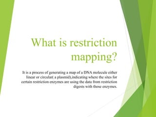
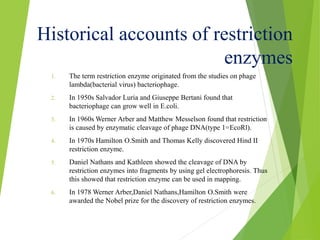
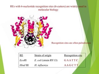
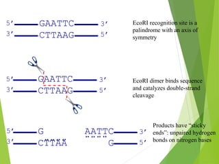
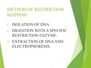
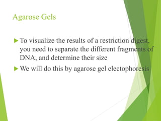
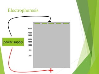
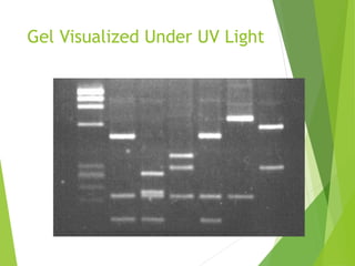
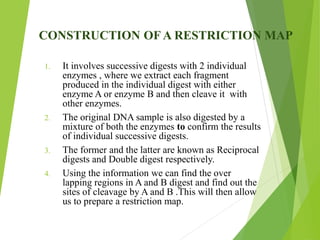
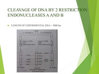
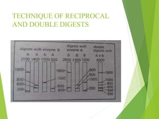
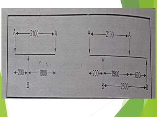
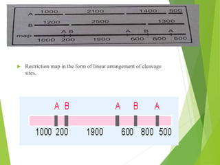
Restriction mapping
By: Minali Singh
What is restriction
mapping?
It is a process of generating a map of a DNA molecule either
linear or circular( a plasmid),indicating where the sites for
certain restriction enzymes are using the data from restriction
digests with those enzymes.
TECHNIQUES INVOLVED :
 USE OF RESTRICTION
ENDONUCLEASES
 GEL ELECTROPHORESIS
What are restriction endonucleases (REs)?
How can REs be used to identify DNA
molecules?
Historical accounts of restriction
enzymes
1. The term restriction enzyme originated from the studies on phage
lambda(bacterial virus) bacteriophage.
2. In 1950s Salvador Luria and Giuseppe Bertani found that
bacteriophage can grow well in E.coli.
3. In 1960s Werner Arber and Matthew Messelson found that restriction
is caused by enzymatic cleavage of phage DNA(type 1=EcoRl).
4. In 1970s Hamilton O.Smith and Thomas Kelly discovered Hind II
restriction enzyme.
5. Daniel Nathans and Kathleen showed the cleavage of DNA by
restriction enzymes into fragments by using gel electrophoresis. Thus
this showed that restriction enzyme can be used in mapping.
6. In 1978 Werner Arber,Daniel Nathans,Hamilton O.Smith were
awarded the Nobel prize for the discovery of restriction enzymes.
REs with 6-nucleotide recognition sites (6-cutters) are widely used in
molecular biology
RE Strain of origin Recognition site
EcoRI E. coli (strain RY13) G AA T T C
Hind III H. influenza AA G C T T
Recognition sites are often palindromes
GAATTC
CTTAAG
3’
5’
5’
3’
GAATTC
CTTAAG
3’
5’
5’
3’
G AATTC
CTTAA G
3’
5’
5’
3’
EcoRI recognition site is a
palindrome with an axis of
symmetry
EcoRI dimer binds sequence
and catalyzes double-strand
cleavage
Products have “sticky
ends”: unpaired hydrogen
bonds on nitrogen bases
METHOD OF RESTRICTION
MAPPING
1. ISOLATION OF DNA
2. DIGESTION WITH A SPECIFIC
RESTRICTION ENZYME.
3. EXTRACTION OF DNAAND
ELECTROPHORESIS.
Agarose Gels
 To visualize the results of a restriction digest,
you need to separate the different fragments of
DNA, and determine their size
 We will do this by agarose gel electophoresis
Agarose
 Agarose is very water soluble polysaccharide
 Forms porous, aqueous gels after heating and
cooling
Electrophoresis
power supply
-
+
200
500
1000
1500
2000
3000
4000
6000
Gel Visualized Under UV Light
CONSTRUCTION OF A RESTRICTION MAP
1. It involves successive digests with 2 individual
enzymes , where we extract each fragment
produced in the individual digest with either
enzyme A or enzyme B and then cleave it with
other enzymes.
2. The original DNA sample is also digested by a
mixture of both the enzymes to confirm the results
of individual successive digests.
3. The former and the latter are known as Reciprocal
digests and Double digest respectively.
4. Using the information we can find the over
lapping regions in A and B digest and find out the
sites of cleavage by A and B .This will then allow
us to prepare a restriction map.
CLEAVAGE OF DNA BY 2 RESTRICTION
ENDONUCLEASES AAND B
 LENGTH OF EXPERIMENTAL DNA = 5000 bp
TECHNIQUE OF RECIPROCAL
AND DOUBLE DIGESTS
 Restriction map in the form of linear arrangement of cleavage
sites.
SIGNIFICANCE
1. In molecular biology restriction maps are used as a
reference to engineer plasmid or other relatively short
pieces of DNA.
2. It is used to sequence the whole molecule of DNA and
run it through computer program that will find the
recognition sites that are present for every restriction
enzyme known.
Restriction mapping

Restriction mapping

  • 1.
  • 2.
    What is restriction mapping? Itis a process of generating a map of a DNA molecule either linear or circular( a plasmid),indicating where the sites for certain restriction enzymes are using the data from restriction digests with those enzymes.
  • 3.
    TECHNIQUES INVOLVED : USE OF RESTRICTION ENDONUCLEASES  GEL ELECTROPHORESIS
  • 4.
    What are restrictionendonucleases (REs)? How can REs be used to identify DNA molecules?
  • 5.
    Historical accounts ofrestriction enzymes 1. The term restriction enzyme originated from the studies on phage lambda(bacterial virus) bacteriophage. 2. In 1950s Salvador Luria and Giuseppe Bertani found that bacteriophage can grow well in E.coli. 3. In 1960s Werner Arber and Matthew Messelson found that restriction is caused by enzymatic cleavage of phage DNA(type 1=EcoRl). 4. In 1970s Hamilton O.Smith and Thomas Kelly discovered Hind II restriction enzyme. 5. Daniel Nathans and Kathleen showed the cleavage of DNA by restriction enzymes into fragments by using gel electrophoresis. Thus this showed that restriction enzyme can be used in mapping. 6. In 1978 Werner Arber,Daniel Nathans,Hamilton O.Smith were awarded the Nobel prize for the discovery of restriction enzymes.
  • 6.
    REs with 6-nucleotiderecognition sites (6-cutters) are widely used in molecular biology RE Strain of origin Recognition site EcoRI E. coli (strain RY13) G AA T T C Hind III H. influenza AA G C T T Recognition sites are often palindromes
  • 7.
    GAATTC CTTAAG 3’ 5’ 5’ 3’ GAATTC CTTAAG 3’ 5’ 5’ 3’ G AATTC CTTAA G 3’ 5’ 5’ 3’ EcoRIrecognition site is a palindrome with an axis of symmetry EcoRI dimer binds sequence and catalyzes double-strand cleavage Products have “sticky ends”: unpaired hydrogen bonds on nitrogen bases
  • 8.
    METHOD OF RESTRICTION MAPPING 1.ISOLATION OF DNA 2. DIGESTION WITH A SPECIFIC RESTRICTION ENZYME. 3. EXTRACTION OF DNAAND ELECTROPHORESIS.
  • 9.
    Agarose Gels  Tovisualize the results of a restriction digest, you need to separate the different fragments of DNA, and determine their size  We will do this by agarose gel electophoresis
  • 10.
    Agarose  Agarose isvery water soluble polysaccharide  Forms porous, aqueous gels after heating and cooling
  • 11.
  • 12.
  • 13.
    CONSTRUCTION OF ARESTRICTION MAP 1. It involves successive digests with 2 individual enzymes , where we extract each fragment produced in the individual digest with either enzyme A or enzyme B and then cleave it with other enzymes. 2. The original DNA sample is also digested by a mixture of both the enzymes to confirm the results of individual successive digests. 3. The former and the latter are known as Reciprocal digests and Double digest respectively. 4. Using the information we can find the over lapping regions in A and B digest and find out the sites of cleavage by A and B .This will then allow us to prepare a restriction map.
  • 14.
    CLEAVAGE OF DNABY 2 RESTRICTION ENDONUCLEASES AAND B  LENGTH OF EXPERIMENTAL DNA = 5000 bp
  • 15.
  • 17.
     Restriction mapin the form of linear arrangement of cleavage sites.
  • 18.
    SIGNIFICANCE 1. In molecularbiology restriction maps are used as a reference to engineer plasmid or other relatively short pieces of DNA. 2. It is used to sequence the whole molecule of DNA and run it through computer program that will find the recognition sites that are present for every restriction enzyme known.