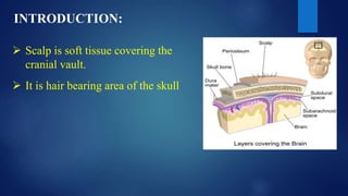
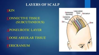
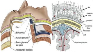
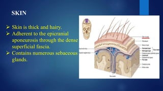
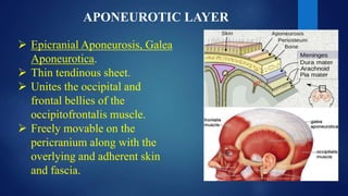
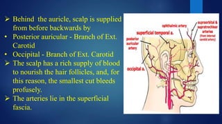
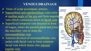
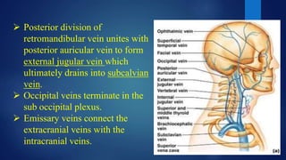
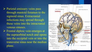
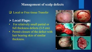
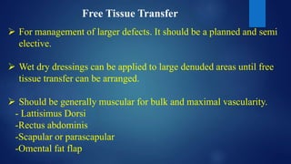
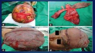
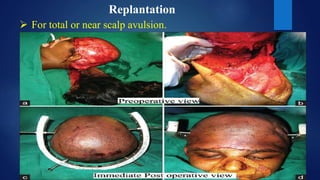
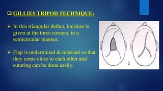
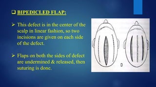
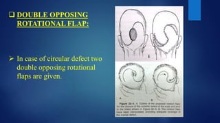
SCALP
- Dr. RATNA DEEPIKA SESHAGIRI
MDS PART I
CONTENTS :
Layers of scalp
Arterial supply
Venous drainage
Lymphatic drainage
Nerve supply
Clinical conditions related to
scalp
Treatment of scalp wound
Defects of scalp
Management of scalp defects
Scalp incisions
Various flaps of scalp
 Scalp is soft tissue covering the
cranial vault.
INTRODUCTION:
 It is hair bearing area of the skull
EXTENT:
 Anteriorly- Supraorbital
Margins
 Posteriorly- External
Occipital protuberance and
superior nuchal lines.
 Laterally-Superior temporal
lines.
LAYERS OF SCALP
SKIN
CONNECTIVE TISSUE
(SUBCUTANEOUS)
APONEUROTIC LAYER
LOOSE AREOLAR TISSUE
PERICRANIUM
SKIN
 Skin is thick and hairy.
 Adherent to the epicranial
aponeurosis through the dense
superficial fascia.
 Contains numerous sebaceous
glands.
CONNECTIVE TISSUE
 Fibrous and dense containing blood
vessels and nerves.
 It has the richest cutaneous blood
supply in the body.
 Binds skin to subjacent aponeurosis
 Wounds bleed profusely as blood
vessels are prevented from
retraction by fibrous tissue.
Bleeding is stopped by applying
pressure against the bone.
APONEUROTIC LAYER
 Epicranial Aponeurosis, Galea
Aponeurotica.
 Thin tendinous sheet.
 Unites the occipital and
frontal bellies of the
occipitofrontalis muscle.
 Freely movable on the
pericranium along with the
overlying and adherent skin
and fascia.
 Anteriorly, it receives
insertion of Frontalis and
posteriorly Occipitalis and is
attached posteriorly to
external occipital
protuberance and to highest
nuchal lines between the
occipital bellies.
 On each side aponeurosis is
attached to superior temporal
lines.
THE OCCIPITO-FRONTALIS MUSCLE
 Origin: It consists of four bellies,
two occipital and two frontal,
connected by an aponeurosis.
 The occipitalis are smaller and
separately arise from the superior
nuchal line on the occipital bone
and pass forward to be attached to
the aponeurosis.
 The frontal bellies are larger and
closer to each other in the middle
line.
 They arise from the skin and
superficial fascia of the eyebrow
and pass backward to be attached
to the aponeurosis.
FRONTALIS PART
Arises from the front of
aponenurosis.
Passes forward to become attached
to the upper part of the orbicularis
oculi and the overlying skin of the
eyebrow.
Right & left frontalis meet in the
midline. Midline fibres blend with
procerus.
Nerve supply- Supplied by the
Temporal branch (of facial nerve)
OCCIPITALIS PART
 Arises from superior nuchal line,
passes forward into aponeurosis.
 Bellies are separated in midline
by aponeurosis which extends
backwards to get attached to the
external occipital protuberance
and the most medial part of the
superior nuchal line.
 Supplied by the Posterior
auricular nerve (Facial nerve)
SUBAPONEUROTIC SPACE
 It is a potential space beneath the epicranial aponeurosis.
 It is limited in front and behind by the origins of the
occipitofrontalis muscle.
 It extends laterally as far as the attachment of the
aponeurosis to the temporal fascia.
 It is occupied by loose areolar tissue.
 It loosely connects the epicranial aponeurosis to the
pericranium. It contains a few small arteries and also some
important emissary veins.
LOOSE AREOLAR TISSUE
 Occupies the subaponeurotic space.
 Extends anteriorly into eyelids as
frontalis muscle has no bony
attachment & Posteriorly it attaches
to highest and superior nuchal lines
 Laterally to superior temporal lines.
 It loosely connects the epicranial
aponeurosis to the pericranium. It
contains a few small arteries and
also some important emissary
veins.
PERICRANIUM
 Loosely attached to surface of bones,but is firmly adherent to their
sutures where the sutural ligaments bind the pericranium to the
endocranium.
ARTERIAL SUPPLY
 In front of the auricle, scalp is
supplied from before backwards by
• Supratrochlear - Branch of
ophthalmic artery.
• Supraorbital- Branch of ophthalmic
artery.
• Superficial temporal arteries-Branch
of external carotid.
 Behind the auricle, scalp is supplied
from before backwards by
• Posterior auricular - Branch of Ext.
Carotid
• Occipital - Branch of Ext. Carotid
 The scalp has a rich supply of blood
to nourish the hair follicles, and, for
this reason, the smallest cut bleeds
profusely.
 The arteries lie in the superficial
fascia.
VENOUS DRAINAGE
 Veins of scalp accompany arteries.
 Supraorbital and supratrochlear veins unite
at median angle of the eye and form angular
vein which continuous down as facial vein.
 Superficial temporal vein descend in front
of tragus, enters the parotid gland and joins
the maxillary vein to form the
retromandibular vein.
 Anterior division of retromandibular vein
unites with facial vein to form common
facial vein which drains into internal
jugular vein.
 Posterior division of
retromandibular vein unites with
posterior auricular vein to form
external jugular vein which
ultimately drains into subcalvian
vein.
 Occipital veins terminate in the
sub occipital plexus.
 Emissary veins connect the
extracranial veins with the
intracranial veins.
 Parietal emissary veins pass
through mastoid foramen to the
sigmoid sinus. Extracranial
infections may spread through
these veins into the intracranial
venous sinuses.
 Frontal diploic vein emerges at
the supraorbital notch and opens
into the occipital vein or into the
transverse sinus near the median
plane.
LYMPHATIC DRAINAGE
 The anterior part of the
scalp drains in to the
preauricular or parotid
lymph nodes, situated on
the surface of the parotid
gland.
 The posterior part of the
scalp drains in to the
posterior auricular or
mastoid and occipital
lymph nodes.
NERVE SUPPLY OF SCALP
 The scalp and temple are
supplied by ten nerves on
each side.
 Out of these five nerves(4
sensory and 1 motor) enter
the scalp in front of the
auricle.
 The remaining five nerves
(4 sensory and 1 motor)
enter the scalp behind the
auricle.
 In front of the auricle-
• Sensory nerves:
 Supratrochlear- branch of frontal nerve ( Opthalmic
division of trigeminal nerve)
 Supraorbital- branch of frontal nerve (opthalmic
division of trigeminal nerve)
 Zygomaticotemporal- branch of Zygomatic nerve
(Maxillary division of trigeminal nerve)
 Auriculotemporal- branch of mandibular nerve
(division of trigeminal nerve)
• Motor nerves:
 temporal branch of facial nerve.
 Behind the auricle
• Sensory nerves:
 Posterior Division of great auricular
nerve– C2,C3 from cervical plexus.
 Lesser occipital nerve –C2 from
cervical plexus.
 Greater occipital nerve –C2, dorsal
ramus.
 Third occipital nerve- C3, dorsal
ramus.
• Motor nerve:
 Posterior auricular branch of facial
nerve.
CONDITIONS RELATED TO SCALP
 Because of the abundance of
sebaceous glands, scalp is
the common site for
sebaceous cysts.
 Main complication of
sebaceous cysts of this area
are infection and ulceration.
 Type of acquired retention
cyst.
Sebaceous cyst:
 Cause:-Obstruction to one of the
sebaceous ducts, resulting in
accumulation of sebaceous material.
 Site:-Scalp, face, back, scrotum. In scalp
& scrotum multiple cyst are found.
 Clinical Features:-
 Slow growing & appear in childhood
or middle age.
 Hemispherical or spherical swelling.
 located in dermis.
 Diagnostic feature of cyst is central
keratin filled punctum
 Punctum indicates blockage of the
duct.
 Smooth surface, round borders, soft &
putty in consistency, non tender.
 Treatment:-Incision and avulsion of
cyst with wall.
 Complications:-
 Infection:-Due to injury or scratch –causing abscess.
• The cyst will be tender, red & hot.
• Treatment- Incision & drainage.
 Sebaceous Horn:-Due to slow drying of contents which
are squeezed out, specially if a patient does not wash the
part.
 Cock’s Peculiar Tumor:-Refers to infected, ulcerated cyst
of scalp with pouting granulation tissue with everted edge
resembling epithelioma.
 Calcification:- Sebum becomes calcified & cyst appears to
be hard.
Dermoid cyst:
 Congenital type of cyst.
 Occurs in the line of embryonic
fusion of these processes.
 Contents are thick, tooth paste-
like i.e. mixture of sweat, sebum
& desquamated epithelial cells &
even hair.
 Clinical Features:-
 Manifests in childhood, becomes
obvious when patient is adult.
 Painless, slow growing swelling.
 Soft, cystic & fluctuant.
 Saucerization of the skull
bone with sclerotic margin
can be felt by tip of finger.
 So X-ray of the skull is
mandatory to detect bony
defect of the skull.
 Treatment:-Excision of the
cyst.
 Pott’s Puffy Tumour:-
Described by Sir Percival Pott in
1760.
 Serious complication of bacterial
frontal sinusitis.
 Subperiosteal abscess and
osteomyelitis of the frontal bone.
 Tender, soft tissue swelling that
causes pitting edema over the
frontal bone.
 Wounds of scalp do not gape
unless epicranial aponeurosis
is divided transversely.
 Because the pericranium is
adherent to sutures, collections
of fluid deep to the
pericranium(Cephalhematoma)
take the shape of the bone
concerned. This is mostly seen
during childbirth.
 Layers of loose areolar tissue is known as dangerous area of scalp
because the emissary veins in this layer may transmit infection from
scalp to cranial venous sinuses.
 Subgaleal layer is
continuous with loose
areolar layer deep to the
orbicularis oculi muscle.
As this layer extends
anteriorly into the eyelids,
blood collects below the
layer & may extend
anteriorly into the eyelids.
This condition is called as
Black Eye or Racoons
Sign.
 Due to the rich supply of scalp,
avulsed portions can be replaced
in position and sutured and
usually take up well and heal.
 Wounds of the scalp bleed
profusely because the vessels are
prevented from retracting by
fibrous fascia. Bleeding can be
arrested by pressing against
bone.
Treatment Of Scalp Wound
1) Preliminary Examination:-
• Extent & Severity of Wound.
• Systemic antibiotics are given depending upon the
contamination of wound.
• X ray Examination.
• Neurologic examination.
• Fractures should be reduced & stabilized.
2) Shaving:-
shaved for a distance of about 5 to 7 cm around wound.
Involved eyebrows should be saved & used as landmark.
3) Debridement:-
under 2% lignocaine solution around the wound with copious
irrigation.
4) Arrest Of Hemorrhage:-
Lack of normal retraction of the vessel causes profuse bleeding.
Pressure should be applied against bone.
5) Scalp Suturing:-
• In case of small wounds-interrupted one layer suturing is
done.
• In case of large wounds-two layer suturing should be done.
6) Dressing
Defects of the scalp
The defects of the scalp can be:
1. Congenital
• Aplasia Cutis Congenita
• Craniopagus
• Premature craniosynostosis
2. Traumatic
• Lacerations
• Partial Scalp defects
• Full thickness scalp defects
• Total scalp Avulsion
Management of scalp defects
 Local or Free tissue Transfer
 Local Flaps:
• For relatively small partial or
full thickness defects (3-5 cm)
• Permit closure of the defect with
hair bearing skin of similar
thickness.
Free Tissue Transfer
 For management of larger defects. It should be a planned and semi
elective.
 Wet dry dressings can be applied to large denuded areas until free
tissue transfer can be arranged.
 Should be generally muscular for bulk and maximal vascularity.
- Lattisimus Dorsi
-Rectus abdominis
-Scapular or parascapular
-Omental fat flap
Replantation
 For total or near scalp avulsion.
SCALP INCISIONS
CORONALAPPROACH
 It is a extremely versatile approach and is routinely used in
craniofacial trauma.
 The incision gives access to the frontal lobe, anterior
cranial fossa, middle cranial fossa, Zygomatic arch,
infratemporal fossa, TMJ.
 It practically exposes a major part midface and when
combined with subciliary/ infraorbital incision, also the
complete orbit.
Advantages:
 Provides excellent access to these areas with
minimal complications.
 Most surgical scars is hidden within the hairline.
 Types of coronal incision-
• Bi-coronal incision- The incision extends from one
superior temporal line to the other.
• Hemi-coronal incision- The incision curves forward at the
midline ending just posterior to the hairline. Curving the
hemi-coronal incision anteriorly provides the relaxation
necessary for retraction of the flap.
• Extension of coronal incision at the level of lobe of the ear
as a preauricular incision
 Locating and marking the incision line
MARKING IN MALE MARKING IN FEMALE
Incision
 Drapes are secured with staples
or sutures
 The incision extends from one
superior temporal line to the
other to the depth of the
pericranium.
 The dissection is in the sub-
galeal plane which is the loose
connective tissue and cleaves
readily.
 Scissor dissection of
the scalp is done in
subgaleal plane
which prevents the
surgeon from
incising the
temporalis fascia
and the muscle
which bleeds freely.
 Finger dissection which
cleaves the areolar tissue
in the sub-galeal plane.
 Scalpel dissection is used
in separating adherent
sub-galeal layer.
 Amount of exposure obtained
with complete dissection of the
upper and middle facial bones
using the coronal approach.
 The infraorbital areas are also
exposed if retraction is
performed from the side of the
orbit.
Harvesting Cranial Bone Graft
 The advantage of coronal
approach is that cranial bone
graft harvesting is facilitated.
 An incision through the
periosteum allows exposure
for harvesting a bone graft.
 The grafts can be of two types:-
 Non vascularized cranial bone graft:-
-Average thickness of adult calvarial bone is 7mm.
-Parietal- Occipital region is nonvascularized cranial graft.
-By splitting of the calvaria, the outer cortex is used as the
graft and inner cortex remains intact to provide a protective
covering of the brain.
 For small areas of bone:-
A bur is used to make initial cuts through the outer cortex of the
calvaria. One side is beveled to allow insertion of curved osteotome
in plane parallel to the outer surface and at the diploic level.
Cross-section of dural striping
by curved elevator
Gigli saw is placed and the
duramater is removed
 For larger block grafts:-
 It may be safer to harvest the bone as
several strips, rather than a single
block.
 In this cases it is often easier to perform
a formal craniotomy, remove a full
thickness segment, and split the bone
on a back table with a mechanical saw
or osteotome.
 After separation, the inner cortex is
used to cover the donor area; the outer
cortex is used as a graft.
b) Vascularized calvarial graft:-
-Temporoparietal flap is used.
-After bicoronal incision is made, scalp skin is elevated in a
subfollicular plane above the superficial musculoaponeurotic
system(SMAS) and the galea to preserve superficial temporal artery.
The outer table of the parietal bone is
harvested with a curved osteotome
 The scalp elevation continues to expose the temporalis
muscle, temporal crest, coronal suture, and superficial
temporal artery.
 Process is same as it was in case of non- vascularised
graft, but after harvesting the graft, sutures should be
passed between the overlying galea, periosteum, and
calvaria are helpful in keeping the periosteum attached
to the bone and preserving blood supply.
 The scalp incision is closed in
two layers using 2-0 slowly
resorbing sutures through the
galea/subcutaneous tissues and
2-0 resorbable or permanent skin
sutures, or staples.
 Use of a suction drain (usually 7
mm flat) is optional.
 The skin sutures/staples are
removed in 7 to 10 days.
SUPRA-ORBITALAPPROACH
 Previously popular incision, to
gain access to the supralateral
orbital rim is the eyebrow
incision.
 Gives simple and rapid access to
the frontozygomatic area.
 ADVANTAGE
• The scar is usually imperceptible, if the incision is
made almost entirely within the confines of the
eyebrow.
 DISADVANTAGE
• Extremely limited access.
VARIOUS FLAPS OF SCALP
 Skin grafts will not take readily on exposed cortical
bone except children.
 Exposed cortical bone will die & sequestrate in time
as its blood supply is obtained through the
periosteum. It is advisable to perform the primary
closure on these complex defects with a flap cover
which may be done using:
• Advancement flap
• Transposition flap
• Rotational flap
 Advancement flaps:
 Advancement of the scalp is hindered by the inelastic nature
of galea aponeurotica. The galea may be therefore, be
released after wide undermining, combined with the scoring
of galea.
 This will usually allow sufficient relaxation for easy closure,
if this maneuver is inadequate relieving incisions through the
full thickness of the scalp may occasionally be made to allow
a bipedicle flap to advance over the affected area.
 Transposition flap:
 A transposition flap is the transfer
of tissue from one place to the
other, except that a secondary
defect will be left in the donar site.
 Transposition of the flaps may be
considered longer than they are
wide but is advisable always to
make these flaps as generous in
proportions as possible & based
proximally or on a known artery
&vein , the secondary defect will
need a skin graft for cover.
 Rotational flaps :
 It can be used to close large defects
which are particularly of triangular type.
 The smallest side of the triangle is
extended by incision , undermined & the
skin flap can be brought over the defect.
 A rotation flap should leave no secondary
defect. The circumference of the rotated
circle must be at least eight times the
size of the defect to be closed.
 GILLIES TRIPOD TECHNIQUE:
 In this triangular defect, incision is
given at the three corners, in a
semicircular manner.
 Flap is undermined & released so that
they come close to each other and
suturing can be done easily.
 BIPEDICLED FLAP:
 This defect is in the center of the
scalp in linear fashion, so two
incisions are given on each side
of the defect.
 Flaps on both the sides of defect
are undermined & released, then
suturing is done.
 DOUBLE OPPOSING
ROTATIONAL FLAP:
 In case of circular defect two
double opposing rotational
flaps are given.
REFERENCES
 Gray’s Anatomy : 35th edition R.WARWICK
&P.L.WILLIAM
 Grant,s Method Of Anatomy : Charles E. Slonecker
 Anatomy with colour atlas : Inderbir Singh
 Surgical approaches to the facial skeleton: Edward Ellis III
 Clinical Anatomy : Richard Snell
Scalp

Scalp

  • 1.
    SCALP - Dr. RATNADEEPIKA SESHAGIRI MDS PART I
  • 2.
    CONTENTS : Layers ofscalp Arterial supply Venous drainage Lymphatic drainage Nerve supply Clinical conditions related to scalp Treatment of scalp wound Defects of scalp Management of scalp defects Scalp incisions Various flaps of scalp
  • 3.
     Scalp issoft tissue covering the cranial vault. INTRODUCTION:  It is hair bearing area of the skull
  • 4.
    EXTENT:  Anteriorly- Supraorbital Margins Posteriorly- External Occipital protuberance and superior nuchal lines.  Laterally-Superior temporal lines.
  • 5.
    LAYERS OF SCALP SKIN CONNECTIVETISSUE (SUBCUTANEOUS) APONEUROTIC LAYER LOOSE AREOLAR TISSUE PERICRANIUM
  • 7.
    SKIN  Skin isthick and hairy.  Adherent to the epicranial aponeurosis through the dense superficial fascia.  Contains numerous sebaceous glands.
  • 8.
    CONNECTIVE TISSUE  Fibrousand dense containing blood vessels and nerves.  It has the richest cutaneous blood supply in the body.  Binds skin to subjacent aponeurosis  Wounds bleed profusely as blood vessels are prevented from retraction by fibrous tissue. Bleeding is stopped by applying pressure against the bone.
  • 9.
    APONEUROTIC LAYER  EpicranialAponeurosis, Galea Aponeurotica.  Thin tendinous sheet.  Unites the occipital and frontal bellies of the occipitofrontalis muscle.  Freely movable on the pericranium along with the overlying and adherent skin and fascia.
  • 10.
     Anteriorly, itreceives insertion of Frontalis and posteriorly Occipitalis and is attached posteriorly to external occipital protuberance and to highest nuchal lines between the occipital bellies.  On each side aponeurosis is attached to superior temporal lines.
  • 11.
    THE OCCIPITO-FRONTALIS MUSCLE Origin: It consists of four bellies, two occipital and two frontal, connected by an aponeurosis.  The occipitalis are smaller and separately arise from the superior nuchal line on the occipital bone and pass forward to be attached to the aponeurosis.  The frontal bellies are larger and closer to each other in the middle line.  They arise from the skin and superficial fascia of the eyebrow and pass backward to be attached to the aponeurosis.
  • 12.
    FRONTALIS PART Arises fromthe front of aponenurosis. Passes forward to become attached to the upper part of the orbicularis oculi and the overlying skin of the eyebrow. Right & left frontalis meet in the midline. Midline fibres blend with procerus. Nerve supply- Supplied by the Temporal branch (of facial nerve)
  • 13.
    OCCIPITALIS PART  Arisesfrom superior nuchal line, passes forward into aponeurosis.  Bellies are separated in midline by aponeurosis which extends backwards to get attached to the external occipital protuberance and the most medial part of the superior nuchal line.  Supplied by the Posterior auricular nerve (Facial nerve)
  • 14.
    SUBAPONEUROTIC SPACE  Itis a potential space beneath the epicranial aponeurosis.  It is limited in front and behind by the origins of the occipitofrontalis muscle.  It extends laterally as far as the attachment of the aponeurosis to the temporal fascia.  It is occupied by loose areolar tissue.  It loosely connects the epicranial aponeurosis to the pericranium. It contains a few small arteries and also some important emissary veins.
  • 15.
    LOOSE AREOLAR TISSUE Occupies the subaponeurotic space.  Extends anteriorly into eyelids as frontalis muscle has no bony attachment & Posteriorly it attaches to highest and superior nuchal lines  Laterally to superior temporal lines.  It loosely connects the epicranial aponeurosis to the pericranium. It contains a few small arteries and also some important emissary veins.
  • 16.
    PERICRANIUM  Loosely attachedto surface of bones,but is firmly adherent to their sutures where the sutural ligaments bind the pericranium to the endocranium.
  • 17.
    ARTERIAL SUPPLY  Infront of the auricle, scalp is supplied from before backwards by • Supratrochlear - Branch of ophthalmic artery. • Supraorbital- Branch of ophthalmic artery. • Superficial temporal arteries-Branch of external carotid.
  • 18.
     Behind theauricle, scalp is supplied from before backwards by • Posterior auricular - Branch of Ext. Carotid • Occipital - Branch of Ext. Carotid  The scalp has a rich supply of blood to nourish the hair follicles, and, for this reason, the smallest cut bleeds profusely.  The arteries lie in the superficial fascia.
  • 19.
    VENOUS DRAINAGE  Veinsof scalp accompany arteries.  Supraorbital and supratrochlear veins unite at median angle of the eye and form angular vein which continuous down as facial vein.  Superficial temporal vein descend in front of tragus, enters the parotid gland and joins the maxillary vein to form the retromandibular vein.  Anterior division of retromandibular vein unites with facial vein to form common facial vein which drains into internal jugular vein.
  • 20.
     Posterior divisionof retromandibular vein unites with posterior auricular vein to form external jugular vein which ultimately drains into subcalvian vein.  Occipital veins terminate in the sub occipital plexus.  Emissary veins connect the extracranial veins with the intracranial veins.
  • 21.
     Parietal emissaryveins pass through mastoid foramen to the sigmoid sinus. Extracranial infections may spread through these veins into the intracranial venous sinuses.  Frontal diploic vein emerges at the supraorbital notch and opens into the occipital vein or into the transverse sinus near the median plane.
  • 22.
    LYMPHATIC DRAINAGE  Theanterior part of the scalp drains in to the preauricular or parotid lymph nodes, situated on the surface of the parotid gland.  The posterior part of the scalp drains in to the posterior auricular or mastoid and occipital lymph nodes.
  • 23.
    NERVE SUPPLY OFSCALP  The scalp and temple are supplied by ten nerves on each side.  Out of these five nerves(4 sensory and 1 motor) enter the scalp in front of the auricle.  The remaining five nerves (4 sensory and 1 motor) enter the scalp behind the auricle.
  • 24.
     In frontof the auricle- • Sensory nerves:  Supratrochlear- branch of frontal nerve ( Opthalmic division of trigeminal nerve)  Supraorbital- branch of frontal nerve (opthalmic division of trigeminal nerve)  Zygomaticotemporal- branch of Zygomatic nerve (Maxillary division of trigeminal nerve)  Auriculotemporal- branch of mandibular nerve (division of trigeminal nerve) • Motor nerves:  temporal branch of facial nerve.
  • 26.
     Behind theauricle • Sensory nerves:  Posterior Division of great auricular nerve– C2,C3 from cervical plexus.  Lesser occipital nerve –C2 from cervical plexus.  Greater occipital nerve –C2, dorsal ramus.  Third occipital nerve- C3, dorsal ramus. • Motor nerve:  Posterior auricular branch of facial nerve.
  • 27.
    CONDITIONS RELATED TOSCALP  Because of the abundance of sebaceous glands, scalp is the common site for sebaceous cysts.  Main complication of sebaceous cysts of this area are infection and ulceration.  Type of acquired retention cyst. Sebaceous cyst:
  • 28.
     Cause:-Obstruction toone of the sebaceous ducts, resulting in accumulation of sebaceous material.  Site:-Scalp, face, back, scrotum. In scalp & scrotum multiple cyst are found.
  • 29.
     Clinical Features:- Slow growing & appear in childhood or middle age.  Hemispherical or spherical swelling.  located in dermis.  Diagnostic feature of cyst is central keratin filled punctum  Punctum indicates blockage of the duct.  Smooth surface, round borders, soft & putty in consistency, non tender.  Treatment:-Incision and avulsion of cyst with wall.
  • 30.
     Complications:-  Infection:-Dueto injury or scratch –causing abscess. • The cyst will be tender, red & hot. • Treatment- Incision & drainage.  Sebaceous Horn:-Due to slow drying of contents which are squeezed out, specially if a patient does not wash the part.  Cock’s Peculiar Tumor:-Refers to infected, ulcerated cyst of scalp with pouting granulation tissue with everted edge resembling epithelioma.  Calcification:- Sebum becomes calcified & cyst appears to be hard.
  • 31.
    Dermoid cyst:  Congenitaltype of cyst.  Occurs in the line of embryonic fusion of these processes.  Contents are thick, tooth paste- like i.e. mixture of sweat, sebum & desquamated epithelial cells & even hair.  Clinical Features:-  Manifests in childhood, becomes obvious when patient is adult.  Painless, slow growing swelling.  Soft, cystic & fluctuant.
  • 32.
     Saucerization ofthe skull bone with sclerotic margin can be felt by tip of finger.  So X-ray of the skull is mandatory to detect bony defect of the skull.  Treatment:-Excision of the cyst.
  • 33.
     Pott’s PuffyTumour:- Described by Sir Percival Pott in 1760.  Serious complication of bacterial frontal sinusitis.  Subperiosteal abscess and osteomyelitis of the frontal bone.  Tender, soft tissue swelling that causes pitting edema over the frontal bone.
  • 34.
     Wounds ofscalp do not gape unless epicranial aponeurosis is divided transversely.  Because the pericranium is adherent to sutures, collections of fluid deep to the pericranium(Cephalhematoma) take the shape of the bone concerned. This is mostly seen during childbirth.
  • 37.
     Layers ofloose areolar tissue is known as dangerous area of scalp because the emissary veins in this layer may transmit infection from scalp to cranial venous sinuses.
  • 38.
     Subgaleal layeris continuous with loose areolar layer deep to the orbicularis oculi muscle. As this layer extends anteriorly into the eyelids, blood collects below the layer & may extend anteriorly into the eyelids. This condition is called as Black Eye or Racoons Sign.
  • 39.
     Due tothe rich supply of scalp, avulsed portions can be replaced in position and sutured and usually take up well and heal.  Wounds of the scalp bleed profusely because the vessels are prevented from retracting by fibrous fascia. Bleeding can be arrested by pressing against bone.
  • 41.
    Treatment Of ScalpWound 1) Preliminary Examination:- • Extent & Severity of Wound. • Systemic antibiotics are given depending upon the contamination of wound. • X ray Examination. • Neurologic examination. • Fractures should be reduced & stabilized.
  • 42.
    2) Shaving:- shaved fora distance of about 5 to 7 cm around wound. Involved eyebrows should be saved & used as landmark. 3) Debridement:- under 2% lignocaine solution around the wound with copious irrigation. 4) Arrest Of Hemorrhage:- Lack of normal retraction of the vessel causes profuse bleeding. Pressure should be applied against bone.
  • 43.
    5) Scalp Suturing:- •In case of small wounds-interrupted one layer suturing is done. • In case of large wounds-two layer suturing should be done. 6) Dressing
  • 44.
    Defects of thescalp The defects of the scalp can be: 1. Congenital • Aplasia Cutis Congenita • Craniopagus • Premature craniosynostosis 2. Traumatic • Lacerations • Partial Scalp defects • Full thickness scalp defects • Total scalp Avulsion
  • 46.
    Management of scalpdefects  Local or Free tissue Transfer  Local Flaps: • For relatively small partial or full thickness defects (3-5 cm) • Permit closure of the defect with hair bearing skin of similar thickness.
  • 47.
    Free Tissue Transfer For management of larger defects. It should be a planned and semi elective.  Wet dry dressings can be applied to large denuded areas until free tissue transfer can be arranged.  Should be generally muscular for bulk and maximal vascularity. - Lattisimus Dorsi -Rectus abdominis -Scapular or parascapular -Omental fat flap
  • 49.
    Replantation  For totalor near scalp avulsion.
  • 50.
  • 51.
    CORONALAPPROACH  It isa extremely versatile approach and is routinely used in craniofacial trauma.  The incision gives access to the frontal lobe, anterior cranial fossa, middle cranial fossa, Zygomatic arch, infratemporal fossa, TMJ.  It practically exposes a major part midface and when combined with subciliary/ infraorbital incision, also the complete orbit.
  • 53.
    Advantages:  Provides excellentaccess to these areas with minimal complications.  Most surgical scars is hidden within the hairline.
  • 54.
     Types ofcoronal incision- • Bi-coronal incision- The incision extends from one superior temporal line to the other. • Hemi-coronal incision- The incision curves forward at the midline ending just posterior to the hairline. Curving the hemi-coronal incision anteriorly provides the relaxation necessary for retraction of the flap. • Extension of coronal incision at the level of lobe of the ear as a preauricular incision
  • 55.
     Locating andmarking the incision line MARKING IN MALE MARKING IN FEMALE
  • 56.
    Incision  Drapes aresecured with staples or sutures  The incision extends from one superior temporal line to the other to the depth of the pericranium.  The dissection is in the sub- galeal plane which is the loose connective tissue and cleaves readily.
  • 57.
     Scissor dissectionof the scalp is done in subgaleal plane which prevents the surgeon from incising the temporalis fascia and the muscle which bleeds freely.
  • 58.
     Finger dissectionwhich cleaves the areolar tissue in the sub-galeal plane.  Scalpel dissection is used in separating adherent sub-galeal layer.
  • 59.
     Amount ofexposure obtained with complete dissection of the upper and middle facial bones using the coronal approach.  The infraorbital areas are also exposed if retraction is performed from the side of the orbit.
  • 60.
    Harvesting Cranial BoneGraft  The advantage of coronal approach is that cranial bone graft harvesting is facilitated.  An incision through the periosteum allows exposure for harvesting a bone graft.
  • 61.
     The graftscan be of two types:-  Non vascularized cranial bone graft:- -Average thickness of adult calvarial bone is 7mm. -Parietal- Occipital region is nonvascularized cranial graft. -By splitting of the calvaria, the outer cortex is used as the graft and inner cortex remains intact to provide a protective covering of the brain.
  • 62.
     For smallareas of bone:- A bur is used to make initial cuts through the outer cortex of the calvaria. One side is beveled to allow insertion of curved osteotome in plane parallel to the outer surface and at the diploic level.
  • 63.
    Cross-section of duralstriping by curved elevator Gigli saw is placed and the duramater is removed
  • 64.
     For largerblock grafts:-  It may be safer to harvest the bone as several strips, rather than a single block.  In this cases it is often easier to perform a formal craniotomy, remove a full thickness segment, and split the bone on a back table with a mechanical saw or osteotome.  After separation, the inner cortex is used to cover the donor area; the outer cortex is used as a graft.
  • 65.
    b) Vascularized calvarialgraft:- -Temporoparietal flap is used. -After bicoronal incision is made, scalp skin is elevated in a subfollicular plane above the superficial musculoaponeurotic system(SMAS) and the galea to preserve superficial temporal artery. The outer table of the parietal bone is harvested with a curved osteotome
  • 66.
     The scalpelevation continues to expose the temporalis muscle, temporal crest, coronal suture, and superficial temporal artery.  Process is same as it was in case of non- vascularised graft, but after harvesting the graft, sutures should be passed between the overlying galea, periosteum, and calvaria are helpful in keeping the periosteum attached to the bone and preserving blood supply.
  • 67.
     The scalpincision is closed in two layers using 2-0 slowly resorbing sutures through the galea/subcutaneous tissues and 2-0 resorbable or permanent skin sutures, or staples.  Use of a suction drain (usually 7 mm flat) is optional.  The skin sutures/staples are removed in 7 to 10 days.
  • 68.
    SUPRA-ORBITALAPPROACH  Previously popularincision, to gain access to the supralateral orbital rim is the eyebrow incision.  Gives simple and rapid access to the frontozygomatic area.
  • 69.
     ADVANTAGE • Thescar is usually imperceptible, if the incision is made almost entirely within the confines of the eyebrow.  DISADVANTAGE • Extremely limited access.
  • 70.
    VARIOUS FLAPS OFSCALP  Skin grafts will not take readily on exposed cortical bone except children.  Exposed cortical bone will die & sequestrate in time as its blood supply is obtained through the periosteum. It is advisable to perform the primary closure on these complex defects with a flap cover which may be done using: • Advancement flap • Transposition flap • Rotational flap
  • 72.
     Advancement flaps: Advancement of the scalp is hindered by the inelastic nature of galea aponeurotica. The galea may be therefore, be released after wide undermining, combined with the scoring of galea.  This will usually allow sufficient relaxation for easy closure, if this maneuver is inadequate relieving incisions through the full thickness of the scalp may occasionally be made to allow a bipedicle flap to advance over the affected area.
  • 74.
     Transposition flap: A transposition flap is the transfer of tissue from one place to the other, except that a secondary defect will be left in the donar site.  Transposition of the flaps may be considered longer than they are wide but is advisable always to make these flaps as generous in proportions as possible & based proximally or on a known artery &vein , the secondary defect will need a skin graft for cover.
  • 76.
     Rotational flaps:  It can be used to close large defects which are particularly of triangular type.  The smallest side of the triangle is extended by incision , undermined & the skin flap can be brought over the defect.  A rotation flap should leave no secondary defect. The circumference of the rotated circle must be at least eight times the size of the defect to be closed.
  • 77.
     GILLIES TRIPODTECHNIQUE:  In this triangular defect, incision is given at the three corners, in a semicircular manner.  Flap is undermined & released so that they come close to each other and suturing can be done easily.
  • 78.
     BIPEDICLED FLAP: This defect is in the center of the scalp in linear fashion, so two incisions are given on each side of the defect.  Flaps on both the sides of defect are undermined & released, then suturing is done.
  • 79.
     DOUBLE OPPOSING ROTATIONALFLAP:  In case of circular defect two double opposing rotational flaps are given.
  • 80.
    REFERENCES  Gray’s Anatomy: 35th edition R.WARWICK &P.L.WILLIAM  Grant,s Method Of Anatomy : Charles E. Slonecker  Anatomy with colour atlas : Inderbir Singh  Surgical approaches to the facial skeleton: Edward Ellis III  Clinical Anatomy : Richard Snell