Downloaded 235 times


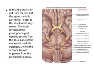
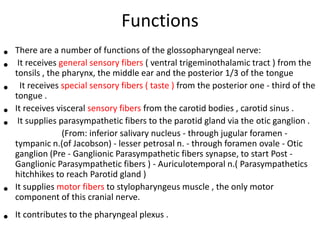
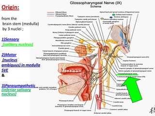
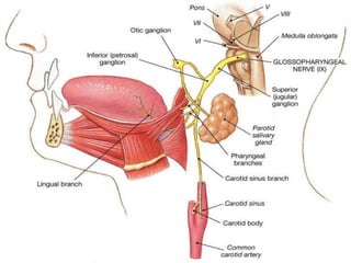
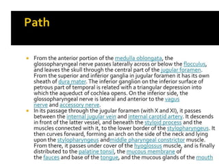
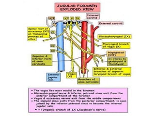
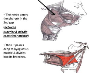
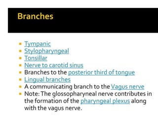
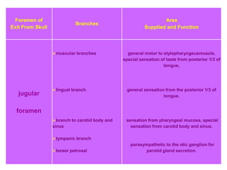


The glossopharyngeal nerve (CN IX) exits the brainstem and has several functions: - It provides general and special sensory innervation to the back third of the tongue (taste sensation), tonsils, middle ear, and pharynx. - It supplies a parasympathetic branch that stimulates saliva production in the parotid gland. - It provides motor innervation to the stylopharyngeus muscle, which elevates the pharynx during swallowing. The nerve exits the skull via the jugular foramen and branches to innervate its target areas.


