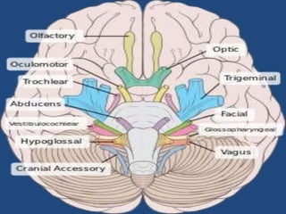
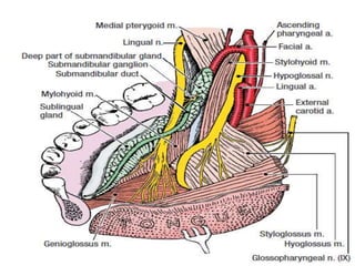
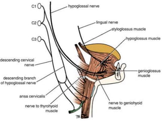
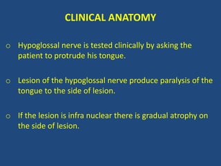
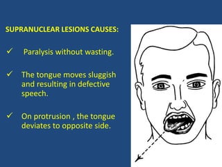
HYPOGLOSSAL NERVE
By
DR.M.MD.MUSTAFA SHARIFF
SENIOR LECTURER
SRMDCH-21
HYPOGLOSSAL NERVE
INTRODUCTION :
• It is twelveth cranial nerve.
• It arises from antrolateral sulcus of medulla oblongata.
• The nerve leaves the skull through hypoglossal canal.
• It supplies the muscles of the tongue.
FUNCTIONAL COMPONENTS:
GENERAL SOMATIC EFFERENT COLUMN:
• The fibers arise from the hypoglossal nucleus which lies
in the medulla.
• In the floor of the fourth ventricle deep to the
hypoglossal triangle.
GENERAL SOMATIC AFFERENT COLUMN:
 Spinal nucleus of cranial nerve 5th.
 where proprioceptive fibers from tongue end
HYPOGLOSSAL NUCLEUS
 It is 2cm long
 It lies in the floor of fourth ventricle beneath
the hypoglossal triangle.
 It is divided into two parts for individual
muscles innervated.
HYPOGLOSSAL NUCLEUS
• Connection of the nucleus with opposite pyramidal
tract forms supranuclear pathway of the nerve.
It is also connection to
 Cerebellum
 Reticular formation of medulla
 Sensory nuclei of 5th nerve
 Nucleus of tractus solitarius
INTRANEURAL COURSE
Fibers pass forwards
Lateral:
 Medial longitudinal bundle
 Medial lemniscus
 Pyramidal tract
Medial:
 Reticular formation
 Olivary nucleus
EXTRACRANIAL COURSE
• The nerve first lies
deep to internal
jugular vein and
descends between
internal jugular vein
and internal carotid
artery.
It is present to deep to the
 Parotid gland
 The styloid process
 The posterior belly
of the digastric and
 Stylohyoid muscle
 At the lower border of posterior belly of the digastric it
curves forwards and crosses internal and external
carotid artery and anterior to loop of lingual artery.
 The nerve then continues forwards on the hyoglossus.
 And Genioglossus muscles, deep to submandibular
gland and mylohyoid muscle and enters the substance
of the tongue to supply its muscles.
BRANCHES AND DISTRIBUTION
Muscular branches :
• It supply Intrinsic and Extrinsic muscles of the tongue .
• Except palatoglossus which is supplied by cranial root of
accessory nerve
• Other branches containing C1 nerve through 12th nerve.
• Meningeal branch
• Ansa cervicalis
• Branches to thyrohyoid and geniohyoid.
CLINICAL ANATOMY
o Hypoglossal nerve is tested clinically by asking the
patient to protrude his tongue.
o Lesion of the hypoglossal nerve produce paralysis of the
tongue to the side of lesion.
o If the lesion is infra nuclear there is gradual atrophy on
the side of lesion.
SUPRANUCLEAR LESIONS CAUSES:
 Paralysis without wasting.
 The tongue moves sluggish
and resulting in defective
speech.
 On protrusion , the tongue
deviates to opposite side.
REFERENCES
 EXAM-ORINTED ANATOMY SHOUKAT N.KAZI
 HEAD & NECK AK DATTA 5TH EDITION
 GRAYS ANATOMY STUDENTS EDITION
 CLINCAL ANATOMY SNELL 8TH EDITION
 CLINICAL ANATOMY MOORE 5TH EDITION
 HEAD & NECK BD CHAURASIA’S 4TH EDITION
 HEAD & NECK DR. AS.MONI
HYPOGLOSSAL NERVE

HYPOGLOSSAL NERVE

  • 1.
  • 2.
    HYPOGLOSSAL NERVE INTRODUCTION : •It is twelveth cranial nerve. • It arises from antrolateral sulcus of medulla oblongata. • The nerve leaves the skull through hypoglossal canal. • It supplies the muscles of the tongue.
  • 4.
    FUNCTIONAL COMPONENTS: GENERAL SOMATICEFFERENT COLUMN: • The fibers arise from the hypoglossal nucleus which lies in the medulla. • In the floor of the fourth ventricle deep to the hypoglossal triangle.
  • 5.
    GENERAL SOMATIC AFFERENTCOLUMN:  Spinal nucleus of cranial nerve 5th.  where proprioceptive fibers from tongue end
  • 6.
    HYPOGLOSSAL NUCLEUS  Itis 2cm long  It lies in the floor of fourth ventricle beneath the hypoglossal triangle.  It is divided into two parts for individual muscles innervated.
  • 8.
    HYPOGLOSSAL NUCLEUS • Connectionof the nucleus with opposite pyramidal tract forms supranuclear pathway of the nerve. It is also connection to  Cerebellum  Reticular formation of medulla  Sensory nuclei of 5th nerve  Nucleus of tractus solitarius
  • 9.
    INTRANEURAL COURSE Fibers passforwards Lateral:  Medial longitudinal bundle  Medial lemniscus  Pyramidal tract Medial:  Reticular formation  Olivary nucleus
  • 10.
    EXTRACRANIAL COURSE • Thenerve first lies deep to internal jugular vein and descends between internal jugular vein and internal carotid artery.
  • 11.
    It is presentto deep to the  Parotid gland  The styloid process  The posterior belly of the digastric and  Stylohyoid muscle
  • 12.
     At thelower border of posterior belly of the digastric it curves forwards and crosses internal and external carotid artery and anterior to loop of lingual artery.  The nerve then continues forwards on the hyoglossus.  And Genioglossus muscles, deep to submandibular gland and mylohyoid muscle and enters the substance of the tongue to supply its muscles.
  • 14.
    BRANCHES AND DISTRIBUTION Muscularbranches : • It supply Intrinsic and Extrinsic muscles of the tongue . • Except palatoglossus which is supplied by cranial root of accessory nerve • Other branches containing C1 nerve through 12th nerve. • Meningeal branch • Ansa cervicalis • Branches to thyrohyoid and geniohyoid.
  • 16.
    CLINICAL ANATOMY o Hypoglossalnerve is tested clinically by asking the patient to protrude his tongue. o Lesion of the hypoglossal nerve produce paralysis of the tongue to the side of lesion. o If the lesion is infra nuclear there is gradual atrophy on the side of lesion.
  • 17.
    SUPRANUCLEAR LESIONS CAUSES: Paralysis without wasting.  The tongue moves sluggish and resulting in defective speech.  On protrusion , the tongue deviates to opposite side.
  • 18.
    REFERENCES  EXAM-ORINTED ANATOMYSHOUKAT N.KAZI  HEAD & NECK AK DATTA 5TH EDITION  GRAYS ANATOMY STUDENTS EDITION  CLINCAL ANATOMY SNELL 8TH EDITION  CLINICAL ANATOMY MOORE 5TH EDITION  HEAD & NECK BD CHAURASIA’S 4TH EDITION  HEAD & NECK DR. AS.MONI