SlideShare a Scribd company logo
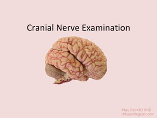
Cranial Nerve Examination Irfan Ziad MD UCD drkupe.blogspot.com
CN2 :Optic CN1 :Olfactory CN3 :Oculomotor CN4 :Trochlear CN5: Trigeminal CN6 :Abducens CN7 :Facial CN8: Vestibulocochlear CN9: Glossoparyngeal CN10: Vagus CN11: Accessory CN12: Hypoglossal midbrain pons medulla Anterior aspect of midbrain Dorsal aspect of midbrain
Inspection Position the patient sitting over the edge of the bed Look for : Scars (eg. craniotomy), pupil equality, facial asymmetry, ptosis, proptosis, neurofibromas Hi, my name is _____. Can I examine you?
CN I CN 1 Olfactory Nerve
Have you ever noticed any change in sense and smell? You ask the patient  “ Have you ever noticed any change in sense and smell?” If the answer is NO, proceed to the next CN Smell CN I CN 1 Olfactory Nerve
If the answer is YES,  Test: occlude one nostril, close eyes, identify smell (mint, coffee) Anosmia-  loss of the sense of smell (eg. flu, nasal polyps) Lesion- nose, cribiform plate of the eythmoid bone, base of skull- eg meningioma, early sign of parkinson. Smell CN I Can you tell me what smell is this? CN 1 Olfactory Nerve
CN II Optic canal CN 2 Optic Nerve
Ask patient do they have any difficulty with their vision. “ Can you see the clock on the wall?” “ Can you read the newspaper?” Ask the pt whether she’s myopic (nearsighted) or hyperopic(farsighted)  Visual Acuity Can you see the clock on the wall? CN II CN 2 Optic Nerve
Snellen chart is hold at arm-length A portable Snellen’s chart will enable you to perform a more formal test A patient who is having visual problems should be asked to count fingers held up in front of each eye in turn, and if this is not possible then perception of hand movement should be assessed. Failing this, light perception only may be present Visual Acuity CN II Test acuity with her glasses on.  Pinhole if she forgets her glasses CN 2 Optic Nerve
Confrontation test : ask the pt to look into your eyes while you place our index finger just outside the outer limits of your temporal fields. Move the fingers in turn and then together. “ Point to the moving finger” In visual inattention (parietal lobe lesions) the patient will only point to one finger when you move both simultaneously. Point to the moving finger Visual Field CN II CN 2 Optic Nerve
Test her peripheral field on each eye separately. “ Can you see the whole of my face?” Can you see the whole of my face? Visual Field CN II CN 2 Optic Nerve
Keep looking at my nose, tell me when you see my finger moves Visual Field Test her left temporal vision against your right temporal vision by moving your wagging finger from the periphery towards the centre “ Tell me when you see my finger moves” The temporal field should be tested in the horizontal plane and in the upper and lower temporal quadrants. Change hands and repeat on the nasal side CN II CN 2 Optic Nerve
Bitemporal hemianopia :  causes: optic chiasm lesion, pituitary tumour, craniopharyngioma Right optic nerve lesion Left Homonymous hemianopia Left Superior Quadrantanopia  Left Inferior Quadrantanopia  Left homonymous scotoma 2a 2b 2b 1 2 3 4 2+2a 5 6 7 2a+2b Binasal hemianopia : Very rare Left Homonymous hemianopia with macular sparing Visual Field Level of lesions CN II CN 2 Optic Nerve
Arcuate scotoma- moderate glaucoma Unilateral defect found with arterial occlusion, branch retinal vein thrombosis and inferior retinal detachment Central scotoma- macular degeneration or macular oedema Lesions at the level of the retina These affect one eye only Visual Field CN II CN 2 Optic Nerve
Tell the pt to look at the tip of your nose. Move the red-headed pin from the temporal periphery through the central field to the nasal periphery, asking the patient “ tell me when the red pin disappears, and reappears.” “ tell me when the red pin disappears and reappears.” The blind spot enlarges with papilloedema e.g. raised intracranial pressure with brain tumour. Demyelination of the optic nerve in multiple sclerosis can cause loss of central vision Blind Spot CN II CN 2 Optic Nerve
This is affected in colour blindness and optic neuritis (loss red colour first). CN II Colour Vision Test is done with an Ishihara plate CN 2 Optic Nerve
Fundoscopy Turn on, set diopters to zero, focus on specific distance, look for red reflex, adjust if pt wear glass, look for blood vessels, follow, look at optic disc- clear or blurred? - Hypertensive, diabetic, papillodema, optic neuropathy, pigmentation (mithocondrial disorder, retinotitis pigmentosa) CN II CN 2 Optic Nerve
Extraocular movements CN III, IV, VI From oculomotor nucleus CN 3,4.6 Oculomotor, Trochlear, Abducens Nerve
Pupillary light reflex Direct and Consensual :  Put your hand in between the patient’s eyes. With a pocket torch shine the light from the side.  Do a swinging-light test. Normally, the pupil into which the light is shone constricts rapidly (Direct light reflex) Simultaneously the other pupil constricts in the same way, (Consensual light reflex) Repeat this procedure on the other side RAPD  (Relative Afferent Pupillary Defect) previous optic neuritis)- swinging light test- damaged nerve dilate in response to light Causes: eg previous optic neuritis Oculomotor Nerve CN III CN 3
Accommodation reflex “ Look at that mark on the wall, now look at my finger” “ Look at that mark on the wall, now look at my finger” Examine the pupils for size, shape, equality and regularity PERRLA :  Pupils Equal, Round, Reactive to Light and Accommodation Pathology Unilateral dilated pupil - drugs- cocaine, eye drops (mydriatic) - 3rd nerve palsy- any associated ptosis, strabismus - Holes-Adie pupil- pupil reacts sluggishly, associated with syphilis -Absent light reflex with an intact accommodation reflex occurs in Argyll Robertson pupil in neurosyphilis Oculomotor Nerve CN III CN 3
Assess for eye movement, diplopia [double vision] and nystagmus Ask the patient to look laterally left and right, continue moving the finger to complete H pattern. Tell the patient to inform you if they see double images [diplopia] Diplopia is an early sign of ocular muscle weakness Without moving your head, follow the pin with your eye. Tell me if you see double Extraocular movements CN 3,4.6 Oculomotor, Trochlear, Abducens Nerve
Extraocular movements SR SR IR IR IO MR LR LR SO LR – Lateral Rectus MR – Medial Rectus SR- Superior Rectus IR- Inferior Rectus IO- Inferior Oblique SO- Superior Oblique CN IV supplies SO CN VI supplies LR CN III supplies all others + levator palpebrae superioris (which elevates the superior eyelids) CN 3,4.6 Oculomotor, Trochlear, Abducens Nerve
Complete ptosis Eye down and out Dilated pupil which is not responsive to light and accommodation. Double vision going down stairs or reading books Ask patient to turn the eye in and then to look down- may cause vertical hypertropia (pic)  Failure of lateral movement Nystagmus.  3 rd  nerve palsy 4 th  nerve palsy 6 th  nerve palsy Extraocular movements Nystagmus The direction of nystagmus is defined as that of the fast [correcting] movement Vestibular lesion – nystagmus away from the side of the lesion Cerebellar lesion – nystagmus to the side of the lesion Internuclear ophthalmoplegia  Abducting eye has greater nystagmus than the adducting eye. Problems btw nuclear, 3rd n 6th connected by medial longitudinal fasciculus (MLF) - MS CN 3,4.6 Oculomotor, Trochlear, Abducens Nerve
Trigeminal Nerve CN V Sensory branch Ophthalmic (V1) Maxillary (V2) Mandibular (V3) Motor Muscle of mastication Masseter Temporalis Medial pterygoid Lateral pterygoid Tensor veli palatini mylohyoid Anterior belly of digastric Tensor tympani Others All involved in biting, chewing, swallowing except for tensor tympani which acts to dampen sound produced from chewing Masseter Temporalis Trigeminal Nerve CN 5
Test for soft touch using cotton wool - sternum first, close eyes in the 3 divisions of the nerve V1- ophthalmic-  forehead up to the top  of the head V2- maxillary V3- mandibular  (up to angle of the jaw) The patient should be instructed to say “yes” each time the touch of the cotton wool is felt. Do not stroke the skin touch it. Test for pain using sharp object. Ask patient does it feel sharp or dull  Causes of sensory problems - MS- MS plaque in the brainstem in young people - Sjogren- dry eyes, dry mouth -Trigeminal neuralgia- older people Facial Sensory Say “yes” if you feel this Trigeminal Nerve CN V CN 5
Ask the pt to look up and away, touch the corneal. Reflex blinking of both eyes is a normal response. Pathology Bell’s palsy- unable to blink due to damage to the efferent limb (CNVII) CNV forms the afferent limb Corneal Reflex I’m going to gently touch your eye with a cotton bud. Trigeminal Nerve CN V CN 5
Inspect for wasting of the temporal and masseter muscles Ask patient to clench their teeth and palpate for contraction of the temporal and masseter muscles Motor Can you grit your teeth, please? Trigeminal Nerve CN V CN 5
Ask patient to open their mouth and hold it open while the examiner attempts to force it shut [pterygoid muscles].  A unilateral weakness of the motor division causes the jaw to deviate towards the weak side.If weakness is suspected patients should be asked to move the jaw laterally against resistance. The jaw can be moved towards the affected muscle but cannot move towards the normal side. Motor Open up your mouth and hold it for me Trigeminal Nerve CN V CN 5
Ask the pt to open her mouth fully, and close halfway, , place index finger on her chin and tap with a patella hammer, if jaw jerk is highly exaggerated.  Help to distinguish btw pseudobulbar palsy (UMN lesion of lower cranial nerve 9, 10,11,12) and a bulbar palsy (LMN lesion of lower cranial nerve 9,10,11,12) The Jaw Jerk I’m going to gently tap your jaw Trigeminal Nerve CN V CN 5
CN VII Facial canal (tortuous course) Internal auditory meatus Geniculate ganglion Stylomastoid foramen Temporal Zygomatic Buccal Mandibular Cervical Major facial branches Inside Skull Outside skull Other Posterior auricular nerve Posterior belly of Digastric Stylohyoid muscle Stapedius Frontalis, orbicularis oculi Z1: Eye & around orbit Z2: Mid face & smile Buccinator, upper lip Lower lip, orbicularis oris Platisma controls scalp muscles around the ear 3.  SENSORY 2.  PARASYMPATHETIC Greater petrosal to Lacrimal gland, sphenoid sinus, frontal sinus, frontal sinus, maxillary sinus, eithmoid sinus, nasal cavity,  The facial nerve has four components: 1.  BRANCHIAL MOTOR 4.  TASTE From facial nerve nucleus Small contribution to external acoustic meatus Palate via greater petrosal Ant 2/3 tongue via chorda tympani  From Nevus Intermedius Petrous temporal bone CN 7 Facial Nerve
Ask the patient to shut the eyes tightly Observe and try to force open each eye. If a lower motor neuron lesion is detected [weakness on one side of face], check for ear and palatal vesicles of herpes zoster of the geniculate ganglion – the Ramsay Hunt syndrome Motor Shut your eyes tightly and don’t let me open them CN VII CN 7 Facial Nerve
Ask patient to look up and wrinkle her forehead. Feel for muscle strength by pushing down on forehead. This movement is preserved on the side of an upper motor neurone lesion [a lesion which occurs above the level of the brainstem nucleus], because of bilateral supranuclear innervation giving some compensation to the upper face which is not the case in LMN lesion (Bells palsy/Ramsay Hunt- Herpes Zoster) The remaining muscles of facial expression are usually affected on the side of an UMN lesion. In a LMN lesion all muscles of facial expression are affected on the side of the lesion. Motor Wrinkle your forehead for me please CN VII CN 7 Facial Nerve
Ask the patient to show their teeth Compare the nasolabial grooves which are smooth on the weak side. Left upper motor neuron seventh nerve lesion leads to drooping of the corner of the mouth, flattened nasolabial fold, and sparing of the forehead on the left side** Motor Show me your teeth CN VII CN 7 Facial Nerve
Ask the patient blow out her cheeks Motor Blow out your cheeks CN VII CN 7 Facial Nerve
CN VII CN 7 Facial Nerve Upper Motor Neurone Lower Motor Neurone Pathway Rt motor cortex-corona-radiata-internal capsule-brainstem (midbrain-pons-medula)-crosses- anterior horn cell-   Anterior horn cell,  intervertebral foramen, lumbar sacral(lower limb)/brachial plexus(upperl imb plexus, runs in peripheral  nerve- stop at NMJ Presentation increased tone, reflex, clonus, upgoing(extensor) plantar - wasting, fasciculation, lose of tone, reflex, flexor plantar ,[object Object],[object Object],[object Object],[object Object],[object Object],[object Object],[object Object],[object Object],[object Object],[object Object],[object Object]
CN VIII Cochlear division- Hearing From organ to Corti in cochlea Hair cells to cell bodies in spiral ganglion (in modiolus) To 2 cochlear nuclei (ventral & dorsal) Vestibular division – Balance From semicircular canals, utricle & saccule Cell bodies in vestibular ganglion in outer part of internal acoustic meatus To 4 vestibular nuclei (medial, lateral, superior, inferior) CN 8 Vestibulo-Cochlear Nerve
Any problem with hearing? Hearing aids? Mask- cover the tragus of the ear and whisper a number, ask pt to repeat If deafness is suspected perform Rinne’s test and Weber’s test Hearing+Balance I’m going to whisper a number. I want you to repeat it. CN VIII CN 8 Vestibulo-Cochlear Nerve
Rinne’s Test Rinne- base of tuning fork on the mastoid process,  “ tell me when it stops”,  then bring it to the ear,  “ Can hear it? “ With nerve deafness the note is audible at the external meatus, as air and bone conduction are reduced equally, so that air conduction is better as is normal. This is termed Rinne-positive. With conduction [middle ear] deafness no note is audible at the external meatus. This is termed Rinne-negative. Can you hear it? CN VIII CN 8 Vestibulo-Cochlear Nerve
Weber’s Test A vibrating tuning fork is placed on the centre of the forehead. Normally the sound is heard in the centre of the forehead. With nerve deafness the sound is transmitted to the normal ear. With conduction deafness the sound is heard louder in the abnormal ear. Patients with defective hearing should be referred for audiometry. This measures the degree of hearing loss at different sound frequencies. Can you hear it? CN VIII CN 8 Vestibulo-Cochlear Nerve
CN IX CN 9 Glossopharyngeal  Nerve
CN  X CN 10 Vagus Nerve
CN IX, X CN 9, 10 Glossopharyngeal and Vagus Nerve
Uvula + Gag Reflex ,[object Object],[object Object],[object Object],[object Object],[object Object],[object Object],[object Object],[object Object],[object Object],CN IX, X Open your mouth and say “ah” CN 9, 10 Glossopharyngeal and Vagus Nerve
CN XII Its nucleus receive Corticonuclear fibers from both cerebral hemispheres,  but the cells supplying the genioglossus muscle receives corticonuclear fibers only from the opposite cerebral hemisphere  It supplies 1. All the intrinsic muscles of the tongue 2. Styloglossus 3. Hyoglossus 4. Genioglossus 5.  Doesn’t supply Palatoglossus – Supplied by the vagus  Function is to control the movement of the tongue  In the upper part, the Hypoglossal nerve is supplied by the C 1  fibers   CN 12 Hypoglossal Nerve
CN XII Motor Nerve of Tongue Observe the tongue at rest- wasting? on one side? fasciculation? Stick out tongue straight- deviate to one side? Tongue deviate to the side of a lesion of CNXII Wiggle tongue side-to-side  - (coordination)altered in cerebellar disorder Wiggle your  tongue side-to-side CN 12 Hypoglossal Nerve
CN XI Cranial  Root Spinal Root Receives corticonuclear fibers from both cerebral hemispheres It joins the spinal root & leaves the skull through jugular foramen Then the roots separate again, cranial root joins the vagus Situated in the anterior grey column of the spinal cord  in the upper 5 cervical segments Nerve fibers emerge from the spinal cord & form a nerve trunk that ascends into the skull through the foramen magnum Spinal part joins the cranial part & pas through the jugular foramen Then they separate again Supply the muscles of: Soft palate ( Except tensor veli palatini ) Pharynx ( Except stylopharyngeus ) Larynx ( Except cricothyroid ) Supplies the  SCM muscle & trapezius muscle CN 11 Accessory Nerve
Trapezius Ask the patient to shrug their shoulders and feel the bulk of the trapezius muscles and attempt to push the shoulders down. CN XI Shrug your shoulder, push up against my hand CN 11 Accessory Nerve
Sternocleidomastoid Ask the patient to turn their head against resistance and feel the bulk of the sternomastoids. Feel for the sternomastoid on the side opposite to the turned head. There will be weakness on turning the head away from the side of a muscle whose strength is impaired. (Optional)Test neck flexors if suspect myasthenia gravis, MND-  “ put chin on chest, I’ll put my hand onto your forehead, push up against my hand” CN XI Turn your head against my hand CN 11 Accessory Nerve
Thank You

More Related Content

What's hot

Superficial reflexes
Superficial reflexesSuperficial reflexes
Superficial reflexes
Kurian Joseph
 
Higher mental function
Higher mental functionHigher mental function
Higher mental function
Pratap Tiwari
 
Muscle Power and Tone Examination
Muscle Power and Tone ExaminationMuscle Power and Tone Examination
Muscle Power and Tone Examination
meducationdotnet
 
Upper and lower motor neuron
Upper and lower motor neuronUpper and lower motor neuron
Upper and lower motor neuron
Muhammad Saim
 

What's hot (20)

Superficial reflexes
Superficial reflexesSuperficial reflexes
Superficial reflexes
 
Bell’s palsy
Bell’s palsyBell’s palsy
Bell’s palsy
 
Sensory Examination
Sensory ExaminationSensory Examination
Sensory Examination
 
Higher mental function
Higher mental functionHigher mental function
Higher mental function
 
Superficial and deep reflexes!
Superficial and deep reflexes!Superficial and deep reflexes!
Superficial and deep reflexes!
 
Cranial nerve assessment..Simple and Easy to perform for medics and Physiothe...
Cranial nerve assessment..Simple and Easy to perform for medics and Physiothe...Cranial nerve assessment..Simple and Easy to perform for medics and Physiothe...
Cranial nerve assessment..Simple and Easy to perform for medics and Physiothe...
 
Cranial Nerve Examination
Cranial Nerve ExaminationCranial Nerve Examination
Cranial Nerve Examination
 
Reflexes
ReflexesReflexes
Reflexes
 
Facial nerve examination . Cranial nerve 7
Facial nerve examination . Cranial nerve 7Facial nerve examination . Cranial nerve 7
Facial nerve examination . Cranial nerve 7
 
Motor and sensory examination, Examination of reflexes
Motor and sensory examination, Examination of reflexesMotor and sensory examination, Examination of reflexes
Motor and sensory examination, Examination of reflexes
 
Facial palsy
Facial palsyFacial palsy
Facial palsy
 
Muscle Power and Tone Examination
Muscle Power and Tone ExaminationMuscle Power and Tone Examination
Muscle Power and Tone Examination
 
Superfacial reflex
Superfacial reflex Superfacial reflex
Superfacial reflex
 
Neuro Proforma
Neuro ProformaNeuro Proforma
Neuro Proforma
 
Torticollis and its P.T. Management
Torticollis and its P.T. Management Torticollis and its P.T. Management
Torticollis and its P.T. Management
 
Facial Nerve Palsy
Facial Nerve PalsyFacial Nerve Palsy
Facial Nerve Palsy
 
Upper and lower motor neuron
Upper and lower motor neuronUpper and lower motor neuron
Upper and lower motor neuron
 
neurological examination ppt
neurological examination pptneurological examination ppt
neurological examination ppt
 
Cranial nerve examination
Cranial nerve examinationCranial nerve examination
Cranial nerve examination
 
Vertigo
VertigoVertigo
Vertigo
 

Viewers also liked (6)

Part 3 cranial nerve examination
Part 3 cranial nerve examinationPart 3 cranial nerve examination
Part 3 cranial nerve examination
 
Vagus nerve
Vagus nerveVagus nerve
Vagus nerve
 
VAGUS NERVE AND ITS DESORDERS AND TREATMENT
VAGUS NERVE AND ITS DESORDERS AND TREATMENTVAGUS NERVE AND ITS DESORDERS AND TREATMENT
VAGUS NERVE AND ITS DESORDERS AND TREATMENT
 
The Vagus Nerve
The Vagus NerveThe Vagus Nerve
The Vagus Nerve
 
3rd, 4th and 6th cranial nerve
3rd, 4th and 6th cranial nerve3rd, 4th and 6th cranial nerve
3rd, 4th and 6th cranial nerve
 
Oculomotor nerve
Oculomotor nerveOculomotor nerve
Oculomotor nerve
 

Similar to Cranial nerve examination

Cns Exam 3rd
Cns Exam 3rdCns Exam 3rd
Cns Exam 3rd
mycomic
 
cranial nerves
cranial nerves cranial nerves
cranial nerves
ayesexy
 

Similar to Cranial nerve examination (20)

Carinal nerve assessment.ppt
Carinal nerve assessment.pptCarinal nerve assessment.ppt
Carinal nerve assessment.ppt
 
CRANIAL NERVE EXAMINATION.pptx
CRANIAL NERVE EXAMINATION.pptxCRANIAL NERVE EXAMINATION.pptx
CRANIAL NERVE EXAMINATION.pptx
 
Cranial Nerve Assessment
Cranial Nerve AssessmentCranial Nerve Assessment
Cranial Nerve Assessment
 
Cns Exam 3rd
Cns Exam 3rdCns Exam 3rd
Cns Exam 3rd
 
CRANIAL NERVE EXAMINATION
CRANIAL NERVE EXAMINATIONCRANIAL NERVE EXAMINATION
CRANIAL NERVE EXAMINATION
 
cranial nerves and their examination ppt
cranial nerves and their examination pptcranial nerves and their examination ppt
cranial nerves and their examination ppt
 
cranial nerves
cranial nerves cranial nerves
cranial nerves
 
Assignment of clinical neurology. topic cranial nerves
Assignment of clinical neurology. topic cranial nervesAssignment of clinical neurology. topic cranial nerves
Assignment of clinical neurology. topic cranial nerves
 
eye assessment.docx
eye assessment.docxeye assessment.docx
eye assessment.docx
 
CN assessment2.ppt
CN assessment2.pptCN assessment2.ppt
CN assessment2.ppt
 
Pares craneales
Pares cranealesPares craneales
Pares craneales
 
Cranial Nerves.ppt
Cranial Nerves.pptCranial Nerves.ppt
Cranial Nerves.ppt
 
Exame Físico Neurológico - Revendo Tópicos Essenciais em Neurologia Clínica
Exame Físico Neurológico - Revendo Tópicos Essenciais em Neurologia ClínicaExame Físico Neurológico - Revendo Tópicos Essenciais em Neurologia Clínica
Exame Físico Neurológico - Revendo Tópicos Essenciais em Neurologia Clínica
 
Undergraduate downloads...How to elicit clinical neurological signs
Undergraduate downloads...How to elicit clinical neurological signsUndergraduate downloads...How to elicit clinical neurological signs
Undergraduate downloads...How to elicit clinical neurological signs
 
Cranial nerve examination
Cranial nerve examinationCranial nerve examination
Cranial nerve examination
 
Neurological examination
Neurological examinationNeurological examination
Neurological examination
 
Cranial nerves
Cranial nervesCranial nerves
Cranial nerves
 
Clinical assessment of trigeminal nerve.pdf
Clinical assessment of trigeminal nerve.pdfClinical assessment of trigeminal nerve.pdf
Clinical assessment of trigeminal nerve.pdf
 
Cranial nerves examination
Cranial nerves examinationCranial nerves examination
Cranial nerves examination
 
cranial nerve examination
cranial nerve examinationcranial nerve examination
cranial nerve examination
 

Recently uploaded

Industrial Training Report- AKTU Industrial Training Report
Industrial Training Report- AKTU Industrial Training ReportIndustrial Training Report- AKTU Industrial Training Report
Industrial Training Report- AKTU Industrial Training Report
Avinash Rai
 

Recently uploaded (20)

INU_CAPSTONEDESIGN_비밀번호486_업로드용 발표자료.pdf
INU_CAPSTONEDESIGN_비밀번호486_업로드용 발표자료.pdfINU_CAPSTONEDESIGN_비밀번호486_업로드용 발표자료.pdf
INU_CAPSTONEDESIGN_비밀번호486_업로드용 발표자료.pdf
 
Benefits and Challenges of Using Open Educational Resources
Benefits and Challenges of Using Open Educational ResourcesBenefits and Challenges of Using Open Educational Resources
Benefits and Challenges of Using Open Educational Resources
 
Industrial Training Report- AKTU Industrial Training Report
Industrial Training Report- AKTU Industrial Training ReportIndustrial Training Report- AKTU Industrial Training Report
Industrial Training Report- AKTU Industrial Training Report
 
Pragya Champions Chalice 2024 Prelims & Finals Q/A set, General Quiz
Pragya Champions Chalice 2024 Prelims & Finals Q/A set, General QuizPragya Champions Chalice 2024 Prelims & Finals Q/A set, General Quiz
Pragya Champions Chalice 2024 Prelims & Finals Q/A set, General Quiz
 
PART A. Introduction to Costumer Service
PART A. Introduction to Costumer ServicePART A. Introduction to Costumer Service
PART A. Introduction to Costumer Service
 
How to Manage Notification Preferences in the Odoo 17
How to Manage Notification Preferences in the Odoo 17How to Manage Notification Preferences in the Odoo 17
How to Manage Notification Preferences in the Odoo 17
 
The Benefits and Challenges of Open Educational Resources
The Benefits and Challenges of Open Educational ResourcesThe Benefits and Challenges of Open Educational Resources
The Benefits and Challenges of Open Educational Resources
 
aaaaaaaaaaaaaaaaaaaaaaaaaaaaaaaaaaaaaaaaaaaaaaaaaaaaaaa
aaaaaaaaaaaaaaaaaaaaaaaaaaaaaaaaaaaaaaaaaaaaaaaaaaaaaaaaaaaaaaaaaaaaaaaaaaaaaaaaaaaaaaaaaaaaaaaaaaaaaaaaaaaaaa
aaaaaaaaaaaaaaaaaaaaaaaaaaaaaaaaaaaaaaaaaaaaaaaaaaaaaaa
 
Operations Management - Book1.p - Dr. Abdulfatah A. Salem
Operations Management - Book1.p  - Dr. Abdulfatah A. SalemOperations Management - Book1.p  - Dr. Abdulfatah A. Salem
Operations Management - Book1.p - Dr. Abdulfatah A. Salem
 
Mattingly "AI & Prompt Design: Limitations and Solutions with LLMs"
Mattingly "AI & Prompt Design: Limitations and Solutions with LLMs"Mattingly "AI & Prompt Design: Limitations and Solutions with LLMs"
Mattingly "AI & Prompt Design: Limitations and Solutions with LLMs"
 
Basic Civil Engg Notes_Chapter-6_Environment Pollution & Engineering
Basic Civil Engg Notes_Chapter-6_Environment Pollution & EngineeringBasic Civil Engg Notes_Chapter-6_Environment Pollution & Engineering
Basic Civil Engg Notes_Chapter-6_Environment Pollution & Engineering
 
Basic_QTL_Marker-assisted_Selection_Sourabh.ppt
Basic_QTL_Marker-assisted_Selection_Sourabh.pptBasic_QTL_Marker-assisted_Selection_Sourabh.ppt
Basic_QTL_Marker-assisted_Selection_Sourabh.ppt
 
Danh sách HSG Bộ môn cấp trường - Cấp THPT.pdf
Danh sách HSG Bộ môn cấp trường - Cấp THPT.pdfDanh sách HSG Bộ môn cấp trường - Cấp THPT.pdf
Danh sách HSG Bộ môn cấp trường - Cấp THPT.pdf
 
Open Educational Resources Primer PowerPoint
Open Educational Resources Primer PowerPointOpen Educational Resources Primer PowerPoint
Open Educational Resources Primer PowerPoint
 
size separation d pharm 1st year pharmaceutics
size separation d pharm 1st year pharmaceuticssize separation d pharm 1st year pharmaceutics
size separation d pharm 1st year pharmaceutics
 
Basic phrases for greeting and assisting costumers
Basic phrases for greeting and assisting costumersBasic phrases for greeting and assisting costumers
Basic phrases for greeting and assisting costumers
 
[GDSC YCCE] Build with AI Online Presentation
[GDSC YCCE] Build with AI Online Presentation[GDSC YCCE] Build with AI Online Presentation
[GDSC YCCE] Build with AI Online Presentation
 
Phrasal Verbs.XXXXXXXXXXXXXXXXXXXXXXXXXX
Phrasal Verbs.XXXXXXXXXXXXXXXXXXXXXXXXXXPhrasal Verbs.XXXXXXXXXXXXXXXXXXXXXXXXXX
Phrasal Verbs.XXXXXXXXXXXXXXXXXXXXXXXXXX
 
How to Break the cycle of negative Thoughts
How to Break the cycle of negative ThoughtsHow to Break the cycle of negative Thoughts
How to Break the cycle of negative Thoughts
 
Telling Your Story_ Simple Steps to Build Your Nonprofit's Brand Webinar.pdf
Telling Your Story_ Simple Steps to Build Your Nonprofit's Brand Webinar.pdfTelling Your Story_ Simple Steps to Build Your Nonprofit's Brand Webinar.pdf
Telling Your Story_ Simple Steps to Build Your Nonprofit's Brand Webinar.pdf
 

Cranial nerve examination

  • 1. Cranial Nerve Examination Irfan Ziad MD UCD drkupe.blogspot.com
  • 2. CN2 :Optic CN1 :Olfactory CN3 :Oculomotor CN4 :Trochlear CN5: Trigeminal CN6 :Abducens CN7 :Facial CN8: Vestibulocochlear CN9: Glossoparyngeal CN10: Vagus CN11: Accessory CN12: Hypoglossal midbrain pons medulla Anterior aspect of midbrain Dorsal aspect of midbrain
  • 3. Inspection Position the patient sitting over the edge of the bed Look for : Scars (eg. craniotomy), pupil equality, facial asymmetry, ptosis, proptosis, neurofibromas Hi, my name is _____. Can I examine you?
  • 4. CN I CN 1 Olfactory Nerve
  • 5. Have you ever noticed any change in sense and smell? You ask the patient “ Have you ever noticed any change in sense and smell?” If the answer is NO, proceed to the next CN Smell CN I CN 1 Olfactory Nerve
  • 6. If the answer is YES, Test: occlude one nostril, close eyes, identify smell (mint, coffee) Anosmia- loss of the sense of smell (eg. flu, nasal polyps) Lesion- nose, cribiform plate of the eythmoid bone, base of skull- eg meningioma, early sign of parkinson. Smell CN I Can you tell me what smell is this? CN 1 Olfactory Nerve
  • 7. CN II Optic canal CN 2 Optic Nerve
  • 8. Ask patient do they have any difficulty with their vision. “ Can you see the clock on the wall?” “ Can you read the newspaper?” Ask the pt whether she’s myopic (nearsighted) or hyperopic(farsighted) Visual Acuity Can you see the clock on the wall? CN II CN 2 Optic Nerve
  • 9. Snellen chart is hold at arm-length A portable Snellen’s chart will enable you to perform a more formal test A patient who is having visual problems should be asked to count fingers held up in front of each eye in turn, and if this is not possible then perception of hand movement should be assessed. Failing this, light perception only may be present Visual Acuity CN II Test acuity with her glasses on. Pinhole if she forgets her glasses CN 2 Optic Nerve
  • 10. Confrontation test : ask the pt to look into your eyes while you place our index finger just outside the outer limits of your temporal fields. Move the fingers in turn and then together. “ Point to the moving finger” In visual inattention (parietal lobe lesions) the patient will only point to one finger when you move both simultaneously. Point to the moving finger Visual Field CN II CN 2 Optic Nerve
  • 11. Test her peripheral field on each eye separately. “ Can you see the whole of my face?” Can you see the whole of my face? Visual Field CN II CN 2 Optic Nerve
  • 12. Keep looking at my nose, tell me when you see my finger moves Visual Field Test her left temporal vision against your right temporal vision by moving your wagging finger from the periphery towards the centre “ Tell me when you see my finger moves” The temporal field should be tested in the horizontal plane and in the upper and lower temporal quadrants. Change hands and repeat on the nasal side CN II CN 2 Optic Nerve
  • 13. Bitemporal hemianopia : causes: optic chiasm lesion, pituitary tumour, craniopharyngioma Right optic nerve lesion Left Homonymous hemianopia Left Superior Quadrantanopia Left Inferior Quadrantanopia Left homonymous scotoma 2a 2b 2b 1 2 3 4 2+2a 5 6 7 2a+2b Binasal hemianopia : Very rare Left Homonymous hemianopia with macular sparing Visual Field Level of lesions CN II CN 2 Optic Nerve
  • 14. Arcuate scotoma- moderate glaucoma Unilateral defect found with arterial occlusion, branch retinal vein thrombosis and inferior retinal detachment Central scotoma- macular degeneration or macular oedema Lesions at the level of the retina These affect one eye only Visual Field CN II CN 2 Optic Nerve
  • 15. Tell the pt to look at the tip of your nose. Move the red-headed pin from the temporal periphery through the central field to the nasal periphery, asking the patient “ tell me when the red pin disappears, and reappears.” “ tell me when the red pin disappears and reappears.” The blind spot enlarges with papilloedema e.g. raised intracranial pressure with brain tumour. Demyelination of the optic nerve in multiple sclerosis can cause loss of central vision Blind Spot CN II CN 2 Optic Nerve
  • 16. This is affected in colour blindness and optic neuritis (loss red colour first). CN II Colour Vision Test is done with an Ishihara plate CN 2 Optic Nerve
  • 17. Fundoscopy Turn on, set diopters to zero, focus on specific distance, look for red reflex, adjust if pt wear glass, look for blood vessels, follow, look at optic disc- clear or blurred? - Hypertensive, diabetic, papillodema, optic neuropathy, pigmentation (mithocondrial disorder, retinotitis pigmentosa) CN II CN 2 Optic Nerve
  • 18. Extraocular movements CN III, IV, VI From oculomotor nucleus CN 3,4.6 Oculomotor, Trochlear, Abducens Nerve
  • 19. Pupillary light reflex Direct and Consensual : Put your hand in between the patient’s eyes. With a pocket torch shine the light from the side. Do a swinging-light test. Normally, the pupil into which the light is shone constricts rapidly (Direct light reflex) Simultaneously the other pupil constricts in the same way, (Consensual light reflex) Repeat this procedure on the other side RAPD (Relative Afferent Pupillary Defect) previous optic neuritis)- swinging light test- damaged nerve dilate in response to light Causes: eg previous optic neuritis Oculomotor Nerve CN III CN 3
  • 20. Accommodation reflex “ Look at that mark on the wall, now look at my finger” “ Look at that mark on the wall, now look at my finger” Examine the pupils for size, shape, equality and regularity PERRLA : Pupils Equal, Round, Reactive to Light and Accommodation Pathology Unilateral dilated pupil - drugs- cocaine, eye drops (mydriatic) - 3rd nerve palsy- any associated ptosis, strabismus - Holes-Adie pupil- pupil reacts sluggishly, associated with syphilis -Absent light reflex with an intact accommodation reflex occurs in Argyll Robertson pupil in neurosyphilis Oculomotor Nerve CN III CN 3
  • 21. Assess for eye movement, diplopia [double vision] and nystagmus Ask the patient to look laterally left and right, continue moving the finger to complete H pattern. Tell the patient to inform you if they see double images [diplopia] Diplopia is an early sign of ocular muscle weakness Without moving your head, follow the pin with your eye. Tell me if you see double Extraocular movements CN 3,4.6 Oculomotor, Trochlear, Abducens Nerve
  • 22. Extraocular movements SR SR IR IR IO MR LR LR SO LR – Lateral Rectus MR – Medial Rectus SR- Superior Rectus IR- Inferior Rectus IO- Inferior Oblique SO- Superior Oblique CN IV supplies SO CN VI supplies LR CN III supplies all others + levator palpebrae superioris (which elevates the superior eyelids) CN 3,4.6 Oculomotor, Trochlear, Abducens Nerve
  • 23. Complete ptosis Eye down and out Dilated pupil which is not responsive to light and accommodation. Double vision going down stairs or reading books Ask patient to turn the eye in and then to look down- may cause vertical hypertropia (pic) Failure of lateral movement Nystagmus. 3 rd nerve palsy 4 th nerve palsy 6 th nerve palsy Extraocular movements Nystagmus The direction of nystagmus is defined as that of the fast [correcting] movement Vestibular lesion – nystagmus away from the side of the lesion Cerebellar lesion – nystagmus to the side of the lesion Internuclear ophthalmoplegia Abducting eye has greater nystagmus than the adducting eye. Problems btw nuclear, 3rd n 6th connected by medial longitudinal fasciculus (MLF) - MS CN 3,4.6 Oculomotor, Trochlear, Abducens Nerve
  • 24. Trigeminal Nerve CN V Sensory branch Ophthalmic (V1) Maxillary (V2) Mandibular (V3) Motor Muscle of mastication Masseter Temporalis Medial pterygoid Lateral pterygoid Tensor veli palatini mylohyoid Anterior belly of digastric Tensor tympani Others All involved in biting, chewing, swallowing except for tensor tympani which acts to dampen sound produced from chewing Masseter Temporalis Trigeminal Nerve CN 5
  • 25. Test for soft touch using cotton wool - sternum first, close eyes in the 3 divisions of the nerve V1- ophthalmic- forehead up to the top  of the head V2- maxillary V3- mandibular (up to angle of the jaw) The patient should be instructed to say “yes” each time the touch of the cotton wool is felt. Do not stroke the skin touch it. Test for pain using sharp object. Ask patient does it feel sharp or dull Causes of sensory problems - MS- MS plaque in the brainstem in young people - Sjogren- dry eyes, dry mouth -Trigeminal neuralgia- older people Facial Sensory Say “yes” if you feel this Trigeminal Nerve CN V CN 5
  • 26. Ask the pt to look up and away, touch the corneal. Reflex blinking of both eyes is a normal response. Pathology Bell’s palsy- unable to blink due to damage to the efferent limb (CNVII) CNV forms the afferent limb Corneal Reflex I’m going to gently touch your eye with a cotton bud. Trigeminal Nerve CN V CN 5
  • 27. Inspect for wasting of the temporal and masseter muscles Ask patient to clench their teeth and palpate for contraction of the temporal and masseter muscles Motor Can you grit your teeth, please? Trigeminal Nerve CN V CN 5
  • 28. Ask patient to open their mouth and hold it open while the examiner attempts to force it shut [pterygoid muscles]. A unilateral weakness of the motor division causes the jaw to deviate towards the weak side.If weakness is suspected patients should be asked to move the jaw laterally against resistance. The jaw can be moved towards the affected muscle but cannot move towards the normal side. Motor Open up your mouth and hold it for me Trigeminal Nerve CN V CN 5
  • 29. Ask the pt to open her mouth fully, and close halfway, , place index finger on her chin and tap with a patella hammer, if jaw jerk is highly exaggerated. Help to distinguish btw pseudobulbar palsy (UMN lesion of lower cranial nerve 9, 10,11,12) and a bulbar palsy (LMN lesion of lower cranial nerve 9,10,11,12) The Jaw Jerk I’m going to gently tap your jaw Trigeminal Nerve CN V CN 5
  • 30. CN VII Facial canal (tortuous course) Internal auditory meatus Geniculate ganglion Stylomastoid foramen Temporal Zygomatic Buccal Mandibular Cervical Major facial branches Inside Skull Outside skull Other Posterior auricular nerve Posterior belly of Digastric Stylohyoid muscle Stapedius Frontalis, orbicularis oculi Z1: Eye & around orbit Z2: Mid face & smile Buccinator, upper lip Lower lip, orbicularis oris Platisma controls scalp muscles around the ear 3. SENSORY 2. PARASYMPATHETIC Greater petrosal to Lacrimal gland, sphenoid sinus, frontal sinus, frontal sinus, maxillary sinus, eithmoid sinus, nasal cavity, The facial nerve has four components: 1. BRANCHIAL MOTOR 4. TASTE From facial nerve nucleus Small contribution to external acoustic meatus Palate via greater petrosal Ant 2/3 tongue via chorda tympani From Nevus Intermedius Petrous temporal bone CN 7 Facial Nerve
  • 31. Ask the patient to shut the eyes tightly Observe and try to force open each eye. If a lower motor neuron lesion is detected [weakness on one side of face], check for ear and palatal vesicles of herpes zoster of the geniculate ganglion – the Ramsay Hunt syndrome Motor Shut your eyes tightly and don’t let me open them CN VII CN 7 Facial Nerve
  • 32. Ask patient to look up and wrinkle her forehead. Feel for muscle strength by pushing down on forehead. This movement is preserved on the side of an upper motor neurone lesion [a lesion which occurs above the level of the brainstem nucleus], because of bilateral supranuclear innervation giving some compensation to the upper face which is not the case in LMN lesion (Bells palsy/Ramsay Hunt- Herpes Zoster) The remaining muscles of facial expression are usually affected on the side of an UMN lesion. In a LMN lesion all muscles of facial expression are affected on the side of the lesion. Motor Wrinkle your forehead for me please CN VII CN 7 Facial Nerve
  • 33. Ask the patient to show their teeth Compare the nasolabial grooves which are smooth on the weak side. Left upper motor neuron seventh nerve lesion leads to drooping of the corner of the mouth, flattened nasolabial fold, and sparing of the forehead on the left side** Motor Show me your teeth CN VII CN 7 Facial Nerve
  • 34. Ask the patient blow out her cheeks Motor Blow out your cheeks CN VII CN 7 Facial Nerve
  • 35.
  • 36. CN VIII Cochlear division- Hearing From organ to Corti in cochlea Hair cells to cell bodies in spiral ganglion (in modiolus) To 2 cochlear nuclei (ventral & dorsal) Vestibular division – Balance From semicircular canals, utricle & saccule Cell bodies in vestibular ganglion in outer part of internal acoustic meatus To 4 vestibular nuclei (medial, lateral, superior, inferior) CN 8 Vestibulo-Cochlear Nerve
  • 37. Any problem with hearing? Hearing aids? Mask- cover the tragus of the ear and whisper a number, ask pt to repeat If deafness is suspected perform Rinne’s test and Weber’s test Hearing+Balance I’m going to whisper a number. I want you to repeat it. CN VIII CN 8 Vestibulo-Cochlear Nerve
  • 38. Rinne’s Test Rinne- base of tuning fork on the mastoid process, “ tell me when it stops”, then bring it to the ear, “ Can hear it? “ With nerve deafness the note is audible at the external meatus, as air and bone conduction are reduced equally, so that air conduction is better as is normal. This is termed Rinne-positive. With conduction [middle ear] deafness no note is audible at the external meatus. This is termed Rinne-negative. Can you hear it? CN VIII CN 8 Vestibulo-Cochlear Nerve
  • 39. Weber’s Test A vibrating tuning fork is placed on the centre of the forehead. Normally the sound is heard in the centre of the forehead. With nerve deafness the sound is transmitted to the normal ear. With conduction deafness the sound is heard louder in the abnormal ear. Patients with defective hearing should be referred for audiometry. This measures the degree of hearing loss at different sound frequencies. Can you hear it? CN VIII CN 8 Vestibulo-Cochlear Nerve
  • 40. CN IX CN 9 Glossopharyngeal Nerve
  • 41. CN X CN 10 Vagus Nerve
  • 42. CN IX, X CN 9, 10 Glossopharyngeal and Vagus Nerve
  • 43.
  • 44. CN XII Its nucleus receive Corticonuclear fibers from both cerebral hemispheres,  but the cells supplying the genioglossus muscle receives corticonuclear fibers only from the opposite cerebral hemisphere  It supplies 1. All the intrinsic muscles of the tongue 2. Styloglossus 3. Hyoglossus 4. Genioglossus 5.  Doesn’t supply Palatoglossus – Supplied by the vagus Function is to control the movement of the tongue  In the upper part, the Hypoglossal nerve is supplied by the C 1  fibers CN 12 Hypoglossal Nerve
  • 45. CN XII Motor Nerve of Tongue Observe the tongue at rest- wasting? on one side? fasciculation? Stick out tongue straight- deviate to one side? Tongue deviate to the side of a lesion of CNXII Wiggle tongue side-to-side  - (coordination)altered in cerebellar disorder Wiggle your tongue side-to-side CN 12 Hypoglossal Nerve
  • 46. CN XI Cranial Root Spinal Root Receives corticonuclear fibers from both cerebral hemispheres It joins the spinal root & leaves the skull through jugular foramen Then the roots separate again, cranial root joins the vagus Situated in the anterior grey column of the spinal cord  in the upper 5 cervical segments Nerve fibers emerge from the spinal cord & form a nerve trunk that ascends into the skull through the foramen magnum Spinal part joins the cranial part & pas through the jugular foramen Then they separate again Supply the muscles of: Soft palate ( Except tensor veli palatini ) Pharynx ( Except stylopharyngeus ) Larynx ( Except cricothyroid ) Supplies the  SCM muscle & trapezius muscle CN 11 Accessory Nerve
  • 47. Trapezius Ask the patient to shrug their shoulders and feel the bulk of the trapezius muscles and attempt to push the shoulders down. CN XI Shrug your shoulder, push up against my hand CN 11 Accessory Nerve
  • 48. Sternocleidomastoid Ask the patient to turn their head against resistance and feel the bulk of the sternomastoids. Feel for the sternomastoid on the side opposite to the turned head. There will be weakness on turning the head away from the side of a muscle whose strength is impaired. (Optional)Test neck flexors if suspect myasthenia gravis, MND- “ put chin on chest, I’ll put my hand onto your forehead, push up against my hand” CN XI Turn your head against my hand CN 11 Accessory Nerve