Cranial Nerve Assessment is a crucial step in neurological assessment. By following the simple theoretical aspects it can be made on your fingertips....here is an try to make the stuff easier for you....
Provides information concerning gravity, rotation and acceleration
Serves as a reference for the somatosensory & visual systems
Contributes to integration of arousal, conscious awareness of the body via connections with vestibular cortex, thalamus and reticular formation
Provides information concerning gravity, rotation and acceleration
Serves as a reference for the somatosensory & visual systems
Contributes to integration of arousal, conscious awareness of the body via connections with vestibular cortex, thalamus and reticular formation
Mental function examination is a part of Neurologic and Psychiatric examination as an emergency and as an outpatient clinic.
Detail Mental examination is required for cases of Dementia in various neurological diseases.
This set of slides are not for Psychiatric patients with disturbance of thought and mood.
Bell’s palsy
Trigeminal Neuralgia ( Tic Douloreux)
Cranial & spinal neuropathies
Bell’s palsy (facial paralysis) is due to unilateral inflammation of the ( CN VII Facial nerve) seventh cranial nerve, which results in weakness or paralysis of the facial muscles on the affected side.
Cranial Nerve is a very important topics in field of ophthalmology and optometry. a stuent with knowledge with cranial nerves can easily understand muscle palsy and diagnosis of neuroohthalmology
Flu Vaccine Alert in Bangalore Karnatakaaddon Scans
As flu season approaches, health officials in Bangalore, Karnataka, are urging residents to get their flu vaccinations. The seasonal flu, while common, can lead to severe health complications, particularly for vulnerable populations such as young children, the elderly, and those with underlying health conditions.
Dr. Vidisha Kumari, a leading epidemiologist in Bangalore, emphasizes the importance of getting vaccinated. "The flu vaccine is our best defense against the influenza virus. It not only protects individuals but also helps prevent the spread of the virus in our communities," he says.
This year, the flu season is expected to coincide with a potential increase in other respiratory illnesses. The Karnataka Health Department has launched an awareness campaign highlighting the significance of flu vaccinations. They have set up multiple vaccination centers across Bangalore, making it convenient for residents to receive their shots.
To encourage widespread vaccination, the government is also collaborating with local schools, workplaces, and community centers to facilitate vaccination drives. Special attention is being given to ensuring that the vaccine is accessible to all, including marginalized communities who may have limited access to healthcare.
Residents are reminded that the flu vaccine is safe and effective. Common side effects are mild and may include soreness at the injection site, mild fever, or muscle aches. These side effects are generally short-lived and far less severe than the flu itself.
Healthcare providers are also stressing the importance of continuing COVID-19 precautions. Wearing masks, practicing good hand hygiene, and maintaining social distancing are still crucial, especially in crowded places.
Protect yourself and your loved ones by getting vaccinated. Together, we can help keep Bangalore healthy and safe this flu season. For more information on vaccination centers and schedules, residents can visit the Karnataka Health Department’s official website or follow their social media pages.
Stay informed, stay safe, and get your flu shot today!
- Video recording of this lecture in English language: https://youtu.be/lK81BzxMqdo
- Video recording of this lecture in Arabic language: https://youtu.be/Ve4P0COk9OI
- Link to download the book free: https://nephrotube.blogspot.com/p/nephrotube-nephrology-books.html
- Link to NephroTube website: www.NephroTube.com
- Link to NephroTube social media accounts: https://nephrotube.blogspot.com/p/join-nephrotube-on-social-media.html
Prix Galien International 2024 Forum ProgramLevi Shapiro
June 20, 2024, Prix Galien International and Jerusalem Ethics Forum in ROME. Detailed agenda including panels:
- ADVANCES IN CARDIOLOGY: A NEW PARADIGM IS COMING
- WOMEN’S HEALTH: FERTILITY PRESERVATION
- WHAT’S NEW IN THE TREATMENT OF INFECTIOUS,
ONCOLOGICAL AND INFLAMMATORY SKIN DISEASES?
- ARTIFICIAL INTELLIGENCE AND ETHICS
- GENE THERAPY
- BEYOND BORDERS: GLOBAL INITIATIVES FOR DEMOCRATIZING LIFE SCIENCE TECHNOLOGIES AND PROMOTING ACCESS TO HEALTHCARE
- ETHICAL CHALLENGES IN LIFE SCIENCES
- Prix Galien International Awards Ceremony
ARTIFICIAL INTELLIGENCE IN HEALTHCARE.pdfAnujkumaranit
Artificial intelligence (AI) refers to the simulation of human intelligence processes by machines, especially computer systems. It encompasses tasks such as learning, reasoning, problem-solving, perception, and language understanding. AI technologies are revolutionizing various fields, from healthcare to finance, by enabling machines to perform tasks that typically require human intelligence.
The prostate is an exocrine gland of the male mammalian reproductive system
It is a walnut-sized gland that forms part of the male reproductive system and is located in front of the rectum and just below the urinary bladder
Function is to store and secrete a clear, slightly alkaline fluid that constitutes 10-30% of the volume of the seminal fluid that along with the spermatozoa, constitutes semen
A healthy human prostate measures (4cm-vertical, by 3cm-horizontal, 2cm ant-post ).
It surrounds the urethra just below the urinary bladder. It has anterior, median, posterior and two lateral lobes
It’s work is regulated by androgens which are responsible for male sex characteristics
Generalised disease of the prostate due to hormonal derangement which leads to non malignant enlargement of the gland (increase in the number of epithelial cells and stromal tissue)to cause compression of the urethra leading to symptoms (LUTS
Title: Sense of Smell
Presenter: Dr. Faiza, Assistant Professor of Physiology
Qualifications:
MBBS (Best Graduate, AIMC Lahore)
FCPS Physiology
ICMT, CHPE, DHPE (STMU)
MPH (GC University, Faisalabad)
MBA (Virtual University of Pakistan)
Learning Objectives:
Describe the primary categories of smells and the concept of odor blindness.
Explain the structure and location of the olfactory membrane and mucosa, including the types and roles of cells involved in olfaction.
Describe the pathway and mechanisms of olfactory signal transmission from the olfactory receptors to the brain.
Illustrate the biochemical cascade triggered by odorant binding to olfactory receptors, including the role of G-proteins and second messengers in generating an action potential.
Identify different types of olfactory disorders such as anosmia, hyposmia, hyperosmia, and dysosmia, including their potential causes.
Key Topics:
Olfactory Genes:
3% of the human genome accounts for olfactory genes.
400 genes for odorant receptors.
Olfactory Membrane:
Located in the superior part of the nasal cavity.
Medially: Folds downward along the superior septum.
Laterally: Folds over the superior turbinate and upper surface of the middle turbinate.
Total surface area: 5-10 square centimeters.
Olfactory Mucosa:
Olfactory Cells: Bipolar nerve cells derived from the CNS (100 million), with 4-25 olfactory cilia per cell.
Sustentacular Cells: Produce mucus and maintain ionic and molecular environment.
Basal Cells: Replace worn-out olfactory cells with an average lifespan of 1-2 months.
Bowman’s Gland: Secretes mucus.
Stimulation of Olfactory Cells:
Odorant dissolves in mucus and attaches to receptors on olfactory cilia.
Involves a cascade effect through G-proteins and second messengers, leading to depolarization and action potential generation in the olfactory nerve.
Quality of a Good Odorant:
Small (3-20 Carbon atoms), volatile, water-soluble, and lipid-soluble.
Facilitated by odorant-binding proteins in mucus.
Membrane Potential and Action Potential:
Resting membrane potential: -55mV.
Action potential frequency in the olfactory nerve increases with odorant strength.
Adaptation Towards the Sense of Smell:
Rapid adaptation within the first second, with further slow adaptation.
Psychological adaptation greater than receptor adaptation, involving feedback inhibition from the central nervous system.
Primary Sensations of Smell:
Camphoraceous, Musky, Floral, Pepperminty, Ethereal, Pungent, Putrid.
Odor Detection Threshold:
Examples: Hydrogen sulfide (0.0005 ppm), Methyl-mercaptan (0.002 ppm).
Some toxic substances are odorless at lethal concentrations.
Characteristics of Smell:
Odor blindness for single substances due to lack of appropriate receptor protein.
Behavioral and emotional influences of smell.
Transmission of Olfactory Signals:
From olfactory cells to glomeruli in the olfactory bulb, involving lateral inhibition.
Primitive, less old, and new olfactory systems with different path
These simplified slides by Dr. Sidra Arshad present an overview of the non-respiratory functions of the respiratory tract.
Learning objectives:
1. Enlist the non-respiratory functions of the respiratory tract
2. Briefly explain how these functions are carried out
3. Discuss the significance of dead space
4. Differentiate between minute ventilation and alveolar ventilation
5. Describe the cough and sneeze reflexes
Study Resources:
1. Chapter 39, Guyton and Hall Textbook of Medical Physiology, 14th edition
2. Chapter 34, Ganong’s Review of Medical Physiology, 26th edition
3. Chapter 17, Human Physiology by Lauralee Sherwood, 9th edition
4. Non-respiratory functions of the lungs https://academic.oup.com/bjaed/article/13/3/98/278874
NVBDCP.pptx Nation vector borne disease control programSapna Thakur
NVBDCP was launched in 2003-2004 . Vector-Borne Disease: Disease that results from an infection transmitted to humans and other animals by blood-feeding arthropods, such as mosquitoes, ticks, and fleas. Examples of vector-borne diseases include Dengue fever, West Nile Virus, Lyme disease, and malaria.
TEST BANK for Operations Management, 14th Edition by William J. Stevenson, Ve...kevinkariuki227
TEST BANK for Operations Management, 14th Edition by William J. Stevenson, Verified Chapters 1 - 19, Complete Newest Version.pdf
TEST BANK for Operations Management, 14th Edition by William J. Stevenson, Verified Chapters 1 - 19, Complete Newest Version.pdf
Report Back from SGO 2024: What’s the Latest in Cervical Cancer?bkling
Are you curious about what’s new in cervical cancer research or unsure what the findings mean? Join Dr. Emily Ko, a gynecologic oncologist at Penn Medicine, to learn about the latest updates from the Society of Gynecologic Oncology (SGO) 2024 Annual Meeting on Women’s Cancer. Dr. Ko will discuss what the research presented at the conference means for you and answer your questions about the new developments.
New Directions in Targeted Therapeutic Approaches for Older Adults With Mantl...i3 Health
i3 Health is pleased to make the speaker slides from this activity available for use as a non-accredited self-study or teaching resource.
This slide deck presented by Dr. Kami Maddocks, Professor-Clinical in the Division of Hematology and
Associate Division Director for Ambulatory Operations
The Ohio State University Comprehensive Cancer Center, will provide insight into new directions in targeted therapeutic approaches for older adults with mantle cell lymphoma.
STATEMENT OF NEED
Mantle cell lymphoma (MCL) is a rare, aggressive B-cell non-Hodgkin lymphoma (NHL) accounting for 5% to 7% of all lymphomas. Its prognosis ranges from indolent disease that does not require treatment for years to very aggressive disease, which is associated with poor survival (Silkenstedt et al, 2021). Typically, MCL is diagnosed at advanced stage and in older patients who cannot tolerate intensive therapy (NCCN, 2022). Although recent advances have slightly increased remission rates, recurrence and relapse remain very common, leading to a median overall survival between 3 and 6 years (LLS, 2021). Though there are several effective options, progress is still needed towards establishing an accepted frontline approach for MCL (Castellino et al, 2022). Treatment selection and management of MCL are complicated by the heterogeneity of prognosis, advanced age and comorbidities of patients, and lack of an established standard approach for treatment, making it vital that clinicians be familiar with the latest research and advances in this area. In this activity chaired by Michael Wang, MD, Professor in the Department of Lymphoma & Myeloma at MD Anderson Cancer Center, expert faculty will discuss prognostic factors informing treatment, the promising results of recent trials in new therapeutic approaches, and the implications of treatment resistance in therapeutic selection for MCL.
Target Audience
Hematology/oncology fellows, attending faculty, and other health care professionals involved in the treatment of patients with mantle cell lymphoma (MCL).
Learning Objectives
1.) Identify clinical and biological prognostic factors that can guide treatment decision making for older adults with MCL
2.) Evaluate emerging data on targeted therapeutic approaches for treatment-naive and relapsed/refractory MCL and their applicability to older adults
3.) Assess mechanisms of resistance to targeted therapies for MCL and their implications for treatment selection
micro teaching on communication m.sc nursing.pdfAnurag Sharma
Microteaching is a unique model of practice teaching. It is a viable instrument for the. desired change in the teaching behavior or the behavior potential which, in specified types of real. classroom situations, tends to facilitate the achievement of specified types of objectives.
Cranial nerve assessment..Simple and Easy to perform for medics and Physiotherapist
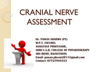
1. CRANIAL NERVE
ASSESSMENT
Dr. PAWAN SHARMA (PT)
M.P.T. (NEURO)
ASSISTANT PROFESSOR,
SHRI U.S.B. COLLEGE OF PHYSIOTHERAPY
ABU-ROAD, RAJASTAHAN
Email- pawan.physio2011@gmail.com
Contact- 07727989353
2. IV Trochlear
III Oculomotor
VII Facial
VI Abducens
V Trigeminal
CEREBRAL
HEMISPHERE
MIDBRAIN
PONS
MEDULLA
CRANIAL NERVES
II Optic
I Olfactory
VIII Vestibulo-
cochlear
XII Hypoglossal
XI Accessory
X Vagus
IX Glossopharyngeal
CRANIAL NERVES
2
3. CRANIAL NERVES
The 12 pairs of cranial nerves are part of the
peripheral nervous system.
The Roman numeral is based on descending
order of the cranial nerve's attachment to the
CNS.
As a rule, cranial nerves do not cross in the
brain.
Cranial nerves may be sensory, motor both
somatic or parasympathetic, or have mixed
function.
General Characteristics:
CRANIAL NERVES
3
4. REMEMBER ME…
SOME
SAYS
MONEY
MATTERS
BUT
MY
BROTHER
SAYS
BIG
BRAIN
MATTERS
MOST
S-SENSORY
M- MOTOR
B- BOTH
All in
sequence
CRANIAL NERVES
4
5. CN I - OLFACTORY
• ORIGIN: Cerebral hemisphere
• INNERVATION: Nasal mucous
membranes.
• FUNCTION: Sense of smell
• DYSFUNCTION: Anosmia
CLINICAL EVALUATION
• Use non-noxious aromatic
substances, i.e. coffee, lemon,
garlic, etc.
• Test each nostril separately.
• Mark if any abnormality noted
CRANIAL NERVES
5
6. CN II – OPTIC NERVE
• VISUAL ACUITY: Snellen
chart for distant vision,
Jaegers chart, newspaper or
fingers for near vision.
• VISUAL FIELDS:
Confrontation.
• FUNDI AND OPTIC DISCS:
Visualization of the termination
of the optic nerve by looking
through pupil with
ophthalmoscope.
CRANIAL NERVES 6
7. CN II – OPTIC NERVE(cont..)
Tested by-
1. Visual acuity
2. Color vision
3. Visual field
CRANIAL NERVES
7
Near field
Far field
Color
matching
Confrontation
test
8. CN II – OPTIC NERVE(cont..)
Visual acuity-
Snellen chart(Far vision)
◦ Chart is placed at 20 feet or 6
meter and patient is asked to
read it
◦ The formula is d/D
Where d is 6 meter and D is
the distance from which he can
read it clearly
Normal is 6/6 or 20/20
Jaegers chart(Near vision)
◦ Paragraphs are printed in
successive coarser type with
0 is finest and 7 is biggest
◦ Patient is asked to read
through the hole
CRANIAL NERVES
8
9. CN II – OPTIC NERVE(cont..)
Color vision-
◦ Checked by asking to
match different colors
• Day or night blindness
can be assessed
• Visual field-
Confrontation test
Peripheral visual fields-
Goldmann Perimeter
CRANIAL NERVES 9
10. SPECIFIC DYSFUNCTIONS
• Blurred vision or complete blindness.
• Ipsilateral vision loss - Optic atrophy, retinal/optic
nerve lesions, trauma.
• Visual loss (one or both eyes) - Optic chiasm or
occipital lobe lesions.
• Hemianopia - (loss of half of visual field in one or
both eyes) - Lesions of optic chiasm, tracts, or
radiations.
• Cortical blindness - Lesion of occipital cortex
bilaterally, pupil reflexes intact.
• Papilledema - Optic nerve tumor, venous
obstruction, chronic increased ICP.
• Optic atrophy - MS, optic neuritis, increased ICP.
• Scotomas- (Abnormal blind spots on visual fields)
- optic neuritis or atrophy.
CRANIAL NERVES
10
11. CN III – OCULOMOTOR NERVE
ORIGIN: Midbrain
INNERVATION: EOM's;
eyelid; ciliary; and sphincter of
iris.
FUNCTION: Eye movement
inward (medially), upward,
downward, and outward; pupil
Constriction, shape and
equality; elevates upper eyelid;
accommodation reflex.
DYSFUNCTION: Unable to
look up, down, or medial
(dysconjugate gaze); ptosis,
pupil dilatation - bilateral or
ipsilateral, and loss of
accommodation reflex.
CRANIAL NERVES
11
12. CN III – OCULOMOTOR
NERVE(cont..)
• Observe for eye opening and
symmetry.
• Direct light response - brisk,
sluggish, or non-reactive.
• Consensual response -
present or absent.
• Pupil size and shape.
• Accommodation.
• Extra ocular movement
(EOM's) (Abducens).
CRANIAL NERVES
12
13. CRANIAL NERVE FUNCTION & MUSCLE
INNERVATION
RELATIVE TO EYE MOVEMENT
Superior rectus
CN III
Inferior oblique
CN III
Lateral rectus
CN VI
Medial rectus
CN III
Superior oblique
CN IV
Inferior rectus
CN III
14. CN IV – TROCHLEAR NERVE
ORIGIN: Midbrain
INNERVATION: Superior
oblique muscle.
FUNCTION: Down and
inward movement of the
eye.
DYSFUNCTION: Loss of
downward, inner
movement of eye,
dysconjugate gaze.
CRANIAL NERVES 14
SUPERIOR OBLIQUE MUSCLE
15. CN VI – ABDUCENS NERVE
ORIGIN: Pons
INNERVATION: Lateral
rectus muscle.
FUNCTION: Outward,
lateral movement of eye.
DYSFUNCTION: Loss of
lateral eye movement,
dysconjugate gaze.
CRANIAL NERVES 15
Clinical evaluation of CN III, IV, VI
•Extraocular movements (EOM's)
•CN IV (Trochlear) and CN VI tested with CN III (Oculomotor)
LATERAL RECTUS
MUSCLE
16. CN V – TRIGEMINAL NERVE
ORIGIN: Pons. The sensory
nucleus extends from the
pons to the midbrain, and also
to the medulla and spinal
cord.
INNERVATION: Three
branches of CN V:
Ophthalmic, maxillary, &
mandibular.
Motor innervation to
masseter & temporal
muscles.
Sensory innervation to skin &
mucous membranes in head;
teeth, tongue, external
auditory canal, and cornea.
CRANIAL NERVES
16
17. CN V – TRIGEMINAL NERVE(cont..)
FUNCTION: Sensation of
pain, touch, hot, & cold; motor
movement of masseter &
temporal muscles.
DYSFUNCTION: Loss of
sensation - if affecting all
three branches, indicative of
peripheral injury.
Brainstem or upper cervical
cord injury may result in loss
of sensation to one or more
branches of the trigeminal
nerve.
Loss of corneal reflex.
CRANIAL NERVES
17
18. CN V – TRIGEMINAL NERVE(cont..)
Paresthesia and/or severe
pain indicative of nerve
compression or irritation
(Trigeminal neuralgia)
Deviation of jaw towards the
same side, loss of sensation.
Inability to bite down and
chew, inability to close jaw.
Chewing, speaking, washing
face, cold water, may
precipitate the
attack…TRIGGER POINT
CRANIAL NERVES
18
19. CN V – TRIGEMINAL
NERVE(cont..)
Tic douloureux or
trigeminal neuralgia
Paroxysmal attacks of
severe, short, sharp, stabbing
pain affecting one or more
branch of the nerve.
Most excruciating pain
known (?)
Caused by inflammation of
nerve
In severe cases, nerve is cut;
relieves agony but results in
loss of sensation on that side
of the face
CRANIAL NERVES 19
20. TESTING TRIGEMINAL NERVE
o Sensation-
o Checked by extroceptive
modalities like superficial pain,
thermal, light touch over jaw,
cheeks, and forehead.
o Motor examination-
o Muscle power of masticatory
muscle namely the masseter
and temporalis.
o Inability to raise, depress,
protrude, retract and deviate
the mandible
o Jaw deflected toward same
side
CRANIAL NERVES
20
21. TESTING TRIGEMINAL NERVE
Jaw jerk-
o Ask the patient to relax
jaw. Place finger on the
chin and tap it with
hammer.
o closing of mouth is the
response
o Brisk is normal
o Exaggerated is
pathological
◦ Corneal reflex-
o Cornea is touched with wisp
of wet cotton
o Response is closing of both
eyes
o Afferent- ophthalmic div of
VI nerve
o Efferent- Facial nerve CRANIAL NERVES
21
22. CN VII- FACIAL NERVE
ORIGIN: Pons & medulla.
INNERVATION: Anterior
two-thirds of tongue; facial
muscles, scalp, ear, and
neck.
FUNCTION:
Control of facial muscles
(expressions)
Motor limb of blink &
corneal reflex
Secretion of salivary &
lacrimal glands
Sensation of taste, anterior
two-thirds tongue.
CRANIAL NERVES
22
23. CN VII- FACIAL NERVE(cont..)
Motor-
◦ Facial asymmetry - Ipsilateral weakness/paralysis, right or left,
indicative of damage to motor nucleus or peripheral component
(lower motor neuron lesion) EX: Bell's palsy
◦ Contralateral weakness/paralysis of lower face indicative of
Contralateral motor cortex damage (upper motor neuron lesion)
or hemispheric lesion, i.e. massive CVA.
◦ Bilateral weakness or paralysis , E.g. myasthenia gravis or
Guillian Barre.
Parasympathetic-
◦ Loss or excessive tearing or salivation
• Sensory-
◦ Loss of taste from anterior 2/3
Combined problem-
◦ speech difficulty and drooling/difficulty handling food
CRANIAL NERVES
23
24. CN VII- FACIAL NERVE(cont..)
CLINICAL EVALUATION
o MOTOR FUNCTION:
o Observe for facial symmetry
o Flattening of nasolabial fold
o Ask patient to wrinkle
forehead, puff cheeks, smile,
show teeth, close eyes
against resistance, and
whistle.
o Wrinkle forehead- Frontalis
o Close eye- orbi oculi
o Purse lip- Buccinator
o Show teeth- Orbi oris
CRANIAL NERVES
24
25. CN VII- FACIAL NERVE(cont..)
SENSORY FUNCTION:
• Test each side of tongue
separately.
• Test for sweet (tip of
tongue); sour (sides of
tongue); salty (over most of
tongue, but concentrated on
sides).
• Give sip of water between
tastes.
• Prevent flowing it to the
posterior aspect of tongue
• Reflex-
• Corneal reflex
• Glabellar reflex- Parkinson's
disease
CRANIAL NERVES
25
27. BELLS PALSY
• Bell’s palsy: paralysis of
facial muscles on affected
side and loss of taste
sensation
• Caused by herpes simplex
I virus, trauma,
• Lower eyelid droops
• Corner of mouth sags
• Eye cannot be completely
closed (dry eye may occur)
• Lacrimation is seldom
affected
• Condition my disappear
spontaneously without
treatment
Bells phenomenon-
Upward and outward
movement of eye
CRANIAL NERVES
27
28. CN VIII – VESTIBULOCOCHLEAR
NERVE
ORIGIN: Pons and medulla
INNERVATION:
◦ Cochlear - ear
◦ Vestibular - ear
FUNCTION:
◦ Cochlear - Hearing
◦ Vestibular - Balance,
maintenance of body
position, and proprioception.
◦ Rule out for presence of
wax, pus, blood or foreign
body Before testing
CRANIAL NERVES
28
29. COCHLEAR NERVE
Rinne’s test-
◦ For comparing bone and air
conduction
◦ Tuning fork placed at the
mastoid till the sound stop
being heard
◦ Then is placed in front of
ear to be tested
◦ +ve Rinne test i.e. air and
bone both are retained
◦ -ve Rinne test i.e. air is lost
but bone is
retained(conductive
deafness)
◦ If both are lost i.e.
sensorineural deafness
◦ BERA TEST CRANIAL NERVES
29
30. COCHLEAR NERVE(cont..)
Weber's test-
◦ Evaluates lateralization
◦ Use vibrating tuning fork on
top of patient's head, ask
patient where he hears it
(one or both sides).
◦ Normally heard equally on
both the sides
◦ If one ear is occluded then
it acts like a resonating
chamber and hear more on
that side
◦ Conductive deafness-
involved side
◦ Sensorineural- Uninvolved
side
CRANIAL NERVES
30
31. VESTIBULAR NERVE
Look for Vertigo,
Nystagmus, loss of balance
NYLEN-BARANY
MANEUVER
◦ Patient lie down supine
with head off the bed
◦ 45 degree extended
◦ Lateral flexion to the
same side produces
Nystagmus
• Other tests are
• caloric test(cows)
• Galvanic test
• Rotation test
CRANIAL NERVES
31
32. CN VIII – VESTIBULOCOCHLEAR
NERVE
DYSFUNCTION (Cochlear)
◦ Unilateral deafness
◦ Loss of sound appreciation
◦ Tinnitus
◦ (Rinne Test) AC >BC is
normal
◦ both diminished
indicative of nerve
damage
◦ BC> AC middle ear
disease.
◦ (Weber Test)
◦ Lateralization to good
ear is nerve damage,
◦ lateralization to bad
ear is, middle ear
CRANIAL NERVES 32
33. CN VIII – VESTIBULOCOCHLEAR
NERVE
DYSFUNCTION
(VESTIBULAR)
◦ Vertigo
◦ Balance disturbances
Vestibular branch normally
not tested unless patient
gives history of vertigo or
balance Disturbance
history is positive, caloric
testing is done by
physician.
CRANIAL NERVES 33
34. CN IX- GLOSSOPHARYNGEAL
NERVE
ORIGIN-
◦ Medulla
INNERVATION:
◦ Mucous membranes of
tonsils, pharynx, posterior
one-third of tongue,
pharyngeal muscles,
carotid sinus and carotid
body
FUNCTION:
◦ Taste from posterior one-
third of tongue - Afferent
limb of gag, swallow, and
cardiac reflexes.
• DYSFUNCTION:
◦ Loss of taste; Neuralgia
CRANIAL NERVES
34
35. CN X – VAGUS NERVE ORIGIN-
◦ Medulla
INNERVATION:
◦ Muscles of larynx, pharynx, and
soft palate.
◦ Parasympathetic innervation of
thoracic and abdominal viscera.
FUNCTION:
◦ Muscles of larynx, pharynx, and
soft palate
◦ Sensation conveyed from the
heart, lungs, digestive tract,
carotid sinus, & carotid body
◦ Efferent limb of gag and swallow
reflex
• DYSFUNCTION:
• Loss of gag & swallow reflex
• Loss of carotid sinus
• oculocardiac reflex; Dysphagia
CRANIAL NERVES 35
36. CN IX- GLOSSOPHARYNGEAL
and CN X - VAGUS
POSSITIVE FINDINGS-
Evaluate voice quality
(hoarseness or dysarthria)
Ask patient to open mouth,
say "ah", observe for
elevation of soft palate,
midline position of uvula.
Gag reflex, bilaterally
Swallowing
Taste (bitter) posterior one-
third tongue
CRANIAL NERVES 36
CN IX and X considered jointly, actions are seldom compared separately; they
are always tested together.
37. CN IX- GLOSSOPHARYNGEAL
and CN X - VAGUS
Negative Findings
Loss of voice quality,
(dysarthria or hoarseness)
Deviation of uvula toward
non-paralyzed side
Swallowing difficulty or
nasal regurgitation
Vagal irritation
(bradycardia)
CRANIAL NERVES 37
38. CN XI - SPINAL ACCESSORY
NERVE
ORIGIN: Medulla
INNERVATION:
Sternocleidomastoid &
trapezius muscles
FUNCTION: Motor
function
Sternocleidomastoid &
trapezius
DYSFUNCTION: Muscle
weakness.
CRANIAL NERVES 38
39. CN XI - SPINAL ACCESSORY
NERVE
• CLINICAL EVALUATION
• Palpate trapezius muscle as
patient shrugs shoulders
against resistance; evaluate
strength.
• Ask patient to turn head to
one side and push against
examiners hand or ask to flex
head against resistance,
palpate and evaluate strength
of sternocleidomastoid
muscle.
• Evaluate both right and left
side, compare for symmetry.
CRANIAL NERVES
39
40. CN XII –HYPOGLOSSAL
NERVE ORIGIN: Medulla
INNERVATION: Muscles of the
tongue except palatoglossus
FUNCTION: Movement of the
tongue
DYSFUNCTION:
◦ Unilateral lesions can cause
paresis, atrophy, furrowing,
fibrillation and fasciculation on
the affected half
◦ On protrusion tongue deviates
towards the affected side due to
unopposed action of the
Contralateral GENIOGLOSSUS
Flaccid paralysis
◦ Dysphagia
◦ Dysarthria
◦ Dyspnea
◦ Difficulty chewing food
CRANIAL NERVES
40
41. PUPILLARY REFLEX
Afferent- Optic
Efferent-
Oculomotor
Yes(T)
Yes(O)
No(T)
No(O)
Yes(T)
No(O)
No(T)
Yes(O)
CRANIAL NERVES 41
Normal
Testing side- A and E = +nt
Opposite side- E +nt
Probable lesion in A of eye
being checked
Probable lesion in E of
Opposite eye
Lesion of E on same side and
E of opposite eye is normal
Afferent- Optic
Efferent-
Oculomotor
Yes(T)
Yes(O)
No(T)
No(O)
Yes(T)
No(O)
No(T)
Yes(O)
Normal
Testing side- A and E = +nt
Opposite side- E +nt
42. CORNEAL REFLEX
CRANIAL NERVES 42
Normal
Testing side- A and E = +nt
Opposite side- E +nt
Probable lesion in A of eye
being checked
Probable lesion in E of
Opposite eye
Lesion of E on same side and
E of opposite eye is normal
Afferent-Trigeminal
Efferent- Facial
Yes(T)
Yes(O)
No(T)
No(O)
Yes(T)
No(O)
No(T)
Yes(O)
Normal
Testing side- A and E = +nt
Opposite side- E +nt