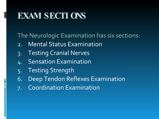
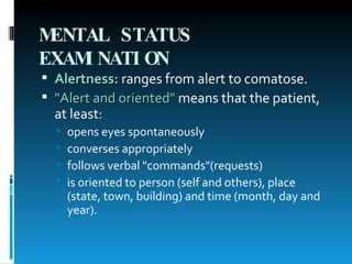
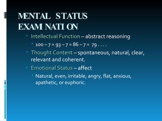
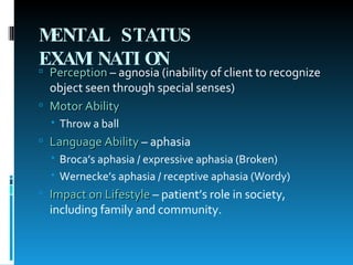
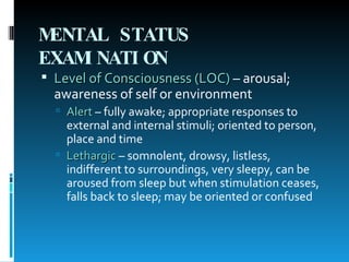
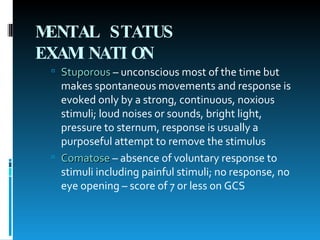
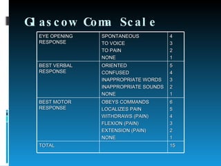
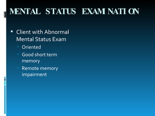
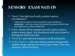
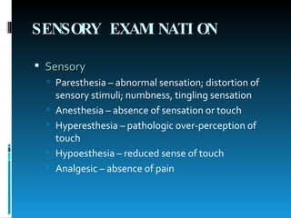
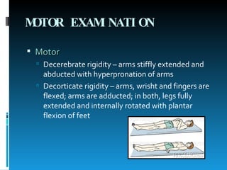
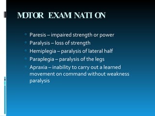
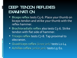
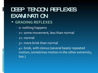
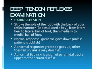
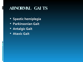
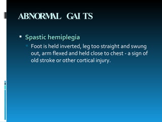
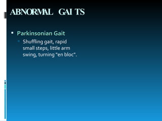
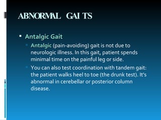
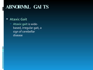
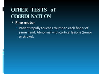
Anthony P. Toledo, MD ,RN, MAN, DPAFP CHAIRMAN, MS2 Professor/Lecturer/Reviewer/Doctor On Call, College of Nursing Our Lady of Fatima University
LEARNING OBJECTIVES   By the end of this session, you will know:  How to test the cranial nerves, and common reasons for abnormalities  How some cranial nerve abnormalities look  How to test touch, sharp, position and vibration sensation  How to grade a patient's strength  How to grade reflexes, and how some abnormal reflexes look  Which nerve roots you are testing when you check reflexes  Several abnormal gaits  How to test coordination, how abnormal tests look and what they mean.
Neurologic Exam
EXAM SECTIONS The Neurologic Examination has six sections: Mental Status Examination  Testing Cranial Nerves  Sensation Examination  Testing Strength  Deep Tendon Reflexes Examination  Coordination Examination
MENTAL STATUS EXAMINATION   Alertness:  ranges from alert to comatose. "Alert and oriented"  means that the patient, at least:  opens eyes spontaneously  converses appropriately  follows verbal "commands"(requests)  is oriented to person (self and others), place (state, town, building) and time (month, day and year).
MENTAL STATUS EXAMINATION   Intellectual Function  – abstract reasoning 100 – 7 = 93 – 7 = 86 – 7 =  79 . . . .  Thought Content  – spontaneous, natural, clear, relevant and coherent. Emotional Status  – affect Natural, even, irritable, angry, flat, anxious, apathetic, or euphoric.
MENTAL STATUS EXAMINATION   Perception  – agnosia (inability of client to recognize object seen through special senses) Motor Ability Throw a ball Language Ability  – aphasia Broca’s aphasia / expressive aphasia (Broken) Wernecke’s aphasia / receptive aphasia (Wordy) Impact on Lifestyle  – patient’s role in society, including family and community.
MENTAL STATUS EXAMINATION   Level of Consciousness (LOC)  – arousal; awareness of self or environment Alert  – fully awake; appropriate responses to external and internal stimuli; oriented to person, place and time Lethargic  – somnolent, drowsy, listless, indifferent to surroundings, very sleepy, can be aroused from sleep but when stimulation ceases, falls back to sleep; may be oriented or confused
MENTAL STATUS EXAMINATION   Stuporous  – unconscious most of the time but makes spontaneous movements and response is evoked only by a strong, continuous, noxious stimuli; loud noises or sounds, bright light, pressure to sternum, response is usually a purposeful attempt to remove the stimulus Comatose  – absence of voluntary response to stimuli including painful stimuli; no response, no eye opening – score of 7 or less on GCS
Glascow Coma Scale EYE OPENING RESPONSE SPONTANEOUS TO VOICE TO PAIN NONE 4 3 2 1 BEST VERBAL RESPONSE ORIENTED CONFUSED INAPPROPRIATE WORDS INAPPROPRIATE SOUNDS NONE 5 4 3 2 1 BEST MOTOR RESPONSE OBEYS COMMANDS LOCALIZES PAIN WITHDRAWS (PAIN) FLEXION (PAIN) EXTENSION (PAIN) NONE 6 5 4 3 2 1 TOTAL 15
MENTAL STATUS EXAMINATION   Client with Abnormal Mental Status Exam Oriented Good short term memory Remote memory impairment
CRANIAL NERVES Cranial Nerve I - THE OLFACTORY NERVES   Test this with odorous things, one nostril at a time. As most physicians don't carry odorants, the screening exam usually omits the first cranial nerve.  Common causes of cranial nerve I dysfunction include:  trauma to the cribriform plate  frontal lobe mass or stroke  nasal problems (e.g. allergic or viral).
CRANIAL NERVES Cranial Nerve I - THE OLFACTORY NERVES   Sensory
CRANIAL NERVES Cranial Nerve II - THE OPTIC NERVE   Test this with field of vision and visual acuity. Many MDs carry a pocket visual screening card. To screen field of vision, test by confrontation (patient looks at your nose while you move fingers).
CRANIAL NERVES Cranial Nerve II - THE OPTIC NERVE  Common causes of optic nerve abnormalities:  Eye disease or injury. Diabetic retinopathy and glaucoma are major causes.  Occipital lobe mass or stroke. This causes loss of visual field in both eyes. Patients can lose ½ or ¼ of a visual field  Optic chiasm mass, such as pituitary tumors. These cause loss of the temporal visual fields bilaterally - bitemporal hemianopsia.
CRANIAL NERVES Cranial Nerve II - THE OPTIC NERVE   Sensory Visual Acuity
CRANIAL NERVES Cranial Nerve II - THE OPTIC NERVE   Sensory Visual Field
CRANIAL NERVES Cranial Nerve II - THE OPTIC NERVE   Fundoscopy
CRANIAL NERVES Cranial Nerve II and III - THE OPTIC NERVE and OCULOMOTOR NERVE Sensory + Motor Pupillary light reflex
CRANIAL NERVES Cranial Nerve III, IV and VI - THE OCULOMOTOR, TROCHLEAR and ABDUCENS NERVES   Test these three nerves with extraocular movements and pupil function (cranial nerve III). To detect subtle abnormalities, ask patient whether they have double vision (diplopia) during extraocular movements.
CRANIAL NERVES Cranial Nerve III, IV and VI - THE OCULOMOTOR, TROCHLEAR and ABDUCENS NERVES   One mnemonic to remember these three nerves is LR6SO4 : all the muscles are innervated by CN III except for the lateral rectus (6) and superior oblique (4).  Some common causes for cranial nerve palsies are: brainstem injury or compression (e.g. tumor, stroke, intracranial bleeding  diabetic neuropathy (can cause temporary palsies).
CRANIAL NERVES Cranial Nerve III, IV and VI - THE OCULOMOTOR, TROCHLEAR and ABDUCENS NERVES   Ocular inspection
CRANIAL NERVES Cranial Nerve III, IV and VI - THE OCULOMOTOR, TROCHLEAR and ABDUCENS NERVES   Motor EOM
CRANIAL NERVES Cranial Nerve V - THE TRIGEMINAL NERVE   Screen this nerve with facial sensation (to light touch, e.g. q-tip) and strength of the masseter muscles.  Common cause for CN V abnormality is stroke in the contralateral sensory cortex.
CRANIAL NERVES Cranial Nerve V - THE TRIGEMINAL NERVE   Sensory
CRANIAL NERVES Cranial Nerve V - THE TRIGEMINAL NERVE   Motor
CRANIAL NERVES Cranial Nerve VII - THE FACIAL NERVE   Test this with facial movements: ask the patient to raise eyebrows, show teeth, smile, puff out cheeks, whistle.  Injuries to facial strength central to the nucleus (in the cortex or corticospinal tracts) - often caused by a stroke - cause weakness of the lower face, with sparing of the forehead, due to cross-innervation of the forehead. We call this a central facial palsy.
CRANIAL NERVES Cranial Nerve VII - THE FACIAL NERVE   Injuries to the facial nerve itself (peripheral facial palsy) cause weakness of the entire side of the face, including the forehead. Common causes of peripheral facial palsy are Bell's palsy (idiopathic - cause is unknown) and Lyme disease (which may cause bilateral peripheral facial palsy).
CRANIAL NERVES Cranial Nerve VII - THE FACIAL NERVE Motor
CRANIAL NERVES Cranial Nerve VII - THE FACIAL NERVE Sensory
CRANIAL NERVES Cranial Nerve VIII - THE ACOUSTIC NERVE   Test the acoustic nerve with hearing test (rub fingers by each ear, or whisper into ear, or use your tuning fork). We do this as part of the ear examination. In patients with vertigo or dizziness, you may test also with positional maneuvers, trying to reproduce vertigo by moving the patient.
CRANIAL NERVES Cranial Nerve VIII - THE ACOUSTIC NERVE   Common causes of acoustic nerve abnormalities:   sensorineural hearing loss due to age or noise exposure  tumors at cerebellopontine angle  acoustic neuroma  earwax or middle ear disease can cause temporary hearing loss.
CRANIAL NERVES Cranial Nerve VIII - THE ACOUSTIC NERVE   Sensory Auditory Acuity Rinne Weber
CRANIAL NERVES Cranial Nerve IX and X - THE GLOSSOPHARINGEAL and VAGUS NERVES   Test this with the gag reflex - put tongue blade on the posterior third of patient's tongue and press down. Many clinicians prefer to have alert patient phonate (say aaah) instead, watching for uvula movement.  A common cause of CN IX and X abnormality is a large stroke. The uvula retracts to the normal side.
CRANIAL NERVES Cranial Nerve IX and X - THE GLOSSOPHARINGEAL and VAGUS NERVES   Sensory + Motor Gag reflex
CRANIAL NERVES Cranial Nerve XI - THE ACCESSORY NERVE Test this nerve by asking patient to shrug shoulders or turn head against resistance.  A common cause of CN XI abnormality is neck injury.
CRANIAL NERVES Cranial Nerve XI - THE ACCESSORY NERVE Motor
CRANIAL NERVES Cranial Nerve XII - THE HYPOGLOSSAL NERVE Test this nerve by asking patient to protrude tongue and move it from side to side.  CN XII function abnormalities are often caused by stroke. The tongue points toward its weak side.
CRANIAL NERVES Cranial Nerve XII - THE HYPOGLOSSAL NERVE Motor
SENSORY EXAMINATION   Touch:   Test light touch with a cotton swab or microfilament.  Subtle abnormality in touch sensation may manifest as  extinction  : with eyes closed, the patient touched on both sides only feels touch on the normal side.  Sharp:   Break off the wooden part of a cotton swab to make a sharp object. Ask the patient with eyes closed to distinguish sharp from dull.  Vibration:   test with low-frequency (128) tuning fork.  Proprioception:   with eyes closed, patient distinguishes whether finger and toe are moved up or down. This tests posterior column function.
SENSORY EXAMINATION Sensory Paresthesia – abnormal sensation; distortion of sensory stimuli; numbness, tingling sensation Anesthesia – absence of sensation or touch Hyperesthesia – pathologic over-perception of touch Hypoesthesia – reduced sense of touch Analgesic – absence of pain
MOTOR EXAMINATION   Test this with resisted motions .  We do this during the extremity examination.  Strength is rated from 0 to 5:  0/5: no motion  1/5: slight muscle motion, but no movement at joint  2/5: full motion parallel to ground, but can't move against gravity  3/5: can move against gravity, but no more  4/5: full strength against some resistance  5/5: full strength against full resistance - normal.  Subtle central weakness (such as with early CNS malignancy) can be tested via  pronator  drift. Ask your patient to hold arms forward with palms up. In mild cortical weakness, patient's hand on the weak side pronates and drifts down.
MOTOR EXAMINATION Motor Decerebrate rigidity – arms stiffly extended and abducted with hyperpronation of arms Decorticate rigidity – arms, wrisht and fingers are flexed; arms are adducted; in both, legs fully extended and internally rotated with plantar flexion of feet
MOTOR EXAMINATION Paresis – impaired strength or power Paralysis – loss of strength Hemiplegia – paralysis of lateral half Paraplegia – paralysis of the legs Apraxia – inability to carry out a learned movement on command without weakness paralysis
DEEP TENDON REFLEXES EXAMINATION Biceps reflex  tests C5-6. Place your thumb on biceps tendon and strike your thumb with the reflex hammer.  Brachioradialis reflex  also tests C5-6. Strike tendon with flat side of hammer.  Triceps reflex   tests C7-8. Tap proximal to olecranon.  Quadriceps reflex  (knee jerk)  tests L2-L4  Achilles reflex  (ankle jerk)  tests L5-S2.
DEEP TENDON REFLEXES EXAMINATION GRADING REFLEXES   0: nothing happens  1+: some movement, less than normal  2+: normal  3+: more brisk than normal  4+: brisk, with clonus (several beats/ repeated motion; sometimes motion in the other extremity, too.)  
DEEP TENDON REFLEXES EXAMINATION BABINSKI's SIGN Stroke the sole of the foot with the back of your reflex hammer (Babinski used a key), from lateral heel to lateral ball of foot, then medially to medial ball of foot.  Normal response: great toe goes down (unless patient is ticklish)  Abnormal response: great toe goes up, other toes fan up, ankle may dorsiflex.  Abnormal Babinski is a sign of pyramidal tract / upper motor neuron disease.
ABNORMAL GAITS   Spastic hemiplegia   Parkinsonian Gait  Antalgic Gait  Ataxic Gait
ABNORMAL GAITS   Spastic hemiplegia   Foot is held inverted, leg too straight and swung out, arm flexed and held close to chest - a sign of old stroke or other cortical injury.
ABNORMAL GAITS   Parkinsonian Gait   Shuffling gait, rapid small steps, little arm swing, turning "en bloc".
ABNORMAL GAITS   Antalgic Gait   Antalgic  (pain-avoiding) gait is not due to neurologic illness. In this gait, patient spends minimal time on the painful leg or side. You can also test coordination with tandem gait: the patient walks heel to toe (the drunk test). It's abnormal in cerebellar or posterior column disease.
ABNORMAL GAITS   Ataxic Gait   Ataxic gait   is wide-based, irregular gait, a sign of cerebellar disease
OTHER TESTS of COORDINATION   Finger to nose   Heel to shin   Rapid alternating movements   Fine motor   Romberg's sign
OTHER TESTS of COORDINATION   Finger to nose   Patient touches nose, then examiner's finger, then goes back and forth rapidly. It's abnormal in cerebellar disease.
OTHER TESTS of COORDINATION   Heel to shin   Patient moves one heel down the other shin. Abnormal jerky motion in cerebellar disease.
OTHER TESTS of COORDINATION   Rapid alternating movements   Ask patient to rapidly pronate and supinate hands. Abnormal (dysdiadochokinesia) in patients with cerebellar disease.
OTHER TESTS of COORDINATION   Fine motor   Patient rapidly touches thumb to each finger of same hand. Abnormal with cortical lesions (tumor or stroke).
OTHER TESTS of COORDINATION   Romberg's sign   Patient stands with feet together and closes eyes. Patient sways and can't hold position with eyes closed. This is abnormal in posterior column disease (with cerebellar disease, patient can't stand with feet together even with eyes open).
 

Cns Exam 3rd

  • 1.
    Anthony P. Toledo,MD ,RN, MAN, DPAFP CHAIRMAN, MS2 Professor/Lecturer/Reviewer/Doctor On Call, College of Nursing Our Lady of Fatima University
  • 2.
    LEARNING OBJECTIVES By the end of this session, you will know: How to test the cranial nerves, and common reasons for abnormalities How some cranial nerve abnormalities look How to test touch, sharp, position and vibration sensation How to grade a patient's strength How to grade reflexes, and how some abnormal reflexes look Which nerve roots you are testing when you check reflexes Several abnormal gaits How to test coordination, how abnormal tests look and what they mean.
  • 3.
  • 4.
    EXAM SECTIONS TheNeurologic Examination has six sections: Mental Status Examination Testing Cranial Nerves Sensation Examination Testing Strength Deep Tendon Reflexes Examination Coordination Examination
  • 5.
    MENTAL STATUS EXAMINATION Alertness: ranges from alert to comatose. "Alert and oriented" means that the patient, at least: opens eyes spontaneously converses appropriately follows verbal "commands"(requests) is oriented to person (self and others), place (state, town, building) and time (month, day and year).
  • 6.
    MENTAL STATUS EXAMINATION Alertness: ranges from alert to comatose. "Alert and oriented" means that the patient, at least: opens eyes spontaneously converses appropriately follows verbal "commands"(requests) is oriented to person (self and others), place (state, town, building) and time (month, day and year).
  • 7.
    MENTAL STATUS EXAMINATION Intellectual Function – abstract reasoning 100 – 7 = 93 – 7 = 86 – 7 = 79 . . . . Thought Content – spontaneous, natural, clear, relevant and coherent. Emotional Status – affect Natural, even, irritable, angry, flat, anxious, apathetic, or euphoric.
  • 8.
    MENTAL STATUS EXAMINATION Perception – agnosia (inability of client to recognize object seen through special senses) Motor Ability Throw a ball Language Ability – aphasia Broca’s aphasia / expressive aphasia (Broken) Wernecke’s aphasia / receptive aphasia (Wordy) Impact on Lifestyle – patient’s role in society, including family and community.
  • 9.
    MENTAL STATUS EXAMINATION Level of Consciousness (LOC) – arousal; awareness of self or environment Alert – fully awake; appropriate responses to external and internal stimuli; oriented to person, place and time Lethargic – somnolent, drowsy, listless, indifferent to surroundings, very sleepy, can be aroused from sleep but when stimulation ceases, falls back to sleep; may be oriented or confused
  • 10.
    MENTAL STATUS EXAMINATION Stuporous – unconscious most of the time but makes spontaneous movements and response is evoked only by a strong, continuous, noxious stimuli; loud noises or sounds, bright light, pressure to sternum, response is usually a purposeful attempt to remove the stimulus Comatose – absence of voluntary response to stimuli including painful stimuli; no response, no eye opening – score of 7 or less on GCS
  • 11.
    Glascow Coma ScaleEYE OPENING RESPONSE SPONTANEOUS TO VOICE TO PAIN NONE 4 3 2 1 BEST VERBAL RESPONSE ORIENTED CONFUSED INAPPROPRIATE WORDS INAPPROPRIATE SOUNDS NONE 5 4 3 2 1 BEST MOTOR RESPONSE OBEYS COMMANDS LOCALIZES PAIN WITHDRAWS (PAIN) FLEXION (PAIN) EXTENSION (PAIN) NONE 6 5 4 3 2 1 TOTAL 15
  • 12.
    MENTAL STATUS EXAMINATION Client with Abnormal Mental Status Exam Oriented Good short term memory Remote memory impairment
  • 13.
    CRANIAL NERVES CranialNerve I - THE OLFACTORY NERVES Test this with odorous things, one nostril at a time. As most physicians don't carry odorants, the screening exam usually omits the first cranial nerve. Common causes of cranial nerve I dysfunction include: trauma to the cribriform plate frontal lobe mass or stroke nasal problems (e.g. allergic or viral).
  • 14.
    CRANIAL NERVES CranialNerve I - THE OLFACTORY NERVES Sensory
  • 15.
    CRANIAL NERVES CranialNerve II - THE OPTIC NERVE Test this with field of vision and visual acuity. Many MDs carry a pocket visual screening card. To screen field of vision, test by confrontation (patient looks at your nose while you move fingers).
  • 16.
    CRANIAL NERVES CranialNerve II - THE OPTIC NERVE Common causes of optic nerve abnormalities: Eye disease or injury. Diabetic retinopathy and glaucoma are major causes. Occipital lobe mass or stroke. This causes loss of visual field in both eyes. Patients can lose ½ or ¼ of a visual field Optic chiasm mass, such as pituitary tumors. These cause loss of the temporal visual fields bilaterally - bitemporal hemianopsia.
  • 17.
    CRANIAL NERVES CranialNerve II - THE OPTIC NERVE Sensory Visual Acuity
  • 18.
    CRANIAL NERVES CranialNerve II - THE OPTIC NERVE Sensory Visual Field
  • 19.
    CRANIAL NERVES CranialNerve II - THE OPTIC NERVE Fundoscopy
  • 20.
    CRANIAL NERVES CranialNerve II and III - THE OPTIC NERVE and OCULOMOTOR NERVE Sensory + Motor Pupillary light reflex
  • 21.
    CRANIAL NERVES CranialNerve III, IV and VI - THE OCULOMOTOR, TROCHLEAR and ABDUCENS NERVES Test these three nerves with extraocular movements and pupil function (cranial nerve III). To detect subtle abnormalities, ask patient whether they have double vision (diplopia) during extraocular movements.
  • 22.
    CRANIAL NERVES CranialNerve III, IV and VI - THE OCULOMOTOR, TROCHLEAR and ABDUCENS NERVES One mnemonic to remember these three nerves is LR6SO4 : all the muscles are innervated by CN III except for the lateral rectus (6) and superior oblique (4). Some common causes for cranial nerve palsies are: brainstem injury or compression (e.g. tumor, stroke, intracranial bleeding diabetic neuropathy (can cause temporary palsies).
  • 23.
    CRANIAL NERVES CranialNerve III, IV and VI - THE OCULOMOTOR, TROCHLEAR and ABDUCENS NERVES Ocular inspection
  • 24.
    CRANIAL NERVES CranialNerve III, IV and VI - THE OCULOMOTOR, TROCHLEAR and ABDUCENS NERVES Motor EOM
  • 25.
    CRANIAL NERVES CranialNerve V - THE TRIGEMINAL NERVE Screen this nerve with facial sensation (to light touch, e.g. q-tip) and strength of the masseter muscles. Common cause for CN V abnormality is stroke in the contralateral sensory cortex.
  • 26.
    CRANIAL NERVES CranialNerve V - THE TRIGEMINAL NERVE Sensory
  • 27.
    CRANIAL NERVES CranialNerve V - THE TRIGEMINAL NERVE Motor
  • 28.
    CRANIAL NERVES CranialNerve VII - THE FACIAL NERVE Test this with facial movements: ask the patient to raise eyebrows, show teeth, smile, puff out cheeks, whistle. Injuries to facial strength central to the nucleus (in the cortex or corticospinal tracts) - often caused by a stroke - cause weakness of the lower face, with sparing of the forehead, due to cross-innervation of the forehead. We call this a central facial palsy.
  • 29.
    CRANIAL NERVES CranialNerve VII - THE FACIAL NERVE Injuries to the facial nerve itself (peripheral facial palsy) cause weakness of the entire side of the face, including the forehead. Common causes of peripheral facial palsy are Bell's palsy (idiopathic - cause is unknown) and Lyme disease (which may cause bilateral peripheral facial palsy).
  • 30.
    CRANIAL NERVES CranialNerve VII - THE FACIAL NERVE Motor
  • 31.
    CRANIAL NERVES CranialNerve VII - THE FACIAL NERVE Sensory
  • 32.
    CRANIAL NERVES CranialNerve VIII - THE ACOUSTIC NERVE Test the acoustic nerve with hearing test (rub fingers by each ear, or whisper into ear, or use your tuning fork). We do this as part of the ear examination. In patients with vertigo or dizziness, you may test also with positional maneuvers, trying to reproduce vertigo by moving the patient.
  • 33.
    CRANIAL NERVES CranialNerve VIII - THE ACOUSTIC NERVE Common causes of acoustic nerve abnormalities: sensorineural hearing loss due to age or noise exposure tumors at cerebellopontine angle acoustic neuroma earwax or middle ear disease can cause temporary hearing loss.
  • 34.
    CRANIAL NERVES CranialNerve VIII - THE ACOUSTIC NERVE Sensory Auditory Acuity Rinne Weber
  • 35.
    CRANIAL NERVES CranialNerve IX and X - THE GLOSSOPHARINGEAL and VAGUS NERVES Test this with the gag reflex - put tongue blade on the posterior third of patient's tongue and press down. Many clinicians prefer to have alert patient phonate (say aaah) instead, watching for uvula movement. A common cause of CN IX and X abnormality is a large stroke. The uvula retracts to the normal side.
  • 36.
    CRANIAL NERVES CranialNerve IX and X - THE GLOSSOPHARINGEAL and VAGUS NERVES Sensory + Motor Gag reflex
  • 37.
    CRANIAL NERVES CranialNerve XI - THE ACCESSORY NERVE Test this nerve by asking patient to shrug shoulders or turn head against resistance. A common cause of CN XI abnormality is neck injury.
  • 38.
    CRANIAL NERVES CranialNerve XI - THE ACCESSORY NERVE Motor
  • 39.
    CRANIAL NERVES CranialNerve XII - THE HYPOGLOSSAL NERVE Test this nerve by asking patient to protrude tongue and move it from side to side. CN XII function abnormalities are often caused by stroke. The tongue points toward its weak side.
  • 40.
    CRANIAL NERVES CranialNerve XII - THE HYPOGLOSSAL NERVE Motor
  • 41.
    SENSORY EXAMINATION Touch: Test light touch with a cotton swab or microfilament. Subtle abnormality in touch sensation may manifest as extinction : with eyes closed, the patient touched on both sides only feels touch on the normal side. Sharp: Break off the wooden part of a cotton swab to make a sharp object. Ask the patient with eyes closed to distinguish sharp from dull. Vibration: test with low-frequency (128) tuning fork. Proprioception: with eyes closed, patient distinguishes whether finger and toe are moved up or down. This tests posterior column function.
  • 42.
    SENSORY EXAMINATION SensoryParesthesia – abnormal sensation; distortion of sensory stimuli; numbness, tingling sensation Anesthesia – absence of sensation or touch Hyperesthesia – pathologic over-perception of touch Hypoesthesia – reduced sense of touch Analgesic – absence of pain
  • 43.
    MOTOR EXAMINATION Test this with resisted motions . We do this during the extremity examination. Strength is rated from 0 to 5: 0/5: no motion 1/5: slight muscle motion, but no movement at joint 2/5: full motion parallel to ground, but can't move against gravity 3/5: can move against gravity, but no more 4/5: full strength against some resistance 5/5: full strength against full resistance - normal. Subtle central weakness (such as with early CNS malignancy) can be tested via pronator drift. Ask your patient to hold arms forward with palms up. In mild cortical weakness, patient's hand on the weak side pronates and drifts down.
  • 44.
    MOTOR EXAMINATION MotorDecerebrate rigidity – arms stiffly extended and abducted with hyperpronation of arms Decorticate rigidity – arms, wrisht and fingers are flexed; arms are adducted; in both, legs fully extended and internally rotated with plantar flexion of feet
  • 45.
    MOTOR EXAMINATION Paresis– impaired strength or power Paralysis – loss of strength Hemiplegia – paralysis of lateral half Paraplegia – paralysis of the legs Apraxia – inability to carry out a learned movement on command without weakness paralysis
  • 46.
    DEEP TENDON REFLEXESEXAMINATION Biceps reflex tests C5-6. Place your thumb on biceps tendon and strike your thumb with the reflex hammer. Brachioradialis reflex also tests C5-6. Strike tendon with flat side of hammer. Triceps reflex tests C7-8. Tap proximal to olecranon. Quadriceps reflex (knee jerk) tests L2-L4 Achilles reflex (ankle jerk) tests L5-S2.
  • 47.
    DEEP TENDON REFLEXESEXAMINATION GRADING REFLEXES 0: nothing happens 1+: some movement, less than normal 2+: normal 3+: more brisk than normal 4+: brisk, with clonus (several beats/ repeated motion; sometimes motion in the other extremity, too.)  
  • 48.
    DEEP TENDON REFLEXESEXAMINATION BABINSKI's SIGN Stroke the sole of the foot with the back of your reflex hammer (Babinski used a key), from lateral heel to lateral ball of foot, then medially to medial ball of foot. Normal response: great toe goes down (unless patient is ticklish) Abnormal response: great toe goes up, other toes fan up, ankle may dorsiflex. Abnormal Babinski is a sign of pyramidal tract / upper motor neuron disease.
  • 49.
    ABNORMAL GAITS Spastic hemiplegia Parkinsonian Gait Antalgic Gait Ataxic Gait
  • 50.
    ABNORMAL GAITS Spastic hemiplegia Foot is held inverted, leg too straight and swung out, arm flexed and held close to chest - a sign of old stroke or other cortical injury.
  • 51.
    ABNORMAL GAITS Parkinsonian Gait Shuffling gait, rapid small steps, little arm swing, turning "en bloc".
  • 52.
    ABNORMAL GAITS Antalgic Gait Antalgic (pain-avoiding) gait is not due to neurologic illness. In this gait, patient spends minimal time on the painful leg or side. You can also test coordination with tandem gait: the patient walks heel to toe (the drunk test). It's abnormal in cerebellar or posterior column disease.
  • 53.
    ABNORMAL GAITS Ataxic Gait Ataxic gait is wide-based, irregular gait, a sign of cerebellar disease
  • 54.
    OTHER TESTS ofCOORDINATION Finger to nose Heel to shin Rapid alternating movements Fine motor Romberg's sign
  • 55.
    OTHER TESTS ofCOORDINATION Finger to nose Patient touches nose, then examiner's finger, then goes back and forth rapidly. It's abnormal in cerebellar disease.
  • 56.
    OTHER TESTS ofCOORDINATION Finger to nose Patient touches nose, then examiner's finger, then goes back and forth rapidly. It's abnormal in cerebellar disease.
  • 57.
    OTHER TESTS ofCOORDINATION Heel to shin Patient moves one heel down the other shin. Abnormal jerky motion in cerebellar disease.
  • 58.
    OTHER TESTS ofCOORDINATION Rapid alternating movements Ask patient to rapidly pronate and supinate hands. Abnormal (dysdiadochokinesia) in patients with cerebellar disease.
  • 59.
    OTHER TESTS ofCOORDINATION Fine motor Patient rapidly touches thumb to each finger of same hand. Abnormal with cortical lesions (tumor or stroke).
  • 60.
    OTHER TESTS ofCOORDINATION Romberg's sign Patient stands with feet together and closes eyes. Patient sways and can't hold position with eyes closed. This is abnormal in posterior column disease (with cerebellar disease, patient can't stand with feet together even with eyes open).
  • 61.