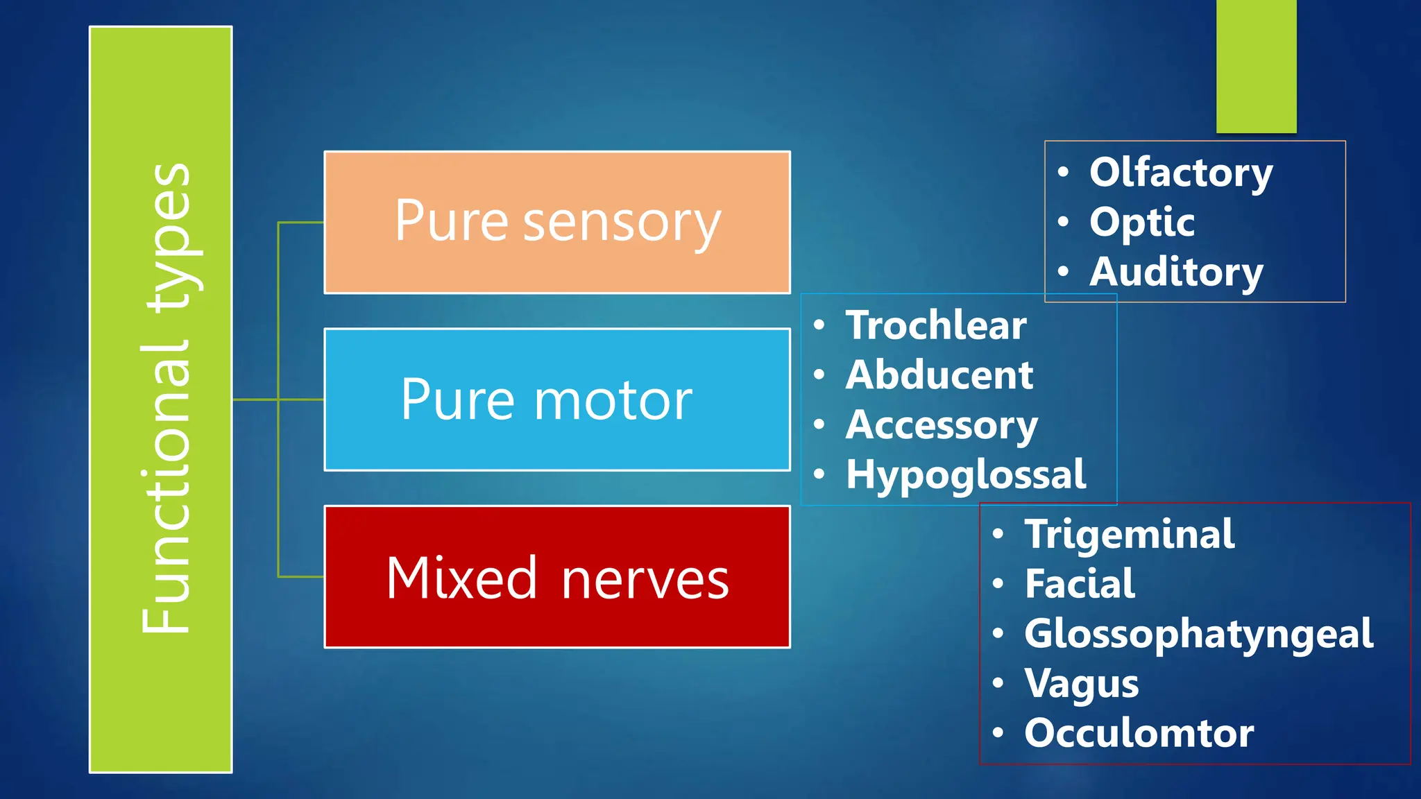
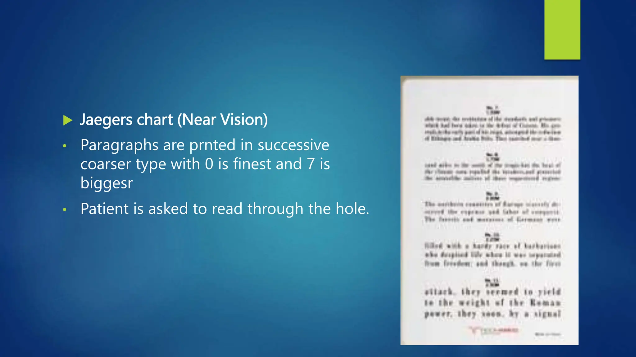
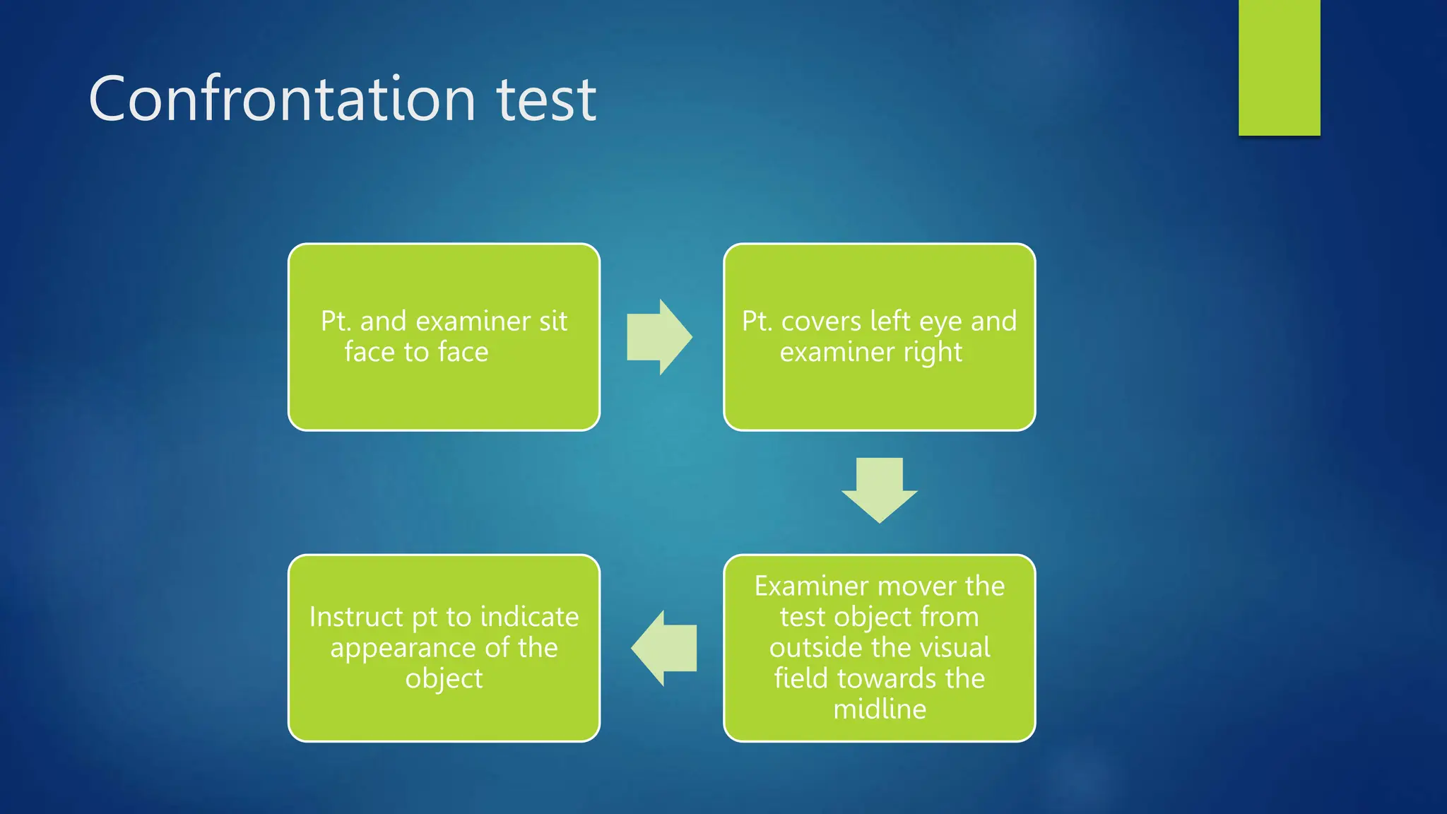
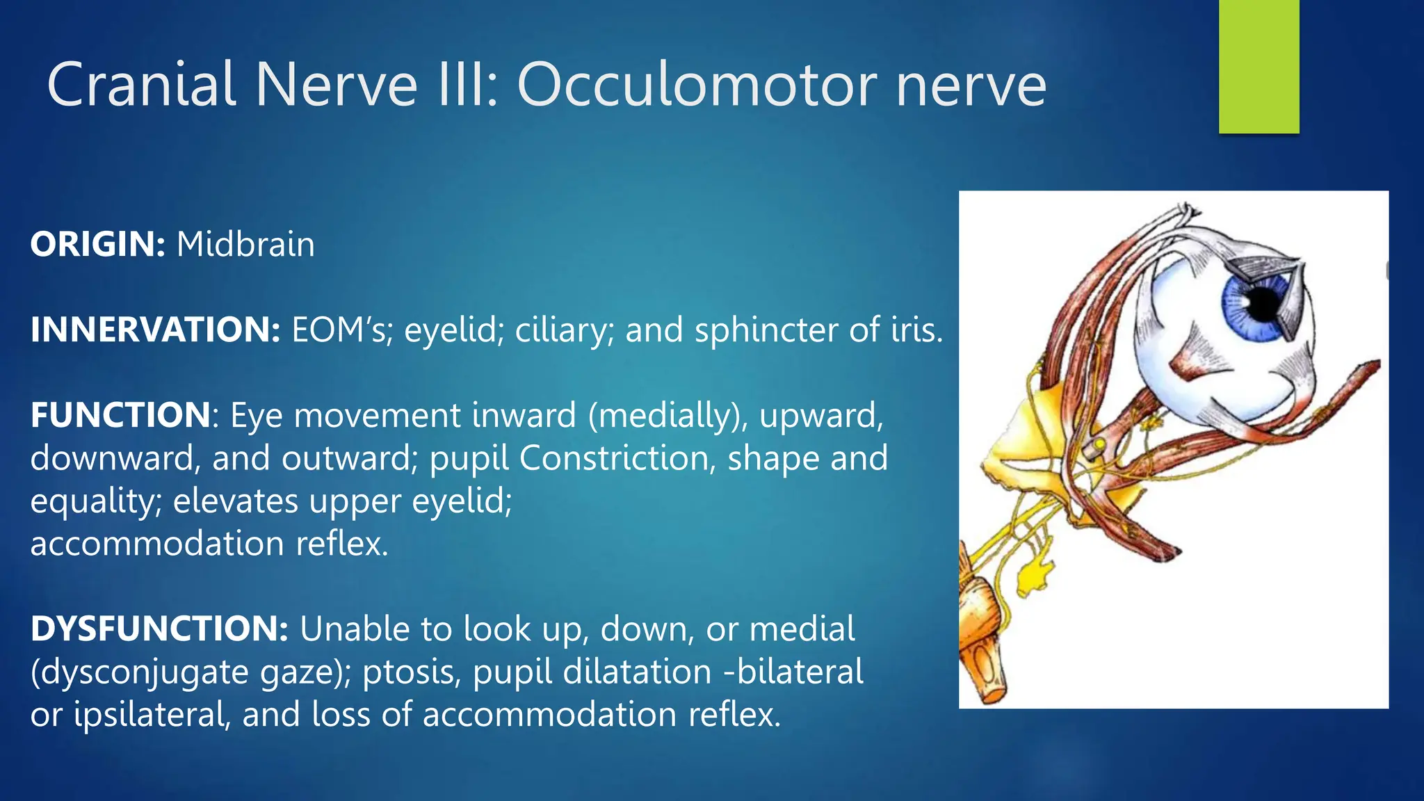
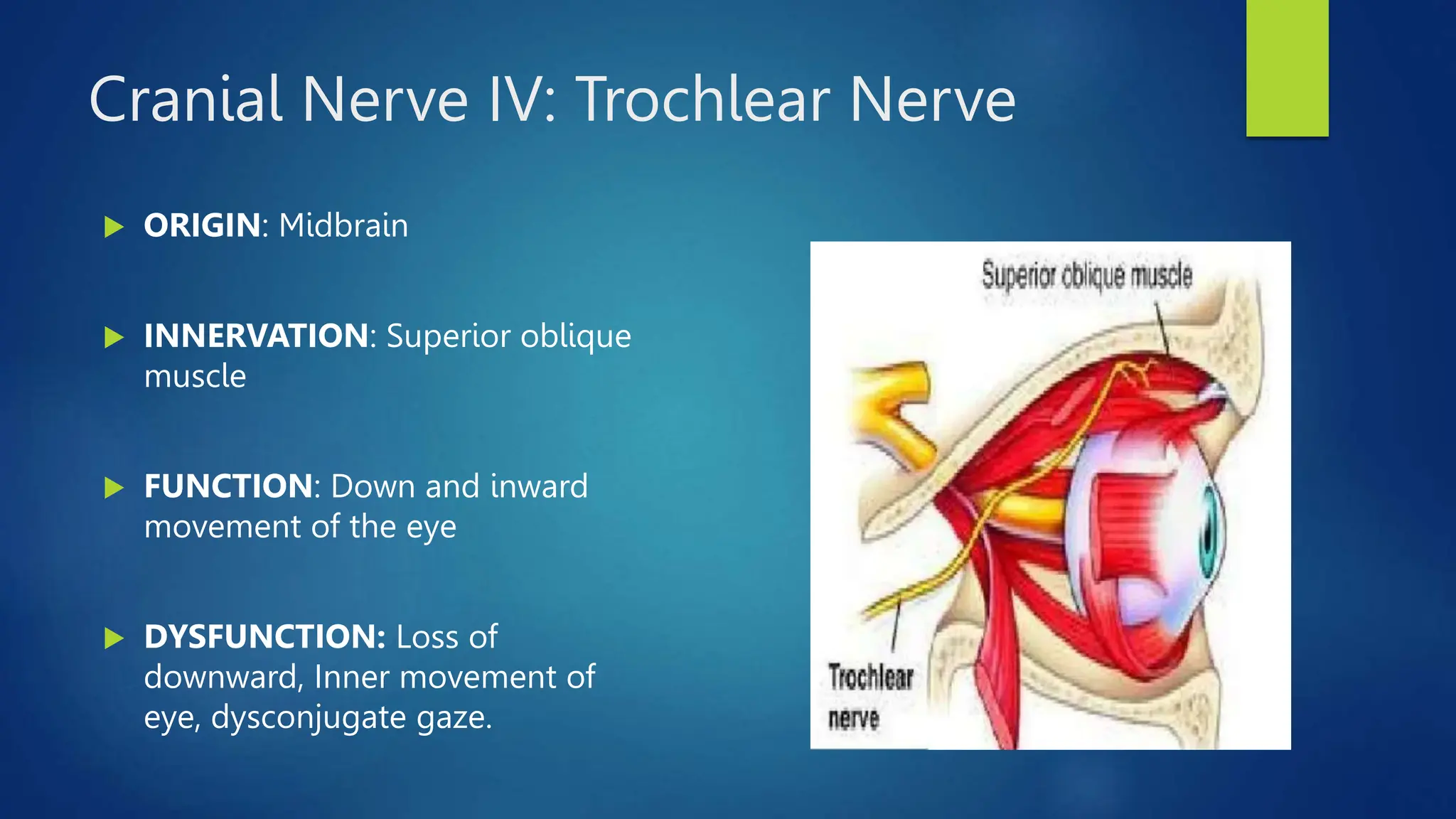
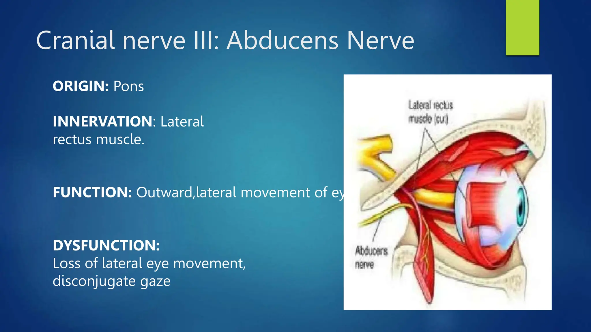
Cranial Nerves Examination
General characteristics
 The 12 pairs of cranial nerves are part of the peripheral nervous system
 As a rule, cranial nerves do not cross in the brain.
 Cranial nerves maybe sensory, motor both somatic or parasympathetic, or
have mixed function
Functional
types
Pure sensory
Pure motor
Mixed nerves
• Olfactory
• Optic
• Auditory
• Trochlear
• Abducent
• Accessory
• Hypoglossal
• Trigeminal
• Facial
• Glossophatyngeal
• Vagus
• Occulomtor
Cranial Nerve 1: Olfactory
ORIGIN: Cerebral
hemisphere
INNERVATION: Nasal
mucous membrane
Function
 Carries the sensation of smell from nasal
mucosa to olfactory bulb
 There is no motor component
Purpose of test
 To determine any impairment of smell ( unilateral/ bilateral)
 Whether impairment is due to any local nasal disease or
neural lesion
Methods of testing
 Small bottles containing essences of very familiar odour are
required
Coffee
Lemon
Chocolate
Asafetida etc
Procedure
Compress 1
nostril and
sniff the atste
odour twice
Ask whether
he/she can
smell or
identify odour
Repeat test on
other nostril and
ask if smell is
similar in both
nostril
Ask whether
he/she can smell
or identify odour
Interpretation of result
 Parosmia: Each odour is similar but distorted and unpleasant
 Anosmia: Cant smell anything
 Impairment will also lead to decreased taste
Common causes of anosmia:
• Mucous blockage of the nose: preventing odours from reaching
the olfactory nerve receptors.
• Head trauma: can result in shearing of the olfactory nerve fibres
leading to anosmia.
• Genetics: some individuals have congenital anosmia.
• Parkinson’s disease: anosmia is an early feature of Parkinson’s
disease.
• COVID-19: transient anosmia is a common feature of COVID-19.
Cranial nerve II: Optic
Function
 Carries the visual impulses from the retina to the optic
chiasma and in the optic tract to the lateral geniculate body
 The impulse acts as an afferent pathway for the pupillary light
reflex
Purpose of test
1. Visual acuity
2. Colour vision
3. Visual field
Near Field
Far field
Colour matching
Confrontation
test
Method of testing
 Visual acuity:
 Snellen’s chart ( Far vision)
• Chart is placed at 20 feet or 6meter and patient is
asked to read it
• The formula is d/D
where d is 6 m and D is distance from which he can
read it clearly
• Normal is 6/6 or 20/20
 Jaegers chart (Near Vision)
• Paragraphs are prnted in successive
coarser type with 0 is finest and 7 is
biggesr
• Patient is asked to read through the hole.
Colour vision
 Checked by asking to match different colours
 Day or night blindeness can be assessed
Visual field
Purpose
 To chart periphery of visual field
 To detect position, size and shape of the blind spot
Confrontation test
Pt. and examiner sit
face to face
Pt. covers left eye and
examiner right
Examiner mover the
test object from
outside the visual
field towards the
midline
Instruct pt to indicate
appearance of the
object
Common Causes
 Total unilateral loss of vision: Optic nerve lesion
 Homonymous hemianopia (loss of half of visual field in one
eye): lesion between optic tact to occipital cortex
 Bitemporal hemianopia: lesion of optic chiasma
Specific Dysfunctions
 Blurred vision or complete blindness
 Ipsilateral vision loss- Optic atrophy, retinal/optic nerve
lesions, trauma
 Cortical blindness: Lesion of occipital cortex B/l, pupil
reflexes intact
 Papilledema: Optic nerve tumour, venous obstruction,
chronic increased ICP
 Optic atrophy- Optic neuritis, increased ICP
 Scotomas (Abnormal blind spots on visual fields) – optic
neuritis or atriophy
Cranial Nerves III, IV, VI: Occulomotor,
Trochlear, Abducent
Cranial Nerve III: Occulomotor nerve
ORIGIN: Midbrain
INNERVATION: EOM’s; eyelid; ciliary; and sphincter of iris.
FUNCTION: Eye movement inward (medially), upward,
downward, and outward; pupil Constriction, shape and
equality; elevates upper eyelid;
accommodation reflex.
DYSFUNCTION: Unable to look up, down, or medial
(dysconjugate gaze); ptosis, pupil dilatation -bilateral
or ipsilateral, and loss of accommodation reflex.
Cranial Nerve IV: Trochlear Nerve
 ORIGIN: Midbrain
 INNERVATION: Superior oblique
muscle
 FUNCTION: Down and inward
movement of the eye
 DYSFUNCTION: Loss of
downward, Inner movement of
eye, dysconjugate gaze.
Cranial nerve III: Abducens Nerve
ORIGIN: Pons
INNERVATION: Lateral
rectus muscle.
FUNCTION: Outward,lateral movement of eye.
DYSFUNCTION:
Loss of lateral eye movement,
disconjugate gaze
Function
 Controls the external ocular muscles and elevators of the lids
 Also regulate the pupillary muscles
Purpose of the test
 Inspect pupils to rule out a local disease, peripheral lesion or a nuclear
involvement
 Examine eye movement and determine if defects is muscular origin or
neural involvement
 To detect nystagmus
Method of testing
Observation
 Presence and absence of ptosis and squint
 Whether unilateral or bilateral
 Constant or variable
 Size, shape, equality and regularity of the pupils
Reaction to light
 Reduce illumination of room and vision should focus on a far object
 A bright beam of light is shone from the side of one eye
 Repeat on the other side (the pupil should constrict briskly)
 Shield one eye and perform test on the other and see for consensual
reaction
Reaction to convergence and
accommodation for near vision
 Fix vision on a distant object and instruct to look in a near object
 Place finger tip in front of the bridge of the nose (22cm)
 Then return to the far object
 Observe pupillary reaction in both
Examination of ocular movement
 Observe lagging of one or both eye
 Observe nystagmus
Analysis of diplopia
 Shield one eye with a transparent red shield
 Object is moved from left to right, up and down
 Ask if-
Pt. sees 1 or 2 objects
Objects lie one above the other or side by side
Rules governing analysis of diplopia
 Separation of image is greatest in the direction in which weak muscle has
its purest action
 False image is displaced farthest in the direction in which the weak muscle
should move the eye
Analyzing nystagmus
 Watch the patients eye while talking
 Ask it look at a definite point and move the
point from left to right and up to down
 Hold each position for 5 sec and assess
nystagmus direction, rate, amplitude)
Common causes of paralysis
 Pontine lesions
 Neoplasms
 Vascular accidents
 Demyelinating disease
 Meningeal inflammation
 Tumour of base of skull
 Increased intra cranial pressure
 Head injury
[Total paralysis of III. IV and VI nerve indicate a lesion in cavernous sinus (carotid
aneurism)]
Cranial Nerve V: Trigeminal
Trigeminal nerve
ORIGIN: Pons. The sensory
nucleus extends from thepons to the midbrain, and also to
the medulla and spinalcord.
INNERVATION:
Three branches of CN V: Ophthalmic, maxillary, &
mandibular.
Motor innervation to masseter & temporal
muscles.
Sensory innervation to skin & mucous membranes in head;
teeth, tongue, external auditory canal, and cornea.
Trigeminal nerve
FUNCTION: Sensation of pain, touch, hot, & cold; motor movement of masseter
&temporal muscles.
DYSFUNCTION:
• Loss of sensation - if affecting all three branches, indicative of peripheral
injury.
• Brainstem or upper cervical cord injury may result in loss of sensation to one
or more branches of the trigeminal nerve.
• Paresthesia and/or severe pain indicative of nerve compression or
irritation(Trigeminal neuralgia)
• Loss of corneal reflex.
Tic douloureux
or trigeminal neuralgia
 Paroxysmal attacks of severe, short, sharp, stabbing pain
affecting one or more branch of the nerve.
 Caused by inflammation of nerve
Purpose of the test
 To determine any sensory impairment
 To determine unilateral or bilateral motor weakness and determine UMN
from LMN
Method of examination
Superifical sensory touch from mainly 6 areas (light touch and
pain)
 Forehead and upper part of the side of nose (ophthalmic)
 Malar and upper lip region (maxillary)
 Chin and anterior part of tongue (mandibular)
Interpretation
 Total loss of sensation: lesion of ganglion or sensory root
 Total sensory loss over 1 division: partial lesion of ganglion root
 Touch only lost: pontine lesion affecting sensory nucleus
 Pain and temp lost: dissociate anesthesia (syringobulia)
Motor examination:
 Muscle power of masticatory muscle namely the masseter and
temporalis
 Inability to raise, depress, protrude, retract and deviate the
mandible
Jaw Jerk
 Ask the patient to relax jaw. Place finger on the chin and tap it
with hammer.
 Closing of mouth is the response
 Brisk is normal
 Exaggerated is pathological
Corneal reflex
 Using a cotton piece, the cornea is teased
 Normal response is a bilateral blink
 Afferent- ophthalmic division of V! nerve
 Efferent- Facial nerve
Interpretation
 If you tap the patient’s sclera and neither eye blinks >> ipsilateral CN V lesion.
 This lesion localization is confirmed if the patient blinks with both eyes when
the opposite eye is stimulated.
 However, if when the opposite eye is stimulated, the patient’s eyes still fail to
blink, the abnormality either localizes to the B/l CN V or B/L CN VII
 If you tap the patient’s sclera and one eye blinks but the other does not >> CN
VII that fails to blink.This same eye should fail to blink when you tap the
patient’s other eye.
Cranial nerve VII: Facial Nerve
Function
 Supplies the muscles of facial expression including platysma and stapedius
 Secretomotor fibers to the lacrimal gland and the salivary gland
 Carries sensation of taste from anterior 2/3rd of tongue and general
sensation from external acoustic meatus
Purpose of the test
 To detect any unilateral or bilateral weakness of facial muscles (UMN or
LMN)
 Detect impairment of taste
Method of testing
 Observation
Symmetry and asymmetry of face
Nasolabial fold and wrinkle on forehead
 Ask the patient to close the eyes, raise the
eyebrows, blow out the cheek, whistle, etc
Examination of taste
 The four primary taste (sweet, salt, sour, bitter) can be carried out by using
sugar, salt, vinegar and quinine.
 The side of the tongue is moistened by the test substance.
 Ask the patient to indicate taste by pointing.
Secretomotor function
 The flow of tears of two side can be compared by giving ammonia to
inhale which will result in tearing of eye
 The flow of saliva can be tested by keeping a spicy substance in the
tongue and the tip is raised to observe the sub mamdibular salivary flow.
Reflexes
 Corneal reflex
 Nasopalpebral reflex: tap on the nasopalpebral ridge will produce closure
of both eyes. In bells palsy there is failure to close on the affected side
Cranial Nerve VIII: Vestibulocochlear
Function
 Carries the impulses of sound from the hair cells of organ of corti to
cochlear nucleus in pons
 Control balance through vestibular nerve
Purpose of the test
 To determine any deafness is bilateral or unilateral
 Whether deafness is due disease of middle ear or cochlear nerve
 To determine the disturbance of vestibular functions
Test of hearing
 Observe if the patient turns one ear
towards you
 Evaluate hearing using a ticking watch,
rub fingers together, whisper.
Rinne’s test
 Strike a tuning fork gently, hold it near one external meatus and ask the
patient if he can hear it
 Place it on the mastoid, ask if he can still hear it and instruct him to say
“NOW” when sound ceases, and keep it on the external meatus again
(normally the notes is still audible)
Interpretation
 In conductive deafness- not be able to hear the tuning fork when it is
moved next to the external auditory meatus
 In sensineural deafness- equally reduced
Weber’s test
 The fork is place on the vertex
 Ask the patient if he can hear the sound all over the head, in both ears or
in one ear
 In sensineural deafness, the sound appear to be heard on the normal ear
 In conductive deafness, sound is louder in the abnormal ear.
Common causes of deafness
 Disease of external and middle ear and eustachian tube
 Prolonged exposure to loud noise
 Old age
 Meningitis
 Demyelinating disease
Test of vestibular function
 Observe equilibrium as patient walks or stands
 Observe abnormal eye movts
 Ask for-
Dizziness
Falling
Nausea and vomiting
Cranial Nerve IX: Glossopharyngeal
Function
Visceral sensory:
subconscious
sensation from
carotid body and
sinus
General Sensory:
posterior 1/3 of
tongue, tonsil, skin
of external ear,
tympanic membrane
and pharynx
Visceral Motor:
parasympathetic
stimulation of
parotid gland, and
control blood
vessels
Branchial Motor:
Supplies
stylopharyngeus
muscle
Special Sensory:
carries taste
from posterior
1/3 tongue
A unilateral lesion:
 loss of the ipsilateral gag reflex
 loss of carotid body and sinus reflex
 loss of taste in the posterior region of the tongue.
Cranial Nerve X: Vagus
Function
 General Sensory: posterior meninges, concha, skin at back of ear, external
tympanic membrane, pharynx and larynx
 Visceral Motor: parasympathetic stimulation to smooth muscle and glands
of pharynx, larynx; thorax and abdominal viscera and cardiac muscle
 Visceral sensory: from larynx, trachea, esophagus, and thoracis and
abdominal viscera, stretch receptors and chemoreceptors
 Motor: superior, middle, inferior constrictors; levator palati,
salpingopharyngeus
Purpose of the test
 To test the elevation of palate and contraction of pharynx
 To examine the movts of vocal cords
Method of testing
 Notice the pitch and quality of voice, cough
 Ask the patient to open his mouth wide, after a few movts ask to say “AH”
while breathing out and “UGH” while in
 The palate should move symmetrically upwards and backwards, the uvula
in midline and two sides of pharynx contract symmetrically
 Unilateral vagus nerve lesions result in hoarseness, dysphagia and dyspnea
 Bilateral vagal nerve injuries result in airway obstruction and asphyxia
Common causes of lesion
 Poliomyelitis
 Syringobulbia
 Posterior fossa tumor
 Advanced parkinsonism
 Myasthenia gravis
 Enlarged cervical glands
 Surgical operation of the neck
Cranial Nerve XI: Accessory
Function
 Supplies sternocleidomastoid and trapezius muscles
Purpose of the test
 To detect wasting and weakness, unilateral or bilateral of the muscles
Method of testing
Common causes of paralysis
 MND
 Poliomyeltis
 Polyneuropathy
 Trauma in neck or base of skull
 Tumour at jugular foramen
 Syringomyelia
Cranial nerve XII: Hypoglossal
Function
 Controls movts of the tongue, hyoid bone and larynx during and after
deglutition
Supplies 3 of 4
extrinsic muscles of
tongue and all
intrinsic muscles of
tongue
Purpose of the test
 To inspect the surface of the tongue
 To detect wasting, weakness and involuntary movts
 To examine voluntary muscle control
Method of testing
Ask the patient to protrude the tongue and observe for
 Reduction in size of affected side
 Excessive ridging and wrinkling
 Restricted protrusion
 Deviation towards one side
Common lesions
 Syringomyelia
 Poliomyelitis
 MND
 Profound hemiplegia
 ALS
References
 Cranial Nerve Assessment: A Concise Guide to Clinical Examination by
OMPRAKASH DAMODARAN ELIAS RIZK, JULIAN RODRIGUEZ, AND
GABRIEL LEE
 Rothwell P. Examination of the cranial nerves. BMJ. 2000 Mar
4;320(7235):655A. PMID: 10698908; PMCID: PMC1117678.
Thank you!

cranial nerves and their examination ppt

  • 1.
  • 2.
    General characteristics  The12 pairs of cranial nerves are part of the peripheral nervous system  As a rule, cranial nerves do not cross in the brain.  Cranial nerves maybe sensory, motor both somatic or parasympathetic, or have mixed function
  • 3.
    Functional types Pure sensory Pure motor Mixednerves • Olfactory • Optic • Auditory • Trochlear • Abducent • Accessory • Hypoglossal • Trigeminal • Facial • Glossophatyngeal • Vagus • Occulomtor
  • 4.
    Cranial Nerve 1:Olfactory ORIGIN: Cerebral hemisphere INNERVATION: Nasal mucous membrane
  • 5.
    Function  Carries thesensation of smell from nasal mucosa to olfactory bulb  There is no motor component
  • 6.
    Purpose of test To determine any impairment of smell ( unilateral/ bilateral)  Whether impairment is due to any local nasal disease or neural lesion
  • 7.
    Methods of testing Small bottles containing essences of very familiar odour are required Coffee Lemon Chocolate Asafetida etc
  • 8.
    Procedure Compress 1 nostril and sniffthe atste odour twice Ask whether he/she can smell or identify odour Repeat test on other nostril and ask if smell is similar in both nostril Ask whether he/she can smell or identify odour
  • 9.
    Interpretation of result Parosmia: Each odour is similar but distorted and unpleasant  Anosmia: Cant smell anything  Impairment will also lead to decreased taste
  • 10.
    Common causes ofanosmia: • Mucous blockage of the nose: preventing odours from reaching the olfactory nerve receptors. • Head trauma: can result in shearing of the olfactory nerve fibres leading to anosmia. • Genetics: some individuals have congenital anosmia. • Parkinson’s disease: anosmia is an early feature of Parkinson’s disease. • COVID-19: transient anosmia is a common feature of COVID-19.
  • 11.
  • 12.
    Function  Carries thevisual impulses from the retina to the optic chiasma and in the optic tract to the lateral geniculate body  The impulse acts as an afferent pathway for the pupillary light reflex
  • 13.
    Purpose of test 1.Visual acuity 2. Colour vision 3. Visual field Near Field Far field Colour matching Confrontation test
  • 14.
    Method of testing Visual acuity:  Snellen’s chart ( Far vision) • Chart is placed at 20 feet or 6meter and patient is asked to read it • The formula is d/D where d is 6 m and D is distance from which he can read it clearly • Normal is 6/6 or 20/20
  • 15.
     Jaegers chart(Near Vision) • Paragraphs are prnted in successive coarser type with 0 is finest and 7 is biggesr • Patient is asked to read through the hole.
  • 16.
    Colour vision  Checkedby asking to match different colours  Day or night blindeness can be assessed
  • 17.
    Visual field Purpose  Tochart periphery of visual field  To detect position, size and shape of the blind spot
  • 18.
    Confrontation test Pt. andexaminer sit face to face Pt. covers left eye and examiner right Examiner mover the test object from outside the visual field towards the midline Instruct pt to indicate appearance of the object
  • 19.
    Common Causes  Totalunilateral loss of vision: Optic nerve lesion  Homonymous hemianopia (loss of half of visual field in one eye): lesion between optic tact to occipital cortex  Bitemporal hemianopia: lesion of optic chiasma
  • 20.
    Specific Dysfunctions  Blurredvision or complete blindness  Ipsilateral vision loss- Optic atrophy, retinal/optic nerve lesions, trauma  Cortical blindness: Lesion of occipital cortex B/l, pupil reflexes intact  Papilledema: Optic nerve tumour, venous obstruction, chronic increased ICP  Optic atrophy- Optic neuritis, increased ICP  Scotomas (Abnormal blind spots on visual fields) – optic neuritis or atriophy
  • 21.
    Cranial Nerves III,IV, VI: Occulomotor, Trochlear, Abducent
  • 22.
    Cranial Nerve III:Occulomotor nerve ORIGIN: Midbrain INNERVATION: EOM’s; eyelid; ciliary; and sphincter of iris. FUNCTION: Eye movement inward (medially), upward, downward, and outward; pupil Constriction, shape and equality; elevates upper eyelid; accommodation reflex. DYSFUNCTION: Unable to look up, down, or medial (dysconjugate gaze); ptosis, pupil dilatation -bilateral or ipsilateral, and loss of accommodation reflex.
  • 23.
    Cranial Nerve IV:Trochlear Nerve  ORIGIN: Midbrain  INNERVATION: Superior oblique muscle  FUNCTION: Down and inward movement of the eye  DYSFUNCTION: Loss of downward, Inner movement of eye, dysconjugate gaze.
  • 24.
    Cranial nerve III:Abducens Nerve ORIGIN: Pons INNERVATION: Lateral rectus muscle. FUNCTION: Outward,lateral movement of eye. DYSFUNCTION: Loss of lateral eye movement, disconjugate gaze
  • 25.
    Function  Controls theexternal ocular muscles and elevators of the lids  Also regulate the pupillary muscles
  • 26.
    Purpose of thetest  Inspect pupils to rule out a local disease, peripheral lesion or a nuclear involvement  Examine eye movement and determine if defects is muscular origin or neural involvement  To detect nystagmus
  • 27.
    Method of testing Observation Presence and absence of ptosis and squint  Whether unilateral or bilateral  Constant or variable  Size, shape, equality and regularity of the pupils
  • 28.
    Reaction to light Reduce illumination of room and vision should focus on a far object  A bright beam of light is shone from the side of one eye  Repeat on the other side (the pupil should constrict briskly)  Shield one eye and perform test on the other and see for consensual reaction
  • 29.
    Reaction to convergenceand accommodation for near vision  Fix vision on a distant object and instruct to look in a near object  Place finger tip in front of the bridge of the nose (22cm)  Then return to the far object  Observe pupillary reaction in both
  • 30.
    Examination of ocularmovement  Observe lagging of one or both eye  Observe nystagmus
  • 31.
    Analysis of diplopia Shield one eye with a transparent red shield  Object is moved from left to right, up and down  Ask if- Pt. sees 1 or 2 objects Objects lie one above the other or side by side
  • 32.
    Rules governing analysisof diplopia  Separation of image is greatest in the direction in which weak muscle has its purest action  False image is displaced farthest in the direction in which the weak muscle should move the eye
  • 33.
    Analyzing nystagmus  Watchthe patients eye while talking  Ask it look at a definite point and move the point from left to right and up to down  Hold each position for 5 sec and assess nystagmus direction, rate, amplitude)
  • 34.
    Common causes ofparalysis  Pontine lesions  Neoplasms  Vascular accidents  Demyelinating disease  Meningeal inflammation  Tumour of base of skull  Increased intra cranial pressure  Head injury [Total paralysis of III. IV and VI nerve indicate a lesion in cavernous sinus (carotid aneurism)]
  • 35.
    Cranial Nerve V:Trigeminal
  • 36.
    Trigeminal nerve ORIGIN: Pons.The sensory nucleus extends from thepons to the midbrain, and also to the medulla and spinalcord. INNERVATION: Three branches of CN V: Ophthalmic, maxillary, & mandibular. Motor innervation to masseter & temporal muscles. Sensory innervation to skin & mucous membranes in head; teeth, tongue, external auditory canal, and cornea.
  • 37.
    Trigeminal nerve FUNCTION: Sensationof pain, touch, hot, & cold; motor movement of masseter &temporal muscles. DYSFUNCTION: • Loss of sensation - if affecting all three branches, indicative of peripheral injury. • Brainstem or upper cervical cord injury may result in loss of sensation to one or more branches of the trigeminal nerve. • Paresthesia and/or severe pain indicative of nerve compression or irritation(Trigeminal neuralgia) • Loss of corneal reflex.
  • 38.
    Tic douloureux or trigeminalneuralgia  Paroxysmal attacks of severe, short, sharp, stabbing pain affecting one or more branch of the nerve.  Caused by inflammation of nerve
  • 39.
    Purpose of thetest  To determine any sensory impairment  To determine unilateral or bilateral motor weakness and determine UMN from LMN
  • 40.
    Method of examination Superificalsensory touch from mainly 6 areas (light touch and pain)  Forehead and upper part of the side of nose (ophthalmic)  Malar and upper lip region (maxillary)  Chin and anterior part of tongue (mandibular)
  • 41.
    Interpretation  Total lossof sensation: lesion of ganglion or sensory root  Total sensory loss over 1 division: partial lesion of ganglion root  Touch only lost: pontine lesion affecting sensory nucleus  Pain and temp lost: dissociate anesthesia (syringobulia)
  • 42.
    Motor examination:  Musclepower of masticatory muscle namely the masseter and temporalis  Inability to raise, depress, protrude, retract and deviate the mandible
  • 43.
    Jaw Jerk  Askthe patient to relax jaw. Place finger on the chin and tap it with hammer.  Closing of mouth is the response  Brisk is normal  Exaggerated is pathological
  • 44.
    Corneal reflex  Usinga cotton piece, the cornea is teased  Normal response is a bilateral blink  Afferent- ophthalmic division of V! nerve  Efferent- Facial nerve
  • 45.
    Interpretation  If youtap the patient’s sclera and neither eye blinks >> ipsilateral CN V lesion.  This lesion localization is confirmed if the patient blinks with both eyes when the opposite eye is stimulated.  However, if when the opposite eye is stimulated, the patient’s eyes still fail to blink, the abnormality either localizes to the B/l CN V or B/L CN VII  If you tap the patient’s sclera and one eye blinks but the other does not >> CN VII that fails to blink.This same eye should fail to blink when you tap the patient’s other eye.
  • 46.
    Cranial nerve VII:Facial Nerve
  • 47.
    Function  Supplies themuscles of facial expression including platysma and stapedius  Secretomotor fibers to the lacrimal gland and the salivary gland  Carries sensation of taste from anterior 2/3rd of tongue and general sensation from external acoustic meatus
  • 48.
    Purpose of thetest  To detect any unilateral or bilateral weakness of facial muscles (UMN or LMN)  Detect impairment of taste
  • 49.
    Method of testing Observation Symmetry and asymmetry of face Nasolabial fold and wrinkle on forehead  Ask the patient to close the eyes, raise the eyebrows, blow out the cheek, whistle, etc
  • 50.
    Examination of taste The four primary taste (sweet, salt, sour, bitter) can be carried out by using sugar, salt, vinegar and quinine.  The side of the tongue is moistened by the test substance.  Ask the patient to indicate taste by pointing.
  • 51.
    Secretomotor function  Theflow of tears of two side can be compared by giving ammonia to inhale which will result in tearing of eye  The flow of saliva can be tested by keeping a spicy substance in the tongue and the tip is raised to observe the sub mamdibular salivary flow.
  • 52.
    Reflexes  Corneal reflex Nasopalpebral reflex: tap on the nasopalpebral ridge will produce closure of both eyes. In bells palsy there is failure to close on the affected side
  • 53.
    Cranial Nerve VIII:Vestibulocochlear
  • 54.
    Function  Carries theimpulses of sound from the hair cells of organ of corti to cochlear nucleus in pons  Control balance through vestibular nerve
  • 55.
    Purpose of thetest  To determine any deafness is bilateral or unilateral  Whether deafness is due disease of middle ear or cochlear nerve  To determine the disturbance of vestibular functions
  • 56.
    Test of hearing Observe if the patient turns one ear towards you  Evaluate hearing using a ticking watch, rub fingers together, whisper.
  • 57.
    Rinne’s test  Strikea tuning fork gently, hold it near one external meatus and ask the patient if he can hear it  Place it on the mastoid, ask if he can still hear it and instruct him to say “NOW” when sound ceases, and keep it on the external meatus again (normally the notes is still audible)
  • 58.
    Interpretation  In conductivedeafness- not be able to hear the tuning fork when it is moved next to the external auditory meatus  In sensineural deafness- equally reduced
  • 59.
    Weber’s test  Thefork is place on the vertex  Ask the patient if he can hear the sound all over the head, in both ears or in one ear  In sensineural deafness, the sound appear to be heard on the normal ear  In conductive deafness, sound is louder in the abnormal ear.
  • 60.
    Common causes ofdeafness  Disease of external and middle ear and eustachian tube  Prolonged exposure to loud noise  Old age  Meningitis  Demyelinating disease
  • 61.
    Test of vestibularfunction  Observe equilibrium as patient walks or stands  Observe abnormal eye movts  Ask for- Dizziness Falling Nausea and vomiting
  • 62.
    Cranial Nerve IX:Glossopharyngeal
  • 63.
    Function Visceral sensory: subconscious sensation from carotidbody and sinus General Sensory: posterior 1/3 of tongue, tonsil, skin of external ear, tympanic membrane and pharynx Visceral Motor: parasympathetic stimulation of parotid gland, and control blood vessels Branchial Motor: Supplies stylopharyngeus muscle Special Sensory: carries taste from posterior 1/3 tongue
  • 64.
    A unilateral lesion: loss of the ipsilateral gag reflex  loss of carotid body and sinus reflex  loss of taste in the posterior region of the tongue.
  • 65.
  • 66.
    Function  General Sensory:posterior meninges, concha, skin at back of ear, external tympanic membrane, pharynx and larynx  Visceral Motor: parasympathetic stimulation to smooth muscle and glands of pharynx, larynx; thorax and abdominal viscera and cardiac muscle  Visceral sensory: from larynx, trachea, esophagus, and thoracis and abdominal viscera, stretch receptors and chemoreceptors  Motor: superior, middle, inferior constrictors; levator palati, salpingopharyngeus
  • 67.
    Purpose of thetest  To test the elevation of palate and contraction of pharynx  To examine the movts of vocal cords
  • 68.
    Method of testing Notice the pitch and quality of voice, cough  Ask the patient to open his mouth wide, after a few movts ask to say “AH” while breathing out and “UGH” while in  The palate should move symmetrically upwards and backwards, the uvula in midline and two sides of pharynx contract symmetrically  Unilateral vagus nerve lesions result in hoarseness, dysphagia and dyspnea  Bilateral vagal nerve injuries result in airway obstruction and asphyxia
  • 69.
    Common causes oflesion  Poliomyelitis  Syringobulbia  Posterior fossa tumor  Advanced parkinsonism  Myasthenia gravis  Enlarged cervical glands  Surgical operation of the neck
  • 70.
  • 71.
  • 72.
    Purpose of thetest  To detect wasting and weakness, unilateral or bilateral of the muscles
  • 73.
  • 74.
    Common causes ofparalysis  MND  Poliomyeltis  Polyneuropathy  Trauma in neck or base of skull  Tumour at jugular foramen  Syringomyelia
  • 75.
    Cranial nerve XII:Hypoglossal
  • 76.
    Function  Controls movtsof the tongue, hyoid bone and larynx during and after deglutition Supplies 3 of 4 extrinsic muscles of tongue and all intrinsic muscles of tongue
  • 77.
    Purpose of thetest  To inspect the surface of the tongue  To detect wasting, weakness and involuntary movts  To examine voluntary muscle control
  • 78.
    Method of testing Askthe patient to protrude the tongue and observe for  Reduction in size of affected side  Excessive ridging and wrinkling  Restricted protrusion  Deviation towards one side
  • 79.
    Common lesions  Syringomyelia Poliomyelitis  MND  Profound hemiplegia  ALS
  • 80.
    References  Cranial NerveAssessment: A Concise Guide to Clinical Examination by OMPRAKASH DAMODARAN ELIAS RIZK, JULIAN RODRIGUEZ, AND GABRIEL LEE  Rothwell P. Examination of the cranial nerves. BMJ. 2000 Mar 4;320(7235):655A. PMID: 10698908; PMCID: PMC1117678.
  • 81.

Editor's Notes

  • #34  an involuntary and repetitive eye moving or tremor
  • #42 neurological disorder characterized by a fluid-filled cavity (syrinx) within the spinal cord that extends to involve the brainstem (medulla)
  • #47 Primarily motor nerve
  • #55 Fibers for hearing originate in the…… And for balance, in the maculae of utricle
  • #58 Air conduction is greater than bone conduction In conductive deafness, not audible near external acoustic meaatus
  • #62 Gait and nystagmus
  • #64 Hering’s nerve- carotid – chemo and baroreceptor inouts to medulla
  • #68 (note: the IX and X nerve are tested together)
  • #69 RLN palsy- hoarse voice Failure of closure of vocal cords- hollow cough – bovine cough
  • #71  cranial and spinal contributions
  • #72 Spinal portion innervates the above The cranial portion innervates the jugular ganglion of vagus nerve and innervates intrinsic muscles of pharynx through RLN
  • #74 Trapezius: to shrug the shoulder while applying resistance
  • #77 Palatoglossus- vagus
  • #79 Inspect for asymmetry and atrophy Protrude (fascicultions) Examine power