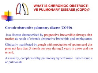
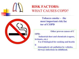
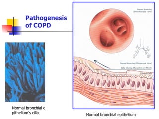
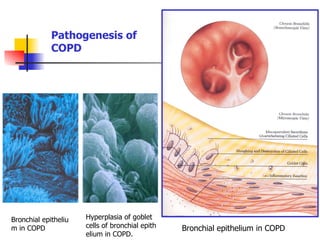
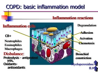
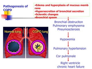
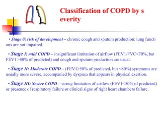
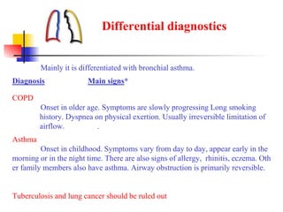
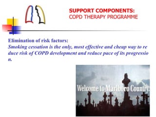
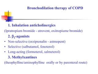
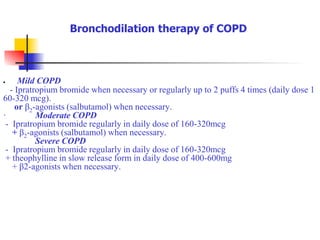
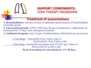
This document discusses chronic obstructive pulmonary disease (COPD). It defines COPD as a progressive lung disease characterized by airway obstruction due to chronic bronchitis and emphysema. The major risk factor is tobacco smoke. Symptoms include cough, sputum production and shortness of breath. Diagnosis involves lung function tests showing irreversible airway obstruction. Treatment focuses on smoking cessation, bronchodilators, antibiotics for exacerbations, oxygen therapy, and pulmonary rehabilitation.
2. WHAT IS CHRRONIOC OBSTRUCTI
VE PULMONARY DISEASE (COPD)?
Chronic obstructive pulmonary disease (COPD) –
-Is a disease characterized by progressive irreversible airways obst
ruction as result of chronic obstructive bronchitis and emphysema;
Clinically manifested by cough with production of sputum and dys
pnea not less than 3 month per year during 2 years in a row and mo
re and,
As usually, complicated by pulmonary hypertension and chronic c
or pulmonale.
3. RISK FACTORS:
WHAT CAUSES COPD?
Tobacco smoke – the
most important risk fac
tor of COPD
Other proven causes of C
OPD:
Industrial dust and chemicals (vapors,
irritants, etc.)
Use if biological for cooking and heatin
g.
Atmospheric air pollution by vehicles.
Airways infections in childhood.
7. Normal lung COPD lung
Bronchial obstruciton
Pulmonary emphysema
Pneumosclerosis
Hypoxemia
Pulmonary hypertension
Cor pulmonale
Right ventricle
chronic heart failure
Pathogenesis of
COPD
•Edema and hyperplasia of mucous memb
rane
•Hypersecretion of bronchial secretion
•Sclerotic changes
•Bronchial spasm
8. • Chronic cough.
• Chronic sputum production.
• Dyspnea: persistent (continues all the time), progressive, increases
in condition of physical exertion and in exacerbations
• Repetitive exacerbations of bronchitis are typical
• Risk factors in anamnesis:
• tobacco smoke (including local types of smoking),
• industrial dust and chemicals
DIAGNOSIS of COPD
9. •Inspection
in early stages there are no significant changes.
Later on:
•Skin cyanosis.
•Emphysematous chest.
• Percussion: bandbox resonance, lowering of lung borders and lim
itation of their mobility.
• Auscultation: diminished breathing, prolonged phase pf expiratio
n, diffuse dry rales, wheezing.
DIAGNOSIS of COPD
11. Instrumental:
Obligatory:
Radiological study of chest organs during first visit, then not less t
han once a year.
Lung function test (FEV1 or peakflowmetry) with bronchodilation
test in every visit.
If indicated:
ECG (for diagnostics of cor pulmonale, arrhythmias and conducti
on impairment).
Echo-CG – if available
DIAGNOSIS of COPD
12. Spirometry measures:
• Forced Vital Capacity (FVC)
• Forced Expiratory Volume on the 1 second (FEV1).
Bronchodilation test:
In COPD growth of FEV1 and PEF less than 15% after 10-30 m
inutes of inhalation of 2 doses of -agonists indicates irreversibi
lity or partial reversibility of bronchial obstruction.
Functional Diagnostics
13. • Stage 0: risk of development – chronic cough and sputum production; lung functi
ons are not impaired.
• Stage I: mild COPD – insignificant limitation of airflow (FEV1/FVC<70%, but
FEV1 >80% of predicted) and cough and sputum production are usual.
• Stage II: Moderate COPD – (FEV150% of predicted, but <80%) symptoms are
usually more severe, accompanied by dyspnea that appears in physical exertion.
• Stage III: Severe COPD – strong limitation of airflow (FEV1<50% of predicted)
or presence of respiratory failure or clinical signs of right heart chambers failure.
Classification of COPD by s
everity
14. Differential diagnostics
Mainly it is differentiated with bronchial asthma.
Diagnosis Main signs*
COPD
Onset in older age. Symptoms are slowly progressing Long smoking
history. Dyspnea on physical exertion. Usually irreversible limitation of
airflow. .
Asthma
Onset in childhood. Symptoms vary from day to day, appear early in the
morning or in the night time. There are also signs of allergy, rhinitis, eczema. Oth
er family members also have asthma. Airway obstruction is primarily reversible.
Tuberculosis and lung cancer should be ruled out
15. «Intersection» of COPD and Asthma
COPD Asthma
Neutrophiles Eosinophiles
No hyperreactivity Hyperreactivity
Steroid therapy
shows no effec
t
Steroid therapy
is effective
ASTHMATIC BRONCHITIS
~ 10%
16. SUPPORT COMPONENTS:
COPD THERAPY PROGRAMME
Elimination of risk factors:
Smoking cessation is the only, most effective and cheap way to re
duce risk of COPD development and reduce pace of its progressio
n.
17. Even brief, 3-minutes interview
with a smoker can stimulate him
for smoking cessation and it sho
uld take place during every visit
of each smoker.
SUPPORT COMPONENTS:
COPD THERAPY PROGRAMME
You have way out of the
cigarette jungle!
18. SUPPORT COMPONENTS:
COPD THERAPY PROGRAMME
Treatment of stable COPD
Long-term drug therapy
Bronchodilators: basic drugs for treatment of COPD symptoms
Mucolytic agents: play subsidiary role.
20. Mild COPD
- Ipratropium bromide when necessary or regularly up to 2 puffs 4 times (daily dose 1
60-320 mcg).
or β2-agonists (salbutamol) when necessary.
· Moderate COPD
- Ipratropium bromide regularly in daily dose of 160-320mcg
+ β2-agonists (salbutamol) when necessary.
Severe COPD
- Ipratropium bromide regularly in daily dose of 160-320mcg
+ theophylline in slow release form in daily dose of 400-600mg
+ β2-agonists when necessary.
Bronchodilation therapy of COPD
21. N-ACETYLCYSTEINE (АЦЦ-100/200, etc.) – derivation of L-cysteine, pro
duced in tablets, capsules, granules solution for inhalation.
CARBOCYSTEIN (Fluditek, etc) – analogue of acetylcysteine. Produced in
tablets, capsules, granules, solution for inhalation, syrup.
AMBROXAL HYDROCHLORIDE (Lasolvan, Ambrosan, etc.) produced i
n tablets by 30 mg., solution for IM, IV, and inhalation administration.
Mucolytic therapy of COPD
22. SUPPORT COMPONENTS:
COPD THERAPY PROGRAMME
Treatment of exacerbations
1. Bronchodilators: increase of dose or additional administration of bronchodilator
of another group.
2. Glucocorticosteroids: if FEV<50% then 30 mg of prednisone is additionally ad
ministered for 7-8 days with subsequent cessation.
3. Antibacterial agents: only if signs of inflammation of bronchial tree are present
.
1 Line drugs: Amoxicillin 0,5g 3 times a day or
Erythromycin 0,5g 4 times a day
2 line drugs: Amoxicillin/clavulanic acid 375-625 mg 3 times or
Doxycyclin 0,2 g once a day
Term of treatment of exacerbation is 5-7-10 days
23. Oxygen therapy: long-term administration of oxygen (more than 1
5 hours a day) in patients with chronic respiratory failure promotes
growth of survival rate and positively influences on pressure in pul
monary artery, polycythemia (haematocrit more than 55%), physica
l tolerance, lung function and emotional status.
Goal of long-term oxygen-therapy:
To increase РаО2 up to 8,0 кpа (60 mmHg)
and/or saturation SaО2 up to 90% (at sea level).
SUPPORT COMPONENTS:
COPD THERAPY PROGRAMME
24. SUPPORT COMPONENTS:
COPD THERAPY PROGRAMME
Non-drug therapy includes
Rehabilitation and oxygen therapy.
Rehabilitation must include at least following::
• Physical exercises (respiratory muscles training)
• Diet
• Education