Skull fractures result from direct impact and indicate a severe blow to the head, with their presence correlating with the likelihood of brain injury. They can be classified into types such as linear, basilar, depressed, and comminuted fractures, each with different clinical implications and complications, including the risk of intracranial infection, bleeding, and post-traumatic epilepsy. Diagnosis often requires imaging techniques like CT scans, and treatment varies based on fracture type and associated injuries.
![SKULL FRACTURES TrH5 (5)
1) open → infection.
2) depressed below level of inner table → underlying brain injury → posttraumatic
epilepsy.
3) overlie major dural venous sinus / middle meningeal artery → bleeding.
4) linear fractures associated with dural tear in young children → leptomeningeal cyst.
N.B. basilar fractures are most serious - deserve closer monitoring than linear vault fractures!
Bleeding
infants may bleed significantly intracranially from skull fractures (skull is very vascular – any
fracture may cause venous epidural hematoma); check hematocrit q 12-24 h.
EPIDURAL HEMATOMA is associated with skull fracture in ≈ 50% cases.
SUBDURAL HEMATOMA is associated with skull fracture in ≈ 33% cases.
Pneumocephalus
CSF leaks - otorrhea and rhinorrhea
Meningitis (via wound or CSF fistula); may extend into brain abscess.
Posttraumatic epilepsy (after depressed skull fractures) – risk factors: loss of consciousness for > 2
hours, associated dural tear, early seizures (within first week).
Superior longitudinal sinus compression (by depressed vertex fractures) → thrombosis.
extrusion (in form of cyst) of leptomeninges and brain
Leptomeningeal cyst (s. growing fracture) –
tissue through dural defect.
etiopathology: skull fracture with separation of fracture edges [depressed or diastatic skull
fracture] and dura laceration → arachnoid and brain are caught between edges of fracture →
brain pulsation forces CSF into cyst → skull erosion.
N.B. seen almost
exclusively in children < 1-3 yrs with fracture accompanied
by
dural
tear
– such children must be followed up closely for several months!!!
most are located in calvarium (rare sites are basiocciput and orbital roof).
underlying brain may herniate through skull defect.
prominent porencephalic cyst or focal dilatation of lateral ventricle usually underlies fracture.
types:
Type I - leptomeningeal cyst herniating through skull defect into subgaleal space.
Type II - damaged or gliotic brain.
Type III - porencephalic cyst is seen.
clinically (manifests several weeks ÷ months after fracture):
1) growing subgaleal mass (slowly expanding pulsatile nontender swelling in area of
previous skull fracture)
2) convulsive seizures or focal neurologic deficit
3) mental retardation
4) mass effect with increased ICP.
diagnosis: serial X-ray (sufficient for diagnosis*) – enlarging oval area of skull erosion
(progressive separation of long edges of seemingly benign linear skull fracture).
*but CT better defines exact pathology; intracranial hypoattenuating
area may be encephalomalacia, arachnoid loculation, or cortical atrophy.
tools for early diagnosis (→ early simpler surgical intervention → prevented long-term
neurologic sequelae):
1) MRI - depicts dural tears early.
2) ultrasound (tool for assessing state of dura).
treatment: cyst excision → dural closure → cranioplasty. See p. Op320 >>
– occasionally, shunt surgery is performed to decompress cyst and treat localized
dilatation of ventricles.
LATERAL SKULL RADIOGRAPH OF LEPTOMENINGEAL
CYST:
AXIAL CT OF LEPTOMENINGEAL CYST: WIDENED
FRACTURE ON LEFT AND FLUID COLLECTION EXTENDING
FROM INTRACRANIAL CAVITY INTO AND THROUGH
FRACTURE SITE:
Dislocation of bones of auricular chain (after temporal bone fracture). see below
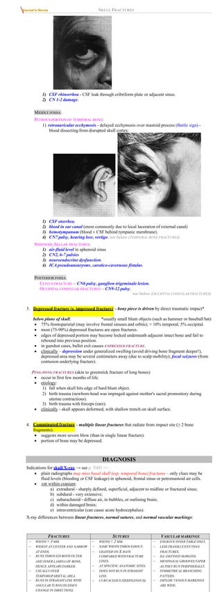
SPECIAL SITUATIONS
TEMPORAL BONE FRACTURES
fractured in 15-48% of all skull fractures (75% of all skull base fractures).
clinical features: Battle's sign, bleeding from ear (hemotympanum or from fracture line in ear
canal), CN7 & 8 damage, ossicular chain & tympanic disruption, CSF otorrhea.
Subtypes (by Ulrich, 1926)
LONGITUDINAL
1. (70-90%) - PARALLEL TO PETROUS PYRAMID:
PARS SQUAMOSA, POSTEROSUPERIOR WALL OF EXTERNAL
AUDITORY CANAL, TEGMEN TYMPANI → RUN EITHER
ANTERIOR OR POSTERIOR TO COCHLEA AND LABYRINTHINE
CAPSULE → END IN MIDDLE CRANIAL FOSSA NEAR FORAMEN
SPINOSUM OR IN MASTOID AIR CELLS, RESPECTIVELY.
CAUSED BY DIRECT LATERAL FORCE OVER MASTOID OR
SQUAMOUS BONE OR BLOW TO MANDIBLE.](https://image.slidesharecdn.com/dkdfcncr02izn07k409-220623191123-08e7231a/85/SKULL-FRACTURES-5-320.jpg)
![SKULL FRACTURES
2. TRANSVERSE (5-30%) - PERPENDICULAR TO PETROUS PYRAMID:
ORIGINATE AT FORAMEN MAGNUM → EXTEND THROUGH
COCHLEA AND LABYRINTH → END IN MIDDLE CRANIAL FOSSA.
CAUSED BY FRONTAL OR PARIETAL BLOW BUT MAY RESULT
FROM OCCIPITAL BLOW.
PNEUMOLABYRINTH MAY BE SIGN.
3. MIXED - components of both LONGITUDINAL and TRANSVERSE fractures.
Complications:
1) facial nerve paralysis (twice more common with transverse fracture):
a) delayed-incomplete – due to neurapraxia (10-20% longitudinal fractures); injury site
is usually horizontal segment distal to geniculate ganglion; H: steroids.
b) immediate-complete – due to nerve transection (50% transverse fractures); injury
site is anywhere from internal auditory canal to horizontal segment distal to
geniculate ganglion; decompression surgery is not always indicated (use
electroneuronography [ENOG] in decision making).
1) hearing loss (hemotympanum and mucosal edema in middle ear may cause temporary deafness -
resolves within ≈ 3 weeks):
a) conductive hearing loss due to hemotympanum, ossicular dislocation / fracture or
tympanic rupture (≈ 50% longitudinal fractures);
– incus (relatively loose ligamentous attachments) is most frequently
dislocated ossicle.
– most tympanic membrane perforations and hemotympanum usually
resolve in 3-4 weeks.
– if conductive hearing loss is present at > 30 dB after 3 months →
tympanoplasty with ossicular chain repair.
a) sensory hearing loss (≈ 80% transverse fractures); H: cochlear implants.
1) vertigo due to:
a) fracture extending into vestibular apparatus (e.g. with transverse fractures).
b) labyrinth concussion (e.g. with longitudinal fractures).
c) development of perilymphatic fistula (paroxysmal vertigo with fluctuating or
progressive hearing loss); H: exploratory tympanotomy.
d) posttraumatic benign paroxysmal positional vertigo.
2) CSF otorrhea (in any subtype of fracture).
3) unusual complications:
carotid injury.
CN6 paralysis (recovery within 6 months is usual).
CN5 damage.
sigmoid sinus thrombosis.
posttraumatic cholesteatoma (can grow undetected for years).
EAGLE syndrome (classically follows tonsillectomy; fracture of ossified styloid and
stylohyoid ligament can cause pressure on ECA or ICA → atypical pain referred to cheek or
eye; treatment is surgical).
sympathic cochleolabyrinthitis (autoimmune inner ear damage - autoantibodies against inner
ear proteins [as in polyarteritis nodosa]; H: immunosuppression).
TRANSVERSE FRACTURES nearly always produce facial
paralysis, permanent hearing loss, severe ablative vertigo.
Diagnosis - high-resolution CT (axial and coronal images) with 1-mm slices and magnified views;
bone windows alone are necessary.
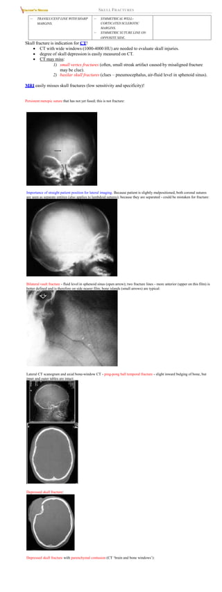
Longitudinal fracture of right temporal bone (axial CT) -
fracture line follows long axis of temporal bone (medium
arrowheads); incus is subluxed laterally (small
arrowhead); mastoid air cells opacified with blood (large
arrowhead):
Longitudinal fracture of right temporal bone (axial CT) -
fracture line follows long axis of temporal bone (small
arrowheads); fracture line is seen to cross area of
geniculate ganglion of CN7 (large arrowhead):
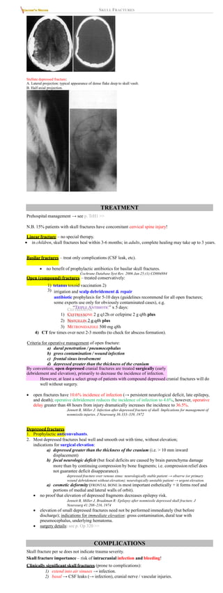
Transverse fracture of temporal bone (axial CT) - fracture
line (arrowheads) crosses petrous pyramid at level of
posterior semicircular canal and posterior genu of CN7
canal (arrow):
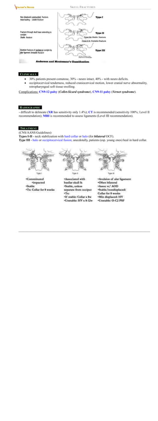
OCCIPITAL CONDYLAR FRACTURES
- very rare and serious injury.
ANDERSON AND MONTESANO TYPES
*preserved alar ligament and tectorial membrane](https://image.slidesharecdn.com/dkdfcncr02izn07k409-220623191123-08e7231a/85/SKULL-FRACTURES-6-320.jpg)