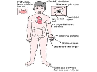
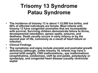
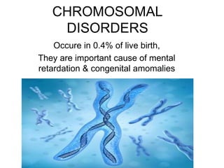
CHROMOSOMAL
DISORDERS
Occure in 0.4% of live birth,
They are important cause of mental
retardation & congenital amomalies
Chromosomal Abnormalities
• 1-Abnormalities of Chromosomal
Number,
When a human cell has 23 chromosomes, such as human ova or sperm,
it is in the haploid state (n). After conception, in cells other than the
reproductive cells, 46 chromosomes are present in the diploid state
(2n).. Trisomy, an example of aneuploidy, is the presence of three of a
particular chromosome rather than two. It results from unequal division,
called nondisjunction, of chromosomes into daughter cells. Trisomies
are the most common numerical chromosomal anomalies found in
humans (eg, trisomy 21 [Down syndrome], trisomy 18, and trisomy 13)..
• Sex Chromosomal Anomalies
Abnormalities involving sex chromosomes, including
aneuploidy and mosaicism, are relatively common in the
general population. The most common sex chromosome
anomalies include 45,X (Turner syndrome), 47,XXX, 47,XXY
(Klinefelter syndrome), 47,XYY, and different mosaic states
TRISOMY 21(DOWN SYNDROME)
• INCIDENCE,
• 1 in 750 of live birth,
• Increases with advanced maternal
age,<35yr.
• Mainly due meiotic nondisjunction,
• Translocation in 4% ,it accounts of 9% of
the children with Down syndrome born to
mother younger than 30 yr of age.
Trisomy 21 (Down Syndrome)
• Essentials of Diagnosis & Typical
Features
Small, brachycephalic head; characteristic facies of
up-slanting palpebral fissures, epicanthal folds,
midface hypoplasia, and small, dysplastic pinnae.
Generalized hypotonia.
Cognitive disabilities (mild to severe).
Associated with congenital heart disease and
gastrointestinal anomalies
TRISOMY 21
– CLINICAL FEATURES
. Cognitive disabilities are characteristic of Down syndrome, with
typical intelligence quotients (IQs) between 20 and 80 (mostly
between 45 and 55). Generalized hypotonia is common. Sexual
development is delayed, especially in males, who are usually sterile.
The affected newborn may have prolonged physiologic jaundice,
polycythemia, and a transient leukemoid reaction. Later, there is an
increased tendency for thyroid dysfunction, hearing loss, celiac
disease, and atlanto-occipital instability. Leukemia is 12–20 times
more common in Down syndrome patients than in unaffected
children
TRISOMY 21
• Clinical Findings
• The principal physical findings include a small,
brachycephalic head, characteristic facies (up-
slanting palpebral fissures, epicanthal folds,
midface hypoplasia, and small, dysplastic
pinnae), and minor limb abnormalities. About
one third to one half of children with Down
syndrome have congenital heart disease, most
often endocardial cushion defects or other septal
defects. Anomalies of the gastrointestinal tract,
including esophageal and duodenal atresias, are
seen in about 15% of cases.
DOWN SYNDROME
Medical Therapy
No convincing documentation is
available. However, interventions for
specific issues such as surgery or
medications for heart problems,
antibiotics for infections and thyroid
function tests, infant stimulation
programs, special education, and
physical, occupational, and speech
therapies are all indicated. The goal of
treatment is to help affected children
develop to their full potential. /.
Trisomy 18 Syndrome
Edwards Syndrome
• The incidence of trisomy 18 syndrome is about 1:4000 live
births, and the ratio of affected males to females is
approximately 1:3. Trisomy 18 is characterized by prenatal and
postnatal growth retardation that is often severe, and
hypertonicity. Complications are related to associated birth
defects. Death is often caused by heart failure or pneumonia
and usually occurs in infancy or early childhood, although a
small percentage of patients reach adulthood. Surviving
children show significant developmental delay.
• Clinical Findings
• Infants with trisomy 18 are often small for gestational age and
have dysmorphic features including a characteristic facies and
extremities (overlapping fingers and rockerbottom feet), and
congenital heart disease (often ventricular septal defect or
patent ductus arteriosus).
Trisomy 13 Syndrome
Patau Syndrome
• The incidence of trisomy 13 is about 1:12,000 live births, and
60% of affected individuals are female. Most infants with
trisomy 13 have congenital anomalies that are incompatible
with survival. Surviving children demonstrate failure to thrive,
developmental retardation, apneic spells, seizures, and
deafness. Death usually occurs in early infancy or by the
second year of life, commonly as a result of heart failure or
infection.
• Clinical Findings
• The symptoms and signs include prenatal and postnatal growth
deficiency (although, unlike trisomy 18, infants may have a
normal birth weight), CNS malformations,, eye malformations
(anophthalmia, colobomas), cleft lip and palate, polydactyly or
syndactyly, and congenital heart disease (usually ventricular
septal
Treatment of Trisomies
• There is no treatment, other than general
supportive care, for trisomy 13 or 18.
Genetic Counseling
• Most parents of trisomic infants have normal karyotypes. The
risk of having a child affected with a trisomy varies with
maternal age. For trisomy 21, age-specific risks are 1:2000 for
mothers younger than 25 years; 1:200 for mothers 35 years of
age; and 1:100 for mothers at age 40. The recurrence risk for
trisomy in future pregnancies is equal to 1:100 plus the age-
specific maternal risk;,.
• If the child has a trisomy resulting from a translocation, and the
parent has an abnormal karyotype, the risks are increased.
• The recurrence risks in other trisomies are same to those for
Down syndrome. The mother's age at the time of conception
and the nature of the chromosomal abnormality are important
in genetic counseling, which is indicated for prevention of all
chromosomal abnormalities. Prenatal diagnosis is available.
Sex Chromosome Abnormalities
• Turner Syndrome (Monosomy X, Gonadal
Dysgenesis)
• Klinefelter Syndrome (XXY)
• XYY Syndrome
• XXX Syndrome
• Chromosomal Abnormalities: Abnormal
Structure
Turner Syndrome (Monosomy X,
Gonadal Dysgenesis
• Essentials of Diagnosis & Typical Features
• Webbed neck, triangular facies, short stature, wide-set
nipples, amenorrhea, and absence of secondary sex
characteristics.
• Associated with coarctation of the aorta and
genitourinary malformations.
• IQ is usually normal but learning disabilities are
common.
• Mosaic individuals may manifest only short stature and
²_x0000__x0000_€
orrhea.
• The incidence of Turner syndrome is 1:10,000 females.
Clinical Findings
• Newborns with Turner syndrome may have webbed neck,
edema of the hands and feet, coarctation of the aorta, and a
characteristic triangular facies. Later symptoms include short
stature, a shield chest with wide-set nipples, streak ovaries,
amenorrhea, absence of secondary sex characteristics, and
infertility. Some affected girls, particularly those with
mosaicism, have only short stature and amenorrhea, without
dysmorphic features.
• Complications relate primarily to coarctation of the aorta, when
present. Rarely, the dysgenetic gonads may become neoplastic
(gonadoblastoma). The incidence of malformations of the
urinary tract is increased. Learning disabilities are common,
secondary to difficulties in perceptual motor integration.
Patients with pseudohypoparathyroidism and Noonan
syndrome have a similar phenotype to patients with Turner
syndrome, but have normal chromosomes.
TURNER SYNDROME
• Treatment
• In Turner syndrome. Teenage patients need counseling to cope
with the stigma of their condition and to understand the need
for hormone therapy. Estrogen replacement therapy will permit
development of secondary sex characteristics and normal
menstruation and prevent osteoporosis. Growth hormone
therapy has been used to increase the height of affected girls.
Females with 45,X or 45,X mosaicism have a low fertility rate,
and those who become pregnant have a high risk (spontaneous
miscarriage, ~30%; stillbirth, 6–10%). Furthermore, their
liveborn offspring have an increased frequency of chromosomal
abnormalities involving either sex chromosomes or autosomes,
and congenital malformations. Thus, prenatal ultrasonography
and chromosome analysis are indicated for the offspring of
females with sex chromosome abnormalities.
Klinefelter Syndrome (XXY)
• Essentials of Diagnosis & Typical Features
• Diagnosis is rarely made before puberty.
• Key findings include microorchidism; lack of libido; minimal
facial hair; and tall, eunuchoid build.
• IQ can vary (normal to borderline or even severely retarded,
depending on karyotype).
• The incidence of Klinefelter syndrome in the newborn
population is roughly 1:1000, but it is about 1% among mentally
retarded males and about 3% among males seen at infertility
clinics. The maternal age at birth is often advanced. Unlike
Turner syndrome, Klinefelter syndrome is rarely the cause of
spontaneous abortions. The diagnosis is seldom made before
puberty except as a result of prenatal diagnosis, because
prepubertal boys have a normal phenotype
Klinefelter syndrome
• .
• Treatment
• Males with Klinefelter syndrome require
testosterone replacement therapy.
Sex chromosome abnormalities
– XYY Syndrome
• Newborns with XYY syndrome in general are normal. Affected
individuals may on occasion exhibit an abnormal behavior
pattern from early childhood and may have mild retardation.
Fertility may be normal. Many males with an XYY karyotype are
normal. There is no treatment. Long-term problems may relate
to low IQ and environmental stress.
• XXX Syndrome
• The incidence of females with an XXX karyotype is
approximately 1:1000. Females with XXX are phenotypically
normal. However, they tend to be taller than usual and to have
lower IQs than their normal siblings. Learning and behavioral
issues are relatively common. This is in contrast to individuals
with XXXX, a much rarer condition causing more severe
developmental issues, and a dysmorphic phenotype
reminiscent of Down syndrome.
Chromosomal Abnormalities:
Abnormal Structure
• Chromosomal abnormalities most often present in
newborns as multiple congenital anomalies in
association with intrauterine growth retardation.. In some
cases, the karyotype is normal, but a subtle
chromosomal rearrangement can be detected by new
technology, called comparative genomic hybridization
array,.
• Although most cases of severe chromosomal
abnormality such as trisomy are lethal, some individuals
may survive if the abnormality exists in mosaic form.
Two examples of this include trisomy 8 and cat eye
syndrome, caused by extra genetic material, which is
derived from a portion of chromosome 22.
Chromosomal Abnormalities:
Abnormal Structure
• Chromosome Deletion Disorders
• Deletion 1p36 Syndrome
• Microcephaly and a large anterior fontanelle are characteristic features of 1p36–
syndrome. Cardiac defects are common, and dilated cardiomyopathy may present in
infancy. Mental retardation, hypotonia, hearing loss, and seizures are usually seen.
• Wolf-Hirschhorn Syndrome
• Also known as 4p– (deletion of 4p16), this syndrome is characterized by
microcephaly and unusual development of the nose and orbits suggesting an ancient.
Other anomalies commonly seen include cleft lip and palate, and cardiac and renal
defects. Seizure disorders are common, and the majority of patients have severe
mental retardation.
• Cri Du Chat Syndrome
• Also known as 5p– (deletion of terminal chromosome 5p), this disorder is
characterized by unique facial features, growth retardation, and microcephaly.
Patients have an unusual catlike cry. Most patients have major organ anomalies and
significant developmental delay.

CHROMOSOMAL DISORDERS

  • 1.
    CHROMOSOMAL DISORDERS Occure in 0.4%of live birth, They are important cause of mental retardation & congenital amomalies
  • 2.
    Chromosomal Abnormalities • 1-Abnormalitiesof Chromosomal Number, When a human cell has 23 chromosomes, such as human ova or sperm, it is in the haploid state (n). After conception, in cells other than the reproductive cells, 46 chromosomes are present in the diploid state (2n).. Trisomy, an example of aneuploidy, is the presence of three of a particular chromosome rather than two. It results from unequal division, called nondisjunction, of chromosomes into daughter cells. Trisomies are the most common numerical chromosomal anomalies found in humans (eg, trisomy 21 [Down syndrome], trisomy 18, and trisomy 13)..
  • 3.
    • Sex ChromosomalAnomalies Abnormalities involving sex chromosomes, including aneuploidy and mosaicism, are relatively common in the general population. The most common sex chromosome anomalies include 45,X (Turner syndrome), 47,XXX, 47,XXY (Klinefelter syndrome), 47,XYY, and different mosaic states
  • 4.
    TRISOMY 21(DOWN SYNDROME) •INCIDENCE, • 1 in 750 of live birth, • Increases with advanced maternal age,<35yr. • Mainly due meiotic nondisjunction, • Translocation in 4% ,it accounts of 9% of the children with Down syndrome born to mother younger than 30 yr of age.
  • 5.
    Trisomy 21 (DownSyndrome) • Essentials of Diagnosis & Typical Features Small, brachycephalic head; characteristic facies of up-slanting palpebral fissures, epicanthal folds, midface hypoplasia, and small, dysplastic pinnae. Generalized hypotonia. Cognitive disabilities (mild to severe). Associated with congenital heart disease and gastrointestinal anomalies
  • 7.
    TRISOMY 21 – CLINICALFEATURES . Cognitive disabilities are characteristic of Down syndrome, with typical intelligence quotients (IQs) between 20 and 80 (mostly between 45 and 55). Generalized hypotonia is common. Sexual development is delayed, especially in males, who are usually sterile. The affected newborn may have prolonged physiologic jaundice, polycythemia, and a transient leukemoid reaction. Later, there is an increased tendency for thyroid dysfunction, hearing loss, celiac disease, and atlanto-occipital instability. Leukemia is 12–20 times more common in Down syndrome patients than in unaffected children
  • 8.
    TRISOMY 21 • ClinicalFindings • The principal physical findings include a small, brachycephalic head, characteristic facies (up- slanting palpebral fissures, epicanthal folds, midface hypoplasia, and small, dysplastic pinnae), and minor limb abnormalities. About one third to one half of children with Down syndrome have congenital heart disease, most often endocardial cushion defects or other septal defects. Anomalies of the gastrointestinal tract, including esophageal and duodenal atresias, are seen in about 15% of cases.
  • 9.
    DOWN SYNDROME Medical Therapy Noconvincing documentation is available. However, interventions for specific issues such as surgery or medications for heart problems, antibiotics for infections and thyroid function tests, infant stimulation programs, special education, and physical, occupational, and speech therapies are all indicated. The goal of treatment is to help affected children develop to their full potential. /.
  • 10.
    Trisomy 18 Syndrome EdwardsSyndrome • The incidence of trisomy 18 syndrome is about 1:4000 live births, and the ratio of affected males to females is approximately 1:3. Trisomy 18 is characterized by prenatal and postnatal growth retardation that is often severe, and hypertonicity. Complications are related to associated birth defects. Death is often caused by heart failure or pneumonia and usually occurs in infancy or early childhood, although a small percentage of patients reach adulthood. Surviving children show significant developmental delay. • Clinical Findings • Infants with trisomy 18 are often small for gestational age and have dysmorphic features including a characteristic facies and extremities (overlapping fingers and rockerbottom feet), and congenital heart disease (often ventricular septal defect or patent ductus arteriosus).
  • 12.
    Trisomy 13 Syndrome PatauSyndrome • The incidence of trisomy 13 is about 1:12,000 live births, and 60% of affected individuals are female. Most infants with trisomy 13 have congenital anomalies that are incompatible with survival. Surviving children demonstrate failure to thrive, developmental retardation, apneic spells, seizures, and deafness. Death usually occurs in early infancy or by the second year of life, commonly as a result of heart failure or infection. • Clinical Findings • The symptoms and signs include prenatal and postnatal growth deficiency (although, unlike trisomy 18, infants may have a normal birth weight), CNS malformations,, eye malformations (anophthalmia, colobomas), cleft lip and palate, polydactyly or syndactyly, and congenital heart disease (usually ventricular septal
  • 13.
    Treatment of Trisomies •There is no treatment, other than general supportive care, for trisomy 13 or 18.
  • 14.
    Genetic Counseling • Mostparents of trisomic infants have normal karyotypes. The risk of having a child affected with a trisomy varies with maternal age. For trisomy 21, age-specific risks are 1:2000 for mothers younger than 25 years; 1:200 for mothers 35 years of age; and 1:100 for mothers at age 40. The recurrence risk for trisomy in future pregnancies is equal to 1:100 plus the age- specific maternal risk;,. • If the child has a trisomy resulting from a translocation, and the parent has an abnormal karyotype, the risks are increased. • The recurrence risks in other trisomies are same to those for Down syndrome. The mother's age at the time of conception and the nature of the chromosomal abnormality are important in genetic counseling, which is indicated for prevention of all chromosomal abnormalities. Prenatal diagnosis is available.
  • 15.
    Sex Chromosome Abnormalities •Turner Syndrome (Monosomy X, Gonadal Dysgenesis) • Klinefelter Syndrome (XXY) • XYY Syndrome • XXX Syndrome • Chromosomal Abnormalities: Abnormal Structure
  • 16.
    Turner Syndrome (MonosomyX, Gonadal Dysgenesis • Essentials of Diagnosis & Typical Features • Webbed neck, triangular facies, short stature, wide-set nipples, amenorrhea, and absence of secondary sex characteristics. • Associated with coarctation of the aorta and genitourinary malformations. • IQ is usually normal but learning disabilities are common. • Mosaic individuals may manifest only short stature and ²_x0000__x0000_€ orrhea. • The incidence of Turner syndrome is 1:10,000 females.
  • 18.
    Clinical Findings • Newbornswith Turner syndrome may have webbed neck, edema of the hands and feet, coarctation of the aorta, and a characteristic triangular facies. Later symptoms include short stature, a shield chest with wide-set nipples, streak ovaries, amenorrhea, absence of secondary sex characteristics, and infertility. Some affected girls, particularly those with mosaicism, have only short stature and amenorrhea, without dysmorphic features. • Complications relate primarily to coarctation of the aorta, when present. Rarely, the dysgenetic gonads may become neoplastic (gonadoblastoma). The incidence of malformations of the urinary tract is increased. Learning disabilities are common, secondary to difficulties in perceptual motor integration. Patients with pseudohypoparathyroidism and Noonan syndrome have a similar phenotype to patients with Turner syndrome, but have normal chromosomes.
  • 19.
    TURNER SYNDROME • Treatment •In Turner syndrome. Teenage patients need counseling to cope with the stigma of their condition and to understand the need for hormone therapy. Estrogen replacement therapy will permit development of secondary sex characteristics and normal menstruation and prevent osteoporosis. Growth hormone therapy has been used to increase the height of affected girls. Females with 45,X or 45,X mosaicism have a low fertility rate, and those who become pregnant have a high risk (spontaneous miscarriage, ~30%; stillbirth, 6–10%). Furthermore, their liveborn offspring have an increased frequency of chromosomal abnormalities involving either sex chromosomes or autosomes, and congenital malformations. Thus, prenatal ultrasonography and chromosome analysis are indicated for the offspring of females with sex chromosome abnormalities.
  • 20.
    Klinefelter Syndrome (XXY) •Essentials of Diagnosis & Typical Features • Diagnosis is rarely made before puberty. • Key findings include microorchidism; lack of libido; minimal facial hair; and tall, eunuchoid build. • IQ can vary (normal to borderline or even severely retarded, depending on karyotype). • The incidence of Klinefelter syndrome in the newborn population is roughly 1:1000, but it is about 1% among mentally retarded males and about 3% among males seen at infertility clinics. The maternal age at birth is often advanced. Unlike Turner syndrome, Klinefelter syndrome is rarely the cause of spontaneous abortions. The diagnosis is seldom made before puberty except as a result of prenatal diagnosis, because prepubertal boys have a normal phenotype
  • 22.
    Klinefelter syndrome • . •Treatment • Males with Klinefelter syndrome require testosterone replacement therapy.
  • 23.
    Sex chromosome abnormalities –XYY Syndrome • Newborns with XYY syndrome in general are normal. Affected individuals may on occasion exhibit an abnormal behavior pattern from early childhood and may have mild retardation. Fertility may be normal. Many males with an XYY karyotype are normal. There is no treatment. Long-term problems may relate to low IQ and environmental stress. • XXX Syndrome • The incidence of females with an XXX karyotype is approximately 1:1000. Females with XXX are phenotypically normal. However, they tend to be taller than usual and to have lower IQs than their normal siblings. Learning and behavioral issues are relatively common. This is in contrast to individuals with XXXX, a much rarer condition causing more severe developmental issues, and a dysmorphic phenotype reminiscent of Down syndrome.
  • 24.
    Chromosomal Abnormalities: Abnormal Structure •Chromosomal abnormalities most often present in newborns as multiple congenital anomalies in association with intrauterine growth retardation.. In some cases, the karyotype is normal, but a subtle chromosomal rearrangement can be detected by new technology, called comparative genomic hybridization array,. • Although most cases of severe chromosomal abnormality such as trisomy are lethal, some individuals may survive if the abnormality exists in mosaic form. Two examples of this include trisomy 8 and cat eye syndrome, caused by extra genetic material, which is derived from a portion of chromosome 22.
  • 25.
    Chromosomal Abnormalities: Abnormal Structure •Chromosome Deletion Disorders • Deletion 1p36 Syndrome • Microcephaly and a large anterior fontanelle are characteristic features of 1p36– syndrome. Cardiac defects are common, and dilated cardiomyopathy may present in infancy. Mental retardation, hypotonia, hearing loss, and seizures are usually seen. • Wolf-Hirschhorn Syndrome • Also known as 4p– (deletion of 4p16), this syndrome is characterized by microcephaly and unusual development of the nose and orbits suggesting an ancient. Other anomalies commonly seen include cleft lip and palate, and cardiac and renal defects. Seizure disorders are common, and the majority of patients have severe mental retardation. • Cri Du Chat Syndrome • Also known as 5p– (deletion of terminal chromosome 5p), this disorder is characterized by unique facial features, growth retardation, and microcephaly. Patients have an unusual catlike cry. Most patients have major organ anomalies and significant developmental delay.