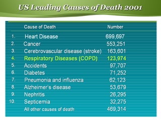
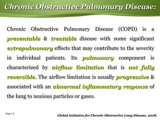
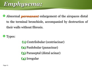
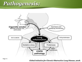
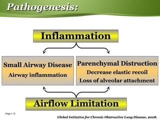
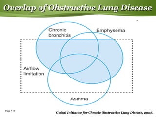
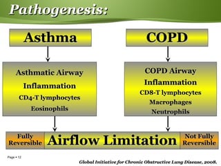
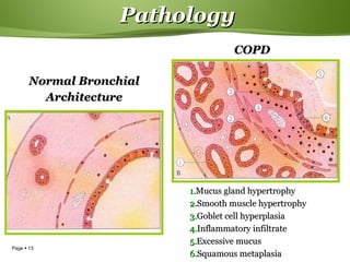
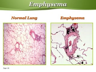
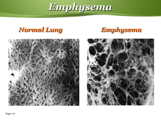
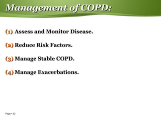
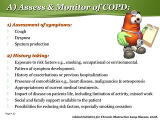
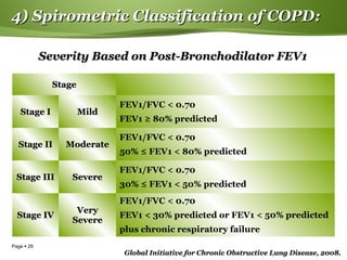
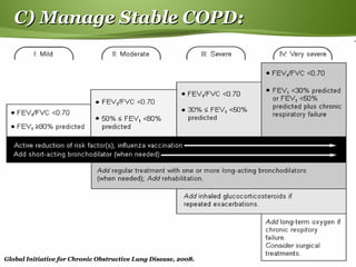
This document provides information on chronic obstructive pulmonary disease (COPD). It discusses the epidemiology, definition, risk factors, pathogenesis, pathology, classification, management, and exacerbations of COPD. Key points include: cigarette smoking is the primary cause of COPD worldwide; the disease involves inflammation in the lungs from noxious particles leading to airflow limitation; emphysema and chronic bronchitis are the major pathological changes; severity is classified based on lung function tests; and management involves reducing risk factors, treating stable COPD, and managing exacerbations.
Chronic Obstructive Pulmonary Disease (COPD) is an umbrella term used to describe progressive lung diseases including emphysema, chronic bronchitis, and refractory (non-reversible) asthma. This disease is characterized by increasing breathlessness
Pneumonia is an inflammation of the lung parenchyma caused by various microorganisms, including bacteria, mycobacteria, fungi, and viruses.
Pneumonitis is a more general term that describes the inflammatory process in the lung tissue that may predispose and Pneumonia is an inflammation of the lung parenchyma that is caused by a microbial agent.
place the patient at risk for microbial invasion.
Pneumonia is classified into four: community-acquired pneumonia (CAP) and hospital-acquired pneumonia (HAP), pneumonia in the immunocompromised host, and aspiration pneumonia.
Pneumonia is an inflammatory condition of the lung affecting primarily the small air sacs known as alveoli. Typically symptoms include some combination of productive or dry cough, chest pain, fever, and trouble breathing. Severity is variable.
Pneumonia is usually caused by infection with viruses or bacteria and less commonly by other microorganisms, certain medications and conditions such as autoimmune diseases. Risk factors include cystic fibrosis, chronic obstructive pulmonary disease (COPD), asthma, diabetes, heart failure, a history of smoking, a poor ability to cough such as following a stroke, and a weak immune system. Diagnosis is often based on the symptoms and physical examination. Chest X-ray, blood tests, and culture of the sputum may help confirm the diagnosis. The disease may be classified by where it was acquired with community, hospital, or health care associated pneumonia.
Vaccines to prevent certain types of pneumonia are available. Other methods of prevention include handwashing and not smoking. Treatment depends on the underlying cause. Pneumonia believed to be due to bacteria is treated with antibiotics. If the pneumonia is severe, the affected person is generally hospitalized. Oxygen therapy may be used if oxygen levels are low.
Pneumonia affects approximately 450 million people globally (7% of the population) and results in about four million deaths per year. Pneumonia was regarded by William Osler in the 19th century as "the captain of the men of death". With the introduction of antibiotics and vaccines in the 20th century, survival improved. Nevertheless, in developing countries, and among the very old, the very young, and the chronically ill, pneumonia remains a leading cause of death. Pneumonia often shortens suffering among those already close to death and has thus been called "the old man's friend"
Chronic Obstructive Pulmonary Disease (COPD) is an umbrella term used to describe progressive lung diseases including emphysema, chronic bronchitis, and refractory (non-reversible) asthma. This disease is characterized by increasing breathlessness
This lecture elucidates in detail the important occupational health diseases, the all-important concept of work absenteeism and, lastly, the prevention of occupational diseases.
Abstract Lung Abscess is a liquefactive necrosis of the lung tissue and arrangement of cavitation (in excess of 2 cm) containing necrotic debris and liquid brought about by parenchymal infection. It very well may be brought about by yearning, which may happen during changed cognizance and it for the most part causes a discharge filled depression. In addition, liquor addiction is the most widely recognized condition inclining to lung abscesses. Lung abscess is viewed as essential (60%) when it comes about because of existing lung parenchymal process and is named auxiliary when it entangles another procedure, e.g., vascular emboli or follows rupture of extrapulmonary abscess into lung. There are a few imaging strategies which can distinguish the material inside the thorax, for example, electronic tomography (CT) output of the thorax and ultrasound of the thorax. Broad Spectrum anti-biotics to cover blended vegetation is the pillar of treatment. Pneumonic physiotherapy and postural drainage are additionally significant. Surgeries are required in specific patients for pneumonic resection Keywords: Lung abscess, anti-bodies, video-assissted thoracoscopic medical procedure (VATS), thoracoscopy
Similar to Chronic obstructive pulmonary disease (20)
Introduction:
RNA interference (RNAi) or Post-Transcriptional Gene Silencing (PTGS) is an important biological process for modulating eukaryotic gene expression.
It is highly conserved process of posttranscriptional gene silencing by which double stranded RNA (dsRNA) causes sequence-specific degradation of mRNA sequences.
dsRNA-induced gene silencing (RNAi) is reported in a wide range of eukaryotes ranging from worms, insects, mammals and plants.
This process mediates resistance to both endogenous parasitic and exogenous pathogenic nucleic acids, and regulates the expression of protein-coding genes.
What are small ncRNAs?
micro RNA (miRNA)
short interfering RNA (siRNA)
Properties of small non-coding RNA:
Involved in silencing mRNA transcripts.
Called “small” because they are usually only about 21-24 nucleotides long.
Synthesized by first cutting up longer precursor sequences (like the 61nt one that Lee discovered).
Silence an mRNA by base pairing with some sequence on the mRNA.
Discovery of siRNA?
The first small RNA:
In 1993 Rosalind Lee (Victor Ambros lab) was studying a non- coding gene in C. elegans, lin-4, that was involved in silencing of another gene, lin-14, at the appropriate time in the
development of the worm C. elegans.
Two small transcripts of lin-4 (22nt and 61nt) were found to be complementary to a sequence in the 3' UTR of lin-14.
Because lin-4 encoded no protein, she deduced that it must be these transcripts that are causing the silencing by RNA-RNA interactions.
Types of RNAi ( non coding RNA)
MiRNA
Length (23-25 nt)
Trans acting
Binds with target MRNA in mismatch
Translation inhibition
Si RNA
Length 21 nt.
Cis acting
Bind with target Mrna in perfect complementary sequence
Piwi-RNA
Length ; 25 to 36 nt.
Expressed in Germ Cells
Regulates trnasposomes activity
MECHANISM OF RNAI:
First the double-stranded RNA teams up with a protein complex named Dicer, which cuts the long RNA into short pieces.
Then another protein complex called RISC (RNA-induced silencing complex) discards one of the two RNA strands.
The RISC-docked, single-stranded RNA then pairs with the homologous mRNA and destroys it.
THE RISC COMPLEX:
RISC is large(>500kD) RNA multi- protein Binding complex which triggers MRNA degradation in response to MRNA
Unwinding of double stranded Si RNA by ATP independent Helicase
Active component of RISC is Ago proteins( ENDONUCLEASE) which cleave target MRNA.
DICER: endonuclease (RNase Family III)
Argonaute: Central Component of the RNA-Induced Silencing Complex (RISC)
One strand of the dsRNA produced by Dicer is retained in the RISC complex in association with Argonaute
ARGONAUTE PROTEIN :
1.PAZ(PIWI/Argonaute/ Zwille)- Recognition of target MRNA
2.PIWI (p-element induced wimpy Testis)- breaks Phosphodiester bond of mRNA.)RNAse H activity.
MiRNA:
The Double-stranded RNAs are naturally produced in eukaryotic cells during development, and they have a key role in regulating gene expression .
THE IMPORTANCE OF MARTIAN ATMOSPHERE SAMPLE RETURN.Sérgio Sacani
The return of a sample of near-surface atmosphere from Mars would facilitate answers to several first-order science questions surrounding the formation and evolution of the planet. One of the important aspects of terrestrial planet formation in general is the role that primary atmospheres played in influencing the chemistry and structure of the planets and their antecedents. Studies of the martian atmosphere can be used to investigate the role of a primary atmosphere in its history. Atmosphere samples would also inform our understanding of the near-surface chemistry of the planet, and ultimately the prospects for life. High-precision isotopic analyses of constituent gases are needed to address these questions, requiring that the analyses are made on returned samples rather than in situ.
Cancer cell metabolism: special Reference to Lactate PathwayAADYARAJPANDEY1
Normal Cell Metabolism:
Cellular respiration describes the series of steps that cells use to break down sugar and other chemicals to get the energy we need to function.
Energy is stored in the bonds of glucose and when glucose is broken down, much of that energy is released.
Cell utilize energy in the form of ATP.
The first step of respiration is called glycolysis. In a series of steps, glycolysis breaks glucose into two smaller molecules - a chemical called pyruvate. A small amount of ATP is formed during this process.
Most healthy cells continue the breakdown in a second process, called the Kreb's cycle. The Kreb's cycle allows cells to “burn” the pyruvates made in glycolysis to get more ATP.
The last step in the breakdown of glucose is called oxidative phosphorylation (Ox-Phos).
It takes place in specialized cell structures called mitochondria. This process produces a large amount of ATP. Importantly, cells need oxygen to complete oxidative phosphorylation.
If a cell completes only glycolysis, only 2 molecules of ATP are made per glucose. However, if the cell completes the entire respiration process (glycolysis - Kreb's - oxidative phosphorylation), about 36 molecules of ATP are created, giving it much more energy to use.
IN CANCER CELL:
Unlike healthy cells that "burn" the entire molecule of sugar to capture a large amount of energy as ATP, cancer cells are wasteful.
Cancer cells only partially break down sugar molecules. They overuse the first step of respiration, glycolysis. They frequently do not complete the second step, oxidative phosphorylation.
This results in only 2 molecules of ATP per each glucose molecule instead of the 36 or so ATPs healthy cells gain. As a result, cancer cells need to use a lot more sugar molecules to get enough energy to survive.
introduction to WARBERG PHENOMENA:
WARBURG EFFECT Usually, cancer cells are highly glycolytic (glucose addiction) and take up more glucose than do normal cells from outside.
Otto Heinrich Warburg (; 8 October 1883 – 1 August 1970) In 1931 was awarded the Nobel Prize in Physiology for his "discovery of the nature and mode of action of the respiratory enzyme.
WARNBURG EFFECT : cancer cells under aerobic (well-oxygenated) conditions to metabolize glucose to lactate (aerobic glycolysis) is known as the Warburg effect. Warburg made the observation that tumor slices consume glucose and secrete lactate at a higher rate than normal tissues.
(May 29th, 2024) Advancements in Intravital Microscopy- Insights for Preclini...Scintica Instrumentation
Intravital microscopy (IVM) is a powerful tool utilized to study cellular behavior over time and space in vivo. Much of our understanding of cell biology has been accomplished using various in vitro and ex vivo methods; however, these studies do not necessarily reflect the natural dynamics of biological processes. Unlike traditional cell culture or fixed tissue imaging, IVM allows for the ultra-fast high-resolution imaging of cellular processes over time and space and were studied in its natural environment. Real-time visualization of biological processes in the context of an intact organism helps maintain physiological relevance and provide insights into the progression of disease, response to treatments or developmental processes.
In this webinar we give an overview of advanced applications of the IVM system in preclinical research. IVIM technology is a provider of all-in-one intravital microscopy systems and solutions optimized for in vivo imaging of live animal models at sub-micron resolution. The system’s unique features and user-friendly software enables researchers to probe fast dynamic biological processes such as immune cell tracking, cell-cell interaction as well as vascularization and tumor metastasis with exceptional detail. This webinar will also give an overview of IVM being utilized in drug development, offering a view into the intricate interaction between drugs/nanoparticles and tissues in vivo and allows for the evaluation of therapeutic intervention in a variety of tissues and organs. This interdisciplinary collaboration continues to drive the advancements of novel therapeutic strategies.
Seminar of U.V. Spectroscopy by SAMIR PANDASAMIR PANDA
Spectroscopy is a branch of science dealing the study of interaction of electromagnetic radiation with matter.
Ultraviolet-visible spectroscopy refers to absorption spectroscopy or reflect spectroscopy in the UV-VIS spectral region.
Ultraviolet-visible spectroscopy is an analytical method that can measure the amount of light received by the analyte.
The increased availability of biomedical data, particularly in the public domain, offers the opportunity to better understand human health and to develop effective therapeutics for a wide range of unmet medical needs. However, data scientists remain stymied by the fact that data remain hard to find and to productively reuse because data and their metadata i) are wholly inaccessible, ii) are in non-standard or incompatible representations, iii) do not conform to community standards, and iv) have unclear or highly restricted terms and conditions that preclude legitimate reuse. These limitations require a rethink on data can be made machine and AI-ready - the key motivation behind the FAIR Guiding Principles. Concurrently, while recent efforts have explored the use of deep learning to fuse disparate data into predictive models for a wide range of biomedical applications, these models often fail even when the correct answer is already known, and fail to explain individual predictions in terms that data scientists can appreciate. These limitations suggest that new methods to produce practical artificial intelligence are still needed.
In this talk, I will discuss our work in (1) building an integrative knowledge infrastructure to prepare FAIR and "AI-ready" data and services along with (2) neurosymbolic AI methods to improve the quality of predictions and to generate plausible explanations. Attention is given to standards, platforms, and methods to wrangle knowledge into simple, but effective semantic and latent representations, and to make these available into standards-compliant and discoverable interfaces that can be used in model building, validation, and explanation. Our work, and those of others in the field, creates a baseline for building trustworthy and easy to deploy AI models in biomedicine.
Bio
Dr. Michel Dumontier is the Distinguished Professor of Data Science at Maastricht University, founder and executive director of the Institute of Data Science, and co-founder of the FAIR (Findable, Accessible, Interoperable and Reusable) data principles. His research explores socio-technological approaches for responsible discovery science, which includes collaborative multi-modal knowledge graphs, privacy-preserving distributed data mining, and AI methods for drug discovery and personalized medicine. His work is supported through the Dutch National Research Agenda, the Netherlands Organisation for Scientific Research, Horizon Europe, the European Open Science Cloud, the US National Institutes of Health, and a Marie-Curie Innovative Training Network. He is the editor-in-chief for the journal Data Science and is internationally recognized for his contributions in bioinformatics, biomedical informatics, and semantic technologies including ontologies and linked data.
Multi-source connectivity as the driver of solar wind variability in the heli...Sérgio Sacani
The ambient solar wind that flls the heliosphere originates from multiple
sources in the solar corona and is highly structured. It is often described
as high-speed, relatively homogeneous, plasma streams from coronal
holes and slow-speed, highly variable, streams whose source regions are
under debate. A key goal of ESA/NASA’s Solar Orbiter mission is to identify
solar wind sources and understand what drives the complexity seen in the
heliosphere. By combining magnetic feld modelling and spectroscopic
techniques with high-resolution observations and measurements, we show
that the solar wind variability detected in situ by Solar Orbiter in March
2022 is driven by spatio-temporal changes in the magnetic connectivity to
multiple sources in the solar atmosphere. The magnetic feld footpoints
connected to the spacecraft moved from the boundaries of a coronal hole
to one active region (12961) and then across to another region (12957). This
is refected in the in situ measurements, which show the transition from fast
to highly Alfvénic then to slow solar wind that is disrupted by the arrival of
a coronal mass ejection. Our results describe solar wind variability at 0.5 au
but are applicable to near-Earth observatories.
2. Page 2
Contents of the Lecture:Contents of the Lecture:
EpidemiologyEpidemiology
DefinitionDefinition
Risk factorsRisk factors
PathogenesisPathogenesis
PathologyPathology
ClassificationClassification
ManagementManagement
COPD exacerbationsCOPD exacerbations
3. Page 3
Epidemiology:Epidemiology:
Cigarette smoking is the primary cause of COPD.Cigarette smoking is the primary cause of COPD.
The WHO estimates 1.3 billion smokers worldwide,The WHO estimates 1.3 billion smokers worldwide,
increasing to 2 billion by 2025.increasing to 2 billion by 2025.
In 2000, the WHO estimated 2.74 million deathsIn 2000, the WHO estimated 2.74 million deaths
worldwide from COPD.worldwide from COPD.
In 1990, COPD was rankedIn 1990, COPD was ranked 1212thth
as a burden of disease.as a burden of disease.
After 25 years of smoking, at least 25% of smokers without
initial disease will have clinically significant COPD
4. Page 4
US Leading Causes of Death 2001US Leading Causes of Death 2001
5. Page 5
Chronic Obstructive Pulmonary Disease:Chronic Obstructive Pulmonary Disease:
Chronic Obstructive Pulmonary Disease (COPD) is aChronic Obstructive Pulmonary Disease (COPD) is a
preventablepreventable && treatabletreatable disease with some significantdisease with some significant
extrapulmonaryextrapulmonary effects that may contribute to the severityeffects that may contribute to the severity
in individual patients. Itsin individual patients. Its pulmonarypulmonary component iscomponent is
characterized bycharacterized by airflow limitationairflow limitation that isthat is not fullynot fully
reversible.reversible. The airflow limitation is usuallyThe airflow limitation is usually progressiveprogressive &&
associated with anassociated with an abnormal inflammatory responseabnormal inflammatory response ofof
the lung to noxious particles or gases.the lung to noxious particles or gases.
Global Initiative for Chronic Obstructive Lung Disease, 2008.Global Initiative for Chronic Obstructive Lung Disease, 2008.
6. Page 6
Emphysema:Emphysema:
AbnormalAbnormal permanentpermanent enlargement of the airspaces distalenlargement of the airspaces distal
to the terminal bronchiole, accompanied by destruction ofto the terminal bronchiole, accompanied by destruction of
their walls without fibrosis.their walls without fibrosis.
Types:Types:
(1)(1) Centrilobular (centriacinar)Centrilobular (centriacinar)
(2)(2) Panlobular (panacinar)Panlobular (panacinar)
(3)(3) Paraseptal (distal acinar)Paraseptal (distal acinar)
(4)(4) IrregularIrregular
7. Page 7
Chronic bronchitis:Chronic bronchitis:
Presence ofPresence of chronic productive coughchronic productive cough forfor
3 months3 months in each ofin each of
2 successive years2 successive years in a patient in whomin a patient in whom otherother
causescauses ofof chronic coughchronic cough have beenhave been
excluded.excluded.
8. Page 8
Risk Factors:Risk Factors:
Genes
Exposure to particles
1. Tobacco smoke
2. Occupational dust, organic & inorganic
3. Indoor air pollution
4. Outdoor air pollution
Lung growth & development
Oxidative stress
Gender
Age
Respiratory infections
Previous TB
Socioeconomic status
Nutrition
Comorbidities
14. Page 14
Healthy Respiratory MucosaHealthy Respiratory Mucosa
This EM shows theThis EM shows the
respiratory mucosa in arespiratory mucosa in a
healthy state.healthy state.
The cells are fully ciliated.The cells are fully ciliated.
The cilia beat in a co-The cilia beat in a co-
ordinated fashion to moveordinated fashion to move
mucus out of the airwaysmucus out of the airways
(mucociliary transport).(mucociliary transport).
Scanning electron micrograph showing aScanning electron micrograph showing a
sheet of mucus being moved along by the ciliasheet of mucus being moved along by the cilia
15. Page 15
Damaged Respiratory MucosaDamaged Respiratory Mucosa
Damage to the cilia & epitheliumDamage to the cilia & epithelium
occur as a result of diseaseoccur as a result of disease
processes in COPD. This can alsoprocesses in COPD. This can also
occur as a result of bacterialoccur as a result of bacterial
damage.damage.
This slide shows the result ofThis slide shows the result of
bacterial infection stripping awaybacterial infection stripping away
the cilia from the mucosa.the cilia from the mucosa.
The damage to the cilia meansThe damage to the cilia means
they are less effective in removingthey are less effective in removing
mucus from the airwaysmucus from the airways
Scanning electron micrograph showing cilialScanning electron micrograph showing cilial
and epithelial damage induced by bacteriaand epithelial damage induced by bacteria
16. Page 16
Centriacinar EmphysemaCentriacinar Emphysema
Characterized by focal destruction limited to the respiratory bronchioles &Characterized by focal destruction limited to the respiratory bronchioles &
the central portions of acinus.the central portions of acinus.
Is is associated with cigarette smoking & is most severe in the upper lobes.Is is associated with cigarette smoking & is most severe in the upper lobes.
17. Page 17
Panacinar EmphysemaPanacinar Emphysema
It involves the entire alveolus distal to the terminal bronchiole.It involves the entire alveolus distal to the terminal bronchiole.
It is most severe in the lower lung zones & generally develops in patientsIt is most severe in the lower lung zones & generally develops in patients
with homozygous alpha1-antitrypsin (AAT) deficiency.with homozygous alpha1-antitrypsin (AAT) deficiency.
18. Page 18
Distal acinar EmphysemaDistal acinar Emphysema
Distal acinar emphysema orDistal acinar emphysema or
paraseptal emphysema, is theparaseptal emphysema, is the
least common form and involvesleast common form and involves
distal airway structures, alveolardistal airway structures, alveolar
ducts, and sacs.ducts, and sacs.
This form of emphysema isThis form of emphysema is
localized to fibrous septa or tolocalized to fibrous septa or to
the pleura & leads to formationthe pleura & leads to formation
of bullae.of bullae.
The apical bullae may causeThe apical bullae may cause
pneumothorax.pneumothorax.
Paraseptal emphysema is notParaseptal emphysema is not
associated with airflowassociated with airflow
obstruction.obstruction.
22. Page 22
Management of COPD:Management of COPD:
(1)(1) Assess and Monitor Disease.Assess and Monitor Disease.
(2)(2) Reduce Risk Factors.Reduce Risk Factors.
(3)(3) Manage Stable COPD.Manage Stable COPD.
(4)(4) Manage Exacerbations.Manage Exacerbations.
23. Page 23
A) Assess & Monitor of COPD:A) Assess & Monitor of COPD:
1) Assessment of symptoms:1) Assessment of symptoms:
CoughCough
DyspneaDyspnea
Sputum productionSputum production
2) History taking:2) History taking:
Exposure to risk factors e.g., smoking, occupational or environmental.Exposure to risk factors e.g., smoking, occupational or environmental.
Pattern of symptom development.Pattern of symptom development.
History of exacerbations or previous hospitalizationsHistory of exacerbations or previous hospitalizations
Presence of comorbidities e.g., heart disease, malignancies & osteoporosisPresence of comorbidities e.g., heart disease, malignancies & osteoporosis
Appropriateness of current medical treatments.Appropriateness of current medical treatments.
Impact of disease on patients life, including limitation of activity, missed workImpact of disease on patients life, including limitation of activity, missed work
Social and family support available to the patientSocial and family support available to the patient
Possibilities for reducing risk factors, especially smoking cessationPossibilities for reducing risk factors, especially smoking cessation
Global Initiative for Chronic Obstructive Lung Disease, 2008.Global Initiative for Chronic Obstructive Lung Disease, 2008.
25. Page 25
Chronic Cough with Normal CXRChronic Cough with Normal CXR
IntrathoracicIntrathoracic
Chronic obstructive pulmonary diseaseChronic obstructive pulmonary disease
Bronchial asthmaBronchial asthma
Central bronchial carcinomaCentral bronchial carcinoma
Endobronchial tuberculosisEndobronchial tuberculosis
BronchiectasisBronchiectasis
Left sided heart failureLeft sided heart failure
Interstitial lung diseaseInterstitial lung disease
Cystic fibrosisCystic fibrosis
ExtrathoracicExtrathoracic
Postnasal dripPostnasal drip
Gastroesophageal refluxGastroesophageal reflux
Drug therapy (e.g., ACE inhibitors)Drug therapy (e.g., ACE inhibitors)
Global Initiative for Chronic Obstructive Lung Disease, 2008.Global Initiative for Chronic Obstructive Lung Disease, 2008.
26. Page 26
Systemic manifestations of COPD:Systemic manifestations of COPD:
Skeletal muscle wastingSkeletal muscle wasting
Cachexia: loss of fat-free massCachexia: loss of fat-free mass
Lung cancer (SCLC & NSCLC)Lung cancer (SCLC & NSCLC)
Pulmonary hypertensionPulmonary hypertension
Ischaemic heart diseaseIschaemic heart disease
Congestive cardiac failureCongestive cardiac failure
OsteoporosisOsteoporosis
Normocytic anaemiaNormocytic anaemia
DiabetesDiabetes
Metabolic syndromeMetabolic syndrome
Obstructive sleep apneaObstructive sleep apnea
DepressionDepression
27. Page 27
Central cyanosisCentral cyanosis
Hyperinflated chestHyperinflated chest
Increased resting respiratory rate with shallow breathingIncreased resting respiratory rate with shallow breathing
Pursed-lip breathingPursed-lip breathing
Respiratory distressRespiratory distress
Lower limb edemaLower limb edema
Difficulty in detection of heart apexDifficulty in detection of heart apex
Resonant bare area of the heartResonant bare area of the heart
Downward displacement of the liverDownward displacement of the liver
Distant breath soundsDistant breath sounds
Wheezy chestWheezy chest
Global Initiative for Chronic Obstructive Lung Disease, 2008.Global Initiative for Chronic Obstructive Lung Disease, 2008.
3) Physical Examination:3) Physical Examination:
29. Page 29
4) Spirometric Classification of COPD:4) Spirometric Classification of COPD:
Severity Based on Post-Bronchodilator FEV1Severity Based on Post-Bronchodilator FEV1
StageStage
Stage IStage I MildMild
FEV1/FVC < 0.70
FEV1 ≥ 80% predicted
Stage IIStage II ModerateModerate
FEV1/FVC < 0.70
50% ≤ FEV1 < 80% predicted
Stage IIIStage III SevereSevere
FEV1/FVC < 0.70
30% ≤ FEV1 < 50% predicted
Stage IVStage IV
VeryVery
SevereSevere
FEV1/FVC < 0.70
FEV1 < 30% predicted or FEV1 < 50% predicted
plus chronic respiratory failure
Global Initiative for Chronic Obstructive Lung Disease, 2008.Global Initiative for Chronic Obstructive Lung Disease, 2008.
30. Page 30
5) Bronchodilator Reversibility Testing:5) Bronchodilator Reversibility Testing:
PreparationPreparation
Patients should be clinically stable & free from respiratory infection.Patients should be clinically stable & free from respiratory infection.
Patients should not have taken inhaled short-acting bronchodilators in thePatients should not have taken inhaled short-acting bronchodilators in the
previous 6 hrs, long-acting bronchodilator in the previous 12 hrs, or sustainedprevious 6 hrs, long-acting bronchodilator in the previous 12 hrs, or sustained
release theophylline in the previous 24 hrs.release theophylline in the previous 24 hrs.
SpirometrySpirometry
FEV1 should be measured before a bronchodilator is given.FEV1 should be measured before a bronchodilator is given.
The bronchodilator should be given by metered dose inhaler through a spacerThe bronchodilator should be given by metered dose inhaler through a spacer
device or by nebulizer.device or by nebulizer.
Possible dosage protocols are 400 gPossible dosage protocols are 400 g ββ2-agonist, up to 160 g anticholinergic, or2-agonist, up to 160 g anticholinergic, or
the two combined.the two combined.
FEV1 should be measured again 10-15 minutes after a short-actingFEV1 should be measured again 10-15 minutes after a short-acting
bronchodilator is given; 30-45 minutes after the combination.bronchodilator is given; 30-45 minutes after the combination.
ResultsResults
Increase in FEV1 both > 200 ml & 12% above pre-bronchodilator FEV1 isIncrease in FEV1 both > 200 ml & 12% above pre-bronchodilator FEV1 is
considered significant.considered significant.
Global Initiative for Chronic Obstructive Lung Disease, 2008.Global Initiative for Chronic Obstructive Lung Disease, 2008.
31. Page 31
6) Lung Volumes:6) Lung Volumes:
The three volumes most relevant to COPD are forced vital
capacity (FVC), residual volume (RV), & total lung capacity
(TLC).
32. Page 32
7) Exercise Testing:7) Exercise Testing:
Measurement COPD
VO2max Decreased
Anaerobic threshold Normal/decreased/indeterminate
Peak HR Decreased, Normal in mild
O2 pulse Normal or decreased
(VE/MVV) X 100 Increased
VE/VCO2 (at AT) Increased
VD/VT Increased
PaO2 Variable
P(A-a)O2 Variable, usually increased
33. Page 33
8) Arterial Blood Gases:8) Arterial Blood Gases:
In advanced COPD, measurement of ABGs should be performed in stableIn advanced COPD, measurement of ABGs should be performed in stable
patients with FEV1 < 50% predicted or with clinical signs of respiratorypatients with FEV1 < 50% predicted or with clinical signs of respiratory
failure or right heart failure.failure or right heart failure.
Changes in arterial blood gas tensions take time to occur. Thus, 20-30Changes in arterial blood gas tensions take time to occur. Thus, 20-30
minutes should pass before rechecking the gas tensions when the FIO2minutes should pass before rechecking the gas tensions when the FIO2
has been changed, e.g., during assessment for domiciliary oxygen therapy.has been changed, e.g., during assessment for domiciliary oxygen therapy.
Adequate pressure must be applied at the arterial puncture site for atAdequate pressure must be applied at the arterial puncture site for at
least one minute, as failure to do so can lead to painful bruising.least one minute, as failure to do so can lead to painful bruising.
Global Initiative for Chronic Obstructive Lung Disease, 2008.Global Initiative for Chronic Obstructive Lung Disease, 2008.
35. Page 35
Differential Diagnosis of COPD:Differential Diagnosis of COPD:
DiagnosisDiagnosis Suggestive FeaturesSuggestive Features
COPDCOPD
Onset in mid-life.Onset in mid-life.
Symptoms slowly progressive.Symptoms slowly progressive.
Long history of tobacco smoking.Long history of tobacco smoking.
Dyspnea during exercise.Dyspnea during exercise.
Largely irreversible airflow limitation.Largely irreversible airflow limitation.
AsthmaAsthma
Onset early in life (often childhood).Onset early in life (often childhood).
Symptoms vary from day to day.Symptoms vary from day to day.
Symptoms at night/early morning.Symptoms at night/early morning.
Allergy, rhinitis, and/or eczema also present.Allergy, rhinitis, and/or eczema also present.
Family history of asthma.Family history of asthma.
Largely reversible airflow limitation.Largely reversible airflow limitation.
CongestiveCongestive
Heart FailureHeart Failure
Fine basilar crackles on auscultation.Fine basilar crackles on auscultation.
Chest X-ray shows dilated heart, pulmonary edemaChest X-ray shows dilated heart, pulmonary edema
Pulmonary function tests indicate volume restriction, not airflowPulmonary function tests indicate volume restriction, not airflow
limitation.limitation.
BronchiectasiBronchiectasi
ss
Onset all agesOnset all ages
Chest X-ray shows lung infiltrate.Chest X-ray shows lung infiltrate.
Microbiological confirmation.Microbiological confirmation.
36. Page 36
B) Reduce Risk Factors:B) Reduce Risk Factors:
1.1. SmokingSmoking
2.2. Occupational exposureOccupational exposure
3.3. Indoor/Outdoor air pollutionIndoor/Outdoor air pollution
37. Page 37
Smoking Cessation:Smoking Cessation:
ASK:ASK: Identify all tobacco users at every visit.Identify all tobacco users at every visit.
ADVISE:ADVISE: Strongly urge all tobacco users to quit.Strongly urge all tobacco users to quit.
ASSESS:ASSESS: Willingness to make a quit attempt.Willingness to make a quit attempt.
ASSIST:ASSIST: Aid the patient in quitting.Aid the patient in quitting.
ARRANGE:ARRANGE: Schedule follow-up contact.Schedule follow-up contact.
38. Page 38
COPD Risk & Smoking CessationCOPD Risk & Smoking Cessation
Adapted from Fletcher C et al. Br Med J. 1977;1:1645–1648.
Stopped smokingStopped smoking
at 45 (mild COPD)at 45 (mild COPD)
Stopped smokingStopped smoking
at 65 (severe COPD)at 65 (severe COPD)
40. Page 40
Global Initiative for Chronic Obstructive Lung Disease, 2008.Global Initiative for Chronic Obstructive Lung Disease, 2008.
C) Manage Stable COPDC) Manage Stable COPD::
41. Page 41
Pharmacological TherapyPharmacological Therapy::
The medications for COPD currently available can reduce
or abolish symptoms, increase exercise capacity, reduce
the number and severity of exacerbations, and improve
health status.
At present, no treatment has been shown to modify the
rate of decline in lung function.
The inhaled route is preferred.
42. Page 42
Pharmacological TherapyPharmacological Therapy:: BronchodilatorsBronchodilators
Three types of bronchodilators are available:Three types of bronchodilators are available:
1.1. β-agonistsβ-agonists
2.2. Anticholinergic drugsAnticholinergic drugs
3.3. Methylxanthines.Methylxanthines.
β-agonistsβ-agonists
Salbutamol (Ventolin)Salbutamol (Ventolin)
Sameterol (Servent)Sameterol (Servent)
Formoterol (Foradil)Formoterol (Foradil)
TerbutalinTerbutalin
AnticholinergicsAnticholinergics
Ibrtropuim bromideIbrtropuim bromide
(Atrovent)(Atrovent)
Tiotropuim bromideTiotropuim bromide
(Spiriva)(Spiriva)
MethylxanthineMethylxanthine
AminophyllineAminophylline
(Uniphylline,(Uniphylline,
Quibron, Theo SR)Quibron, Theo SR)
46. Page 46
C) Manage Stable COPD:C) Manage Stable COPD:
Global Initiative for Chronic Obstructive Lung Disease, 2008.Global Initiative for Chronic Obstructive Lung Disease, 2008.
47. Page 47
Long-term Oxygen Therapy:Long-term Oxygen Therapy:
Long-term oxygen therapy (LTOT) improves survival,
exercise, sleep and cognitive performance.
Physiological indications for oxygen include an arterial
oxygen tension (Pa,O2) <55 mmHg guided by ABGs.
The therapeutic goal is to maintain Sa,O2 >90% during
rest, sleep and exertion.
If oxygen was prescribed during an exacerbation, recheck
ABGs after 30–90 days.
49. Page 49
RehabilitationRehabilitation
For the lungs to get more airFor the lungs to get more air
PURSED-LIP BREATHINGPURSED-LIP BREATHING
(like breathing out slowly into a straw)(like breathing out slowly into a straw)
INHALEINHALE EXHALEEXHALE
50. Page 50
RehabilitationRehabilitation
Sit comfortablySit comfortably
&&
relax your shouldersrelax your shoulders
Put one hand on yourPut one hand on your
abdomen. Now inhaleabdomen. Now inhale
slowly through yourslowly through your
nose. (Push yournose. (Push your
abdomen out while youabdomen out while you
breathe in)breathe in)
Then push in yourThen push in your
abdominal musclesabdominal muscles
and breathe out usingand breathe out using
the pursed-lipthe pursed-lip
techniquetechnique
For the lungs to get more airFor the lungs to get more air
DIAPHRAGMATIC BREATHINGDIAPHRAGMATIC BREATHING
51. Page 51
Nutrition:Nutrition:
Weight loss & depletion of fat-free mass (FFM) may be observed inWeight loss & depletion of fat-free mass (FFM) may be observed in
stable COPD patients.stable COPD patients.
Being underweight is associated with an increased mortality risk.Being underweight is associated with an increased mortality risk.
Criteria to define weight loss are:Criteria to define weight loss are:
Weight loss >10% in the past 6 months or >5% in the past month.Weight loss >10% in the past 6 months or >5% in the past month.
Nutritional therapy may only be effective if combined with exercise orNutritional therapy may only be effective if combined with exercise or
other anabolic stimuli.other anabolic stimuli.
UnderweightUnderweight BMI <21 kgBMI <21 kg··mm-2-2
;age >50 yrs;age >50 yrs
Normal weightNormal weight BMI <21–25 kgBMI <21–25 kg··mm-2-2
OverweightOverweight BMI <30 kgBMI <30 kg··mm-2-2
ObeseObese BMIBMI ≥≥30 kg30 kg··mm-2-2
53. Page 53
What's a COPD Exacerbation?What's a COPD Exacerbation?
An exacerbation of COPD is defined as an event in theAn exacerbation of COPD is defined as an event in the
natural course of the disease characterized by a change innatural course of the disease characterized by a change in
the patients baseline dyspnea, cough, and/or sputum that isthe patients baseline dyspnea, cough, and/or sputum that is
beyond normal day-to-day variations, is acute in onset, andbeyond normal day-to-day variations, is acute in onset, and
may warrant a change in regular medication in a patientmay warrant a change in regular medication in a patient
with underlying COPD.with underlying COPD.
Global Initiative for Chronic Obstructive Lung Disease, 2008.Global Initiative for Chronic Obstructive Lung Disease, 2008.
54. Page 54
Anthonisen's Typing of COPD ExacerbationAnthonisen's Typing of COPD Exacerbation
Cardinal SignsCardinal Signs
Worsening dyspnea, increase in sputum volume & purulenceWorsening dyspnea, increase in sputum volume & purulence
Other SignsOther Signs
Upper respiratory tract infection in past 5 days, fever without otherUpper respiratory tract infection in past 5 days, fever without other
apparent cause, wheezing, increase cough & increase respiratoryapparent cause, wheezing, increase cough & increase respiratory
rate or heart rate by 20% above baselinerate or heart rate by 20% above baseline
All 3 cardinal symptomsAll 3 cardinal symptoms Type 1 (SEVERE)Type 1 (SEVERE)
2 of 3 cardinal symptoms2 of 3 cardinal symptoms Type 2 (MODERATE)Type 2 (MODERATE)
1 of 3 cardinal symptoms1 of 3 cardinal symptoms Type 3 (MILD)Type 3 (MILD)
55. Page 55
Causes of COPD ExacerbationsCauses of COPD Exacerbations
56. Page 56
D) Manage Exacerbations:D) Manage Exacerbations:
Assessment of COPD ExacerbationsAssessment of COPD Exacerbations
Medical HistoryMedical History
Severity of FEV1Severity of FEV1
Duration of worsening or newDuration of worsening or new
symptomssymptoms
Number of previousNumber of previous
(exacerbations/hospitalizations)(exacerbations/hospitalizations)
ComordibitiesComordibities
Present treatment regimenPresent treatment regimen
Signs of SeveritySigns of Severity
Use of accessory respiratoryUse of accessory respiratory
musclesmuscles
Paradoxical chest wall movementsParadoxical chest wall movements
Worsening or new central cyanosisWorsening or new central cyanosis
Peripheral edemaPeripheral edema
Hemodynamic instabilityHemodynamic instability
Signs of right heart failureSigns of right heart failure
Reduced alertnessReduced alertness
57. Page 57
Indications for Hospitalization in AECB:Indications for Hospitalization in AECB:
Marked increase in symptoms, e.g. sudden development of restingMarked increase in symptoms, e.g. sudden development of resting
dyspnea.dyspnea.
Severe underlying COPDSevere underlying COPD
Onset of new physical signs (e.g., cyanosis, peripheral edema)Onset of new physical signs (e.g., cyanosis, peripheral edema)
Failure of exacerbation to respond to initial medical managementFailure of exacerbation to respond to initial medical management
Significant comorbidities.Significant comorbidities.
Frequent exacerbations.Frequent exacerbations.
Newly occurring arrhythmias.Newly occurring arrhythmias.
Diagnostic uncertainty.Diagnostic uncertainty.
Older age.Older age.
Insufficient home support.Insufficient home support.
58. Page 58
Indications for ICU admission in AECB:Indications for ICU admission in AECB:
Severe dyspnea that responds inadequately to initialSevere dyspnea that responds inadequately to initial
emergency therapy.emergency therapy.
Changes in mental status (Confusion, lethargy, coma).Changes in mental status (Confusion, lethargy, coma).
Persistent or worsening hypoxemia (PaO2<40 mmHg),Persistent or worsening hypoxemia (PaO2<40 mmHg),
and/or severe/worsening hypercapnia (PaCO2>60 mmHg),and/or severe/worsening hypercapnia (PaCO2>60 mmHg),
and/or severe/worsening acidosis (pH<7.25) despiteand/or severe/worsening acidosis (pH<7.25) despite
supplemental oxygen & non-invasive ventilation.supplemental oxygen & non-invasive ventilation.
Need for invasive mechanical ventilation.Need for invasive mechanical ventilation.
Hemodynamic instability – need for vasopressors.Hemodynamic instability – need for vasopressors.
Average adult inspires and expires around 6 litres of air from the lungs every minute
The mucociliary escalator is the primary mechanism within the lungs
It beigns in the nose and extends throughout the respiratory tract
This esculator is contiually active, therefore propelling foreign matter out of the respiratory tract
Cigarette smoke compromises the action of the cilla and results in mucus being retained for longer periods
Thus increasing risk of infection, the causing symptoms such as productive cough and thus gives rise to an obstructive pattern.
Dilation of alveolar wall
↓ alveolar capillary network, loss of guy rope effect
↓ lung tissue elasticity
Caused by smoking » irritation » inflammation » neutrophils and macrophages » release neutrophil elastase (type of proteases)
The forced vital capacity is the maximum volume of air which can be forcibly expelled after inhaling as deeply as possible. Not all of the air in the lungs is removed when measuring the vital capacity. The amount remaining is called the residual volume. The total lung capacity is the combination of the forced vital capacity and residual volume. While most of the measured lung volumes or capacities change to some degree with COPD, residual volume usually increases quite markedly. This increase is the result of the weakened airways collapsing before all the normally expired air can leave the lungs. The increased residual volume makes breathing even more difficult and labored.
Remove hyperinflated areas of lung:
Improve V/Q matching
Reduce resting length of respiratory muscles
Reduce Dynamic Hyperinflation