Downloaded 175 times
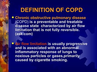


This document provides an overview of Chronic Obstructive Pulmonary Disease (COPD). It defines COPD as a preventable and treatable lung disease characterized by limited airflow. The two main conditions that make up COPD are chronic bronchitis and emphysema. Chronic bronchitis involves long-term inflammation of the bronchial tubes, while emphysema involves breakdown of lung tissue. Cigarette smoking is the primary cause of COPD. Symptoms include shortness of breath, cough, and sputum production. Diagnosis involves patient history, exams, pulmonary function tests, chest x-rays, and blood tests. Management focuses on smoking cessation, medications like bronchodilators, oxygen therapy, pulmonary rehabilitation


