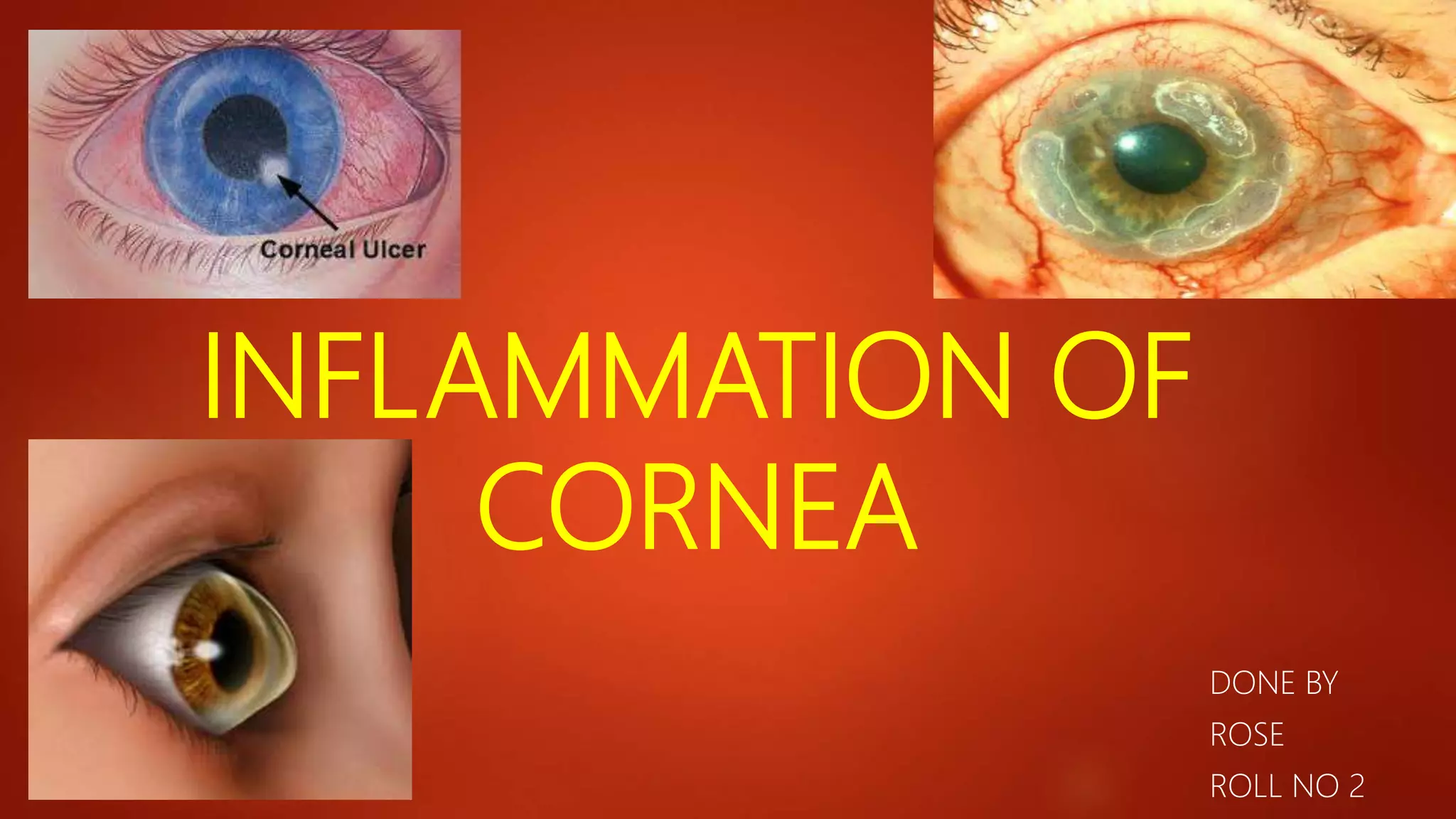
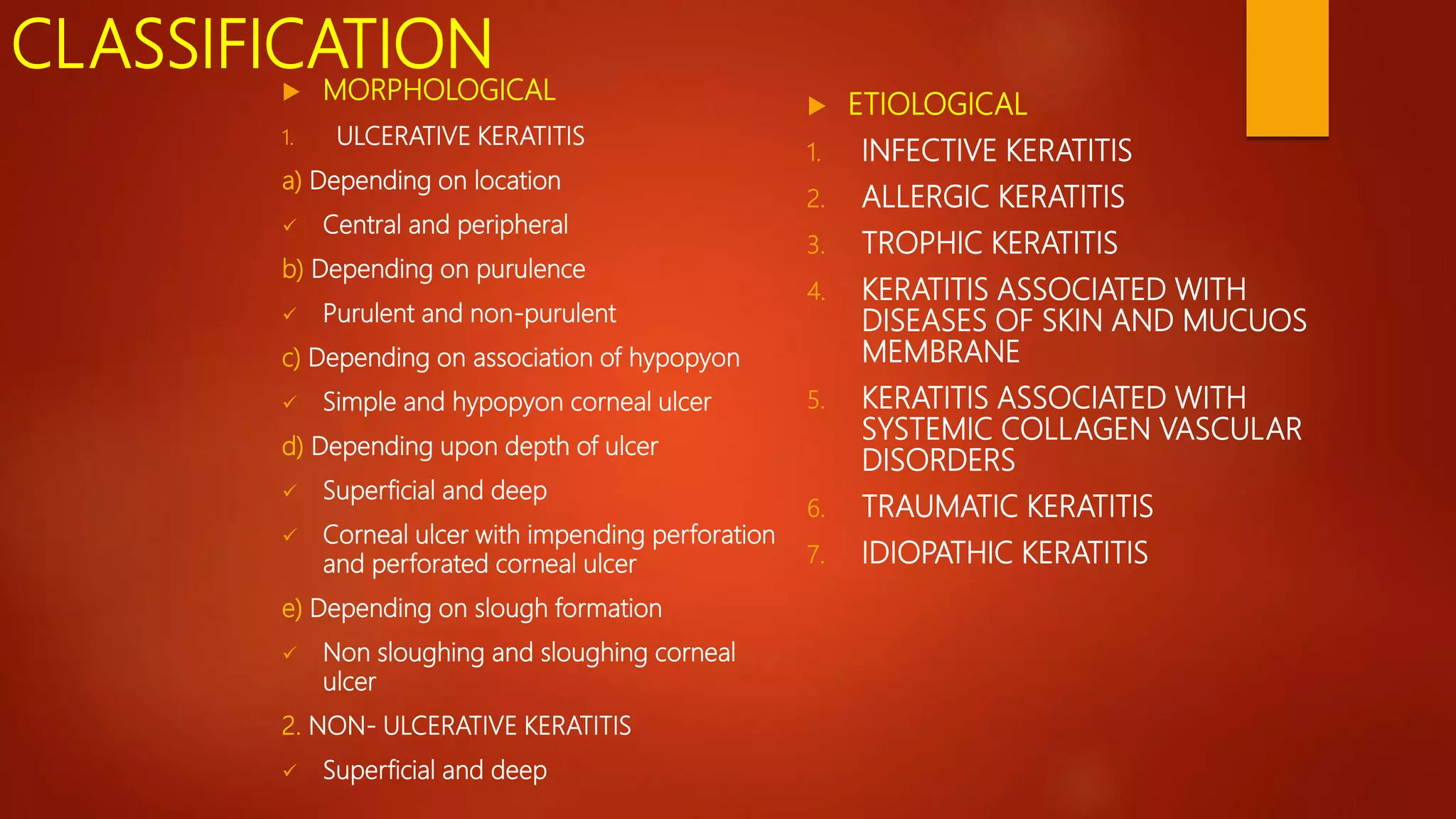
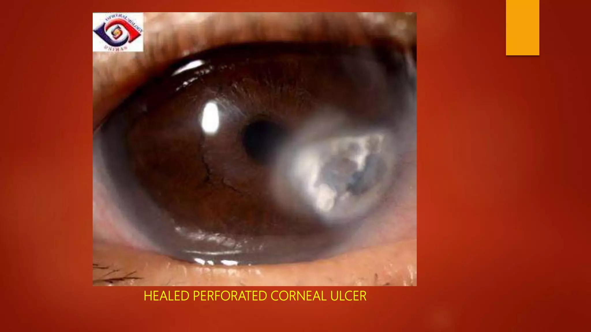
This document describes different types of keratitis, including ulcerative keratitis, non-ulcerative keratitis, and infective keratitis caused by bacteria, fungi, viruses, and protozoa. It discusses the classification, etiology, signs, diagnosis, and management of various forms of corneal inflammation and infection. Key pathogens mentioned include Staphylococcus aureus, Pseudomonas, herpes simplex virus, acanthamoeba, and fungi like Aspergillus and Fusarium.
This presentation describes the background of the cornea and the corneal diseases in general, also it describes in detailed manner how to manage the corneal ulcer with its different causes
BLEPHARITIS
Blepharitis is a chronic inflammation of the lid margins.
Etiology
1. It follows chronic conjunctivitis due to Staphyloccocus in
debilitated children usually who are living in poor hygienic
conditions.
2. Parasites such as Demodex folliculorum, Phthiriasis
palpebrarum, crab louse, head louse also cause blepharitis.
A stye is an infection (abcess) of one of the small oil producing glands lining the eyelid, usually caused by the bacteria that are normally found along the eyelids.
A stye can occur on either the upper or lower eyelid.
There are two types of styes, internal and external hordeola.
An internal hordeolum (stye) is a bacterial infection of the meibomian glands inside the eyelids.
Internal styes tend to be more severe and occur a little less often than an external hordeolum.
An external hordeolum (stye) is a bacterial infection of the Glands of Zeis and/or Glands of Moll inside the eyelids.
This type of stye is more superficial and tends to heal more readily.
It is a chronic lipogranulomatous inflammatory lesion caused by blockage of meibomian gland orifices and stagnation of sebaceous secretion.
Patient with acne rosacea or seborhoeic dermatitis are at increased risk of chalazion formation which may be multiple or recurrent.
If it is recurrent, one should think of sebaceous gland carcinoma
Ectropion
It is an outward turning of the eyelid margin . This more frequently affects the lower eyelid.Upper eyelid ectropion is uncommon.Classified in 5 types
1)Congenital 2) Involutional 3) Paralytic 4) Cicatricial 5) Mechanical
Involutional ectropion is more common.Congenital ectropion is very rare.
Symptoms Epiphora :- excessive tearing.Excessive dryness.
Foreign body sensation Irritation.Burning.Redness.Chronic conjunctivitis KeratinizationCorneal exposure
Grading
Lid margin is out rolled and depending on out rolling ectropion can be classified as under:
Grade I –only punctum is everted
Grade II –lid margin is everted and palpebral conjunctiva is visible
Grade III –fornix is also visible
Etiological factors
Horizontal lid laxity:-can be demonstrated by pulling the central part of the lid 8 mm or more from the globe, with a failure to snap back to its normal position on release without the patient first blinking.
Medial canthal tendon laxity
demonstrated by pulling the lower lid laterally and observing the position of the inferior punctum If the lid is normal the punctum should not be displaced more than 1–2 mm
Lateral canthal tendon laxity
characterized by a rounded appearance of the lateral canthus and the ability to pull the lower lid medially more than 2 mm.
>Normally, the displacement should only be 0-2 mm.
Treatment
1 medical therapy
2 surgical therapy
orneal ulcer, also called keratitis, is an inflammatory or, more seriously, infective condition of the cornea involving disruption of its epithelial layer with involvement of the corneal stroma. It is a common condition in humans particularly in the tropics and in farming. In developing countries, children afflicted by vitamin A deficiency are at high risk for corneal ulcer and may become blind i
The corneal diseases are one of the leading causes of blindness in the world. in most cases, these infections are preventable or treatable.
This seminar provides an overview of the anatomy and physiology of the cornea, as well as an overview of common conditions.
fungal / mycotic corneal ulcer power point presentation for O.A 2nd year stud...Vinitkumar MJ
Belong suborder Acanthopodina and the genus Acanthamoeba
• Family of free-living cyst-forming protozoans that are ubiquitous in air, soil, dust and water.
• 11 species of which A. Castellanii and A. polyphaga are the most common in keratitis
• Life cycle consist of motile trophozoite and cyst dormant stage
Ahmed Abd-Eljalil
4th medical student in Alexandria Uni. - Egypt
Almoroj1994@yahoo.com
References "Kanski_Clinical_Ophthalmology_8th_edition"
"Adler's Physiology of the Eye_ Expert Consult - Online and Print, 11th Edition"
"Atlas Of Clinical Ophthalmology 3rd ed - David J. Spalton et al. (Mosby, 2004)"
Text Book of Alexandria University
This presentation describes all clinical aspects of infectious corneal ulcers
You can watch the illustrated presentation in this link :
https://www.youtube.com/watch?v=okWDPG3C34g&list=PLZ_mM13I_TrhwqZuGjB6M9Z3n7MntrURd
micro teaching on communication m.sc nursing.pdfAnurag Sharma
Microteaching is a unique model of practice teaching. It is a viable instrument for the. desired change in the teaching behavior or the behavior potential which, in specified types of real. classroom situations, tends to facilitate the achievement of specified types of objectives.
Anti ulcer drugs and their Advance pharmacology ||
Anti-ulcer drugs are medications used to prevent and treat ulcers in the stomach and upper part of the small intestine (duodenal ulcers). These ulcers are often caused by an imbalance between stomach acid and the mucosal lining, which protects the stomach lining.
||Scope: Overview of various classes of anti-ulcer drugs, their mechanisms of action, indications, side effects, and clinical considerations.
HOT NEW PRODUCT! BIG SALES FAST SHIPPING NOW FROM CHINA!! EU KU DB BK substit...GL Anaacs
Contact us if you are interested:
Email / Skype : kefaya1771@gmail.com
Threema: PXHY5PDH
New BATCH Ku !!! MUCH IN DEMAND FAST SALE EVERY BATCH HAPPY GOOD EFFECT BIG BATCH !
Contact me on Threema or skype to start big business!!
Hot-sale products:
NEW HOT EUTYLONE WHITE CRYSTAL!!
5cl-adba precursor (semi finished )
5cl-adba raw materials
ADBB precursor (semi finished )
ADBB raw materials
APVP powder
5fadb/4f-adb
Jwh018 / Jwh210
Eutylone crystal
Protonitazene (hydrochloride) CAS: 119276-01-6
Flubrotizolam CAS: 57801-95-3
Metonitazene CAS: 14680-51-4
Payment terms: Western Union,MoneyGram,Bitcoin or USDT.
Deliver Time: Usually 7-15days
Shipping method: FedEx, TNT, DHL,UPS etc.Our deliveries are 100% safe, fast, reliable and discreet.
Samples will be sent for your evaluation!If you are interested in, please contact me, let's talk details.
We specializes in exporting high quality Research chemical, medical intermediate, Pharmaceutical chemicals and so on. Products are exported to USA, Canada, France, Korea, Japan,Russia, Southeast Asia and other countries.
Title: Sense of Taste
Presenter: Dr. Faiza, Assistant Professor of Physiology
Qualifications:
MBBS (Best Graduate, AIMC Lahore)
FCPS Physiology
ICMT, CHPE, DHPE (STMU)
MPH (GC University, Faisalabad)
MBA (Virtual University of Pakistan)
Learning Objectives:
Describe the structure and function of taste buds.
Describe the relationship between the taste threshold and taste index of common substances.
Explain the chemical basis and signal transduction of taste perception for each type of primary taste sensation.
Recognize different abnormalities of taste perception and their causes.
Key Topics:
Significance of Taste Sensation:
Differentiation between pleasant and harmful food
Influence on behavior
Selection of food based on metabolic needs
Receptors of Taste:
Taste buds on the tongue
Influence of sense of smell, texture of food, and pain stimulation (e.g., by pepper)
Primary and Secondary Taste Sensations:
Primary taste sensations: Sweet, Sour, Salty, Bitter, Umami
Chemical basis and signal transduction mechanisms for each taste
Taste Threshold and Index:
Taste threshold values for Sweet (sucrose), Salty (NaCl), Sour (HCl), and Bitter (Quinine)
Taste index relationship: Inversely proportional to taste threshold
Taste Blindness:
Inability to taste certain substances, particularly thiourea compounds
Example: Phenylthiocarbamide
Structure and Function of Taste Buds:
Composition: Epithelial cells, Sustentacular/Supporting cells, Taste cells, Basal cells
Features: Taste pores, Taste hairs/microvilli, and Taste nerve fibers
Location of Taste Buds:
Found in papillae of the tongue (Fungiform, Circumvallate, Foliate)
Also present on the palate, tonsillar pillars, epiglottis, and proximal esophagus
Mechanism of Taste Stimulation:
Interaction of taste substances with receptors on microvilli
Signal transduction pathways for Umami, Sweet, Bitter, Sour, and Salty tastes
Taste Sensitivity and Adaptation:
Decrease in sensitivity with age
Rapid adaptation of taste sensation
Role of Saliva in Taste:
Dissolution of tastants to reach receptors
Washing away the stimulus
Taste Preferences and Aversions:
Mechanisms behind taste preference and aversion
Influence of receptors and neural pathways
Impact of Sensory Nerve Damage:
Degeneration of taste buds if the sensory nerve fiber is cut
Abnormalities of Taste Detection:
Conditions: Ageusia, Hypogeusia, Dysgeusia (parageusia)
Causes: Nerve damage, neurological disorders, infections, poor oral hygiene, adverse drug effects, deficiencies, aging, tobacco use, altered neurotransmitter levels
Neurotransmitters and Taste Threshold:
Effects of serotonin (5-HT) and norepinephrine (NE) on taste sensitivity
Supertasters:
25% of the population with heightened sensitivity to taste, especially bitterness
Increased number of fungiform papillae
New Directions in Targeted Therapeutic Approaches for Older Adults With Mantl...i3 Health
i3 Health is pleased to make the speaker slides from this activity available for use as a non-accredited self-study or teaching resource.
This slide deck presented by Dr. Kami Maddocks, Professor-Clinical in the Division of Hematology and
Associate Division Director for Ambulatory Operations
The Ohio State University Comprehensive Cancer Center, will provide insight into new directions in targeted therapeutic approaches for older adults with mantle cell lymphoma.
STATEMENT OF NEED
Mantle cell lymphoma (MCL) is a rare, aggressive B-cell non-Hodgkin lymphoma (NHL) accounting for 5% to 7% of all lymphomas. Its prognosis ranges from indolent disease that does not require treatment for years to very aggressive disease, which is associated with poor survival (Silkenstedt et al, 2021). Typically, MCL is diagnosed at advanced stage and in older patients who cannot tolerate intensive therapy (NCCN, 2022). Although recent advances have slightly increased remission rates, recurrence and relapse remain very common, leading to a median overall survival between 3 and 6 years (LLS, 2021). Though there are several effective options, progress is still needed towards establishing an accepted frontline approach for MCL (Castellino et al, 2022). Treatment selection and management of MCL are complicated by the heterogeneity of prognosis, advanced age and comorbidities of patients, and lack of an established standard approach for treatment, making it vital that clinicians be familiar with the latest research and advances in this area. In this activity chaired by Michael Wang, MD, Professor in the Department of Lymphoma & Myeloma at MD Anderson Cancer Center, expert faculty will discuss prognostic factors informing treatment, the promising results of recent trials in new therapeutic approaches, and the implications of treatment resistance in therapeutic selection for MCL.
Target Audience
Hematology/oncology fellows, attending faculty, and other health care professionals involved in the treatment of patients with mantle cell lymphoma (MCL).
Learning Objectives
1.) Identify clinical and biological prognostic factors that can guide treatment decision making for older adults with MCL
2.) Evaluate emerging data on targeted therapeutic approaches for treatment-naive and relapsed/refractory MCL and their applicability to older adults
3.) Assess mechanisms of resistance to targeted therapies for MCL and their implications for treatment selection
TEST BANK for Operations Management, 14th Edition by William J. Stevenson, Ve...kevinkariuki227
TEST BANK for Operations Management, 14th Edition by William J. Stevenson, Verified Chapters 1 - 19, Complete Newest Version.pdf
MANAGEMENT OF ATRIOVENTRICULAR CONDUCTION BLOCK.pdfJim Jacob Roy
Cardiac conduction defects can occur due to various causes.
Atrioventricular conduction blocks ( AV blocks ) are classified into 3 types.
This document describes the acute management of AV block.
Flu Vaccine Alert in Bangalore Karnatakaaddon Scans
As flu season approaches, health officials in Bangalore, Karnataka, are urging residents to get their flu vaccinations. The seasonal flu, while common, can lead to severe health complications, particularly for vulnerable populations such as young children, the elderly, and those with underlying health conditions.
Dr. Vidisha Kumari, a leading epidemiologist in Bangalore, emphasizes the importance of getting vaccinated. "The flu vaccine is our best defense against the influenza virus. It not only protects individuals but also helps prevent the spread of the virus in our communities," he says.
This year, the flu season is expected to coincide with a potential increase in other respiratory illnesses. The Karnataka Health Department has launched an awareness campaign highlighting the significance of flu vaccinations. They have set up multiple vaccination centers across Bangalore, making it convenient for residents to receive their shots.
To encourage widespread vaccination, the government is also collaborating with local schools, workplaces, and community centers to facilitate vaccination drives. Special attention is being given to ensuring that the vaccine is accessible to all, including marginalized communities who may have limited access to healthcare.
Residents are reminded that the flu vaccine is safe and effective. Common side effects are mild and may include soreness at the injection site, mild fever, or muscle aches. These side effects are generally short-lived and far less severe than the flu itself.
Healthcare providers are also stressing the importance of continuing COVID-19 precautions. Wearing masks, practicing good hand hygiene, and maintaining social distancing are still crucial, especially in crowded places.
Protect yourself and your loved ones by getting vaccinated. Together, we can help keep Bangalore healthy and safe this flu season. For more information on vaccination centers and schedules, residents can visit the Karnataka Health Department’s official website or follow their social media pages.
Stay informed, stay safe, and get your flu shot today!
2. CLASSIFICATION
MORPHOLOGICAL
1. ULCERATIVE KERATITIS
a) Depending on location
Central and peripheral
b) Depending on purulence
Purulent and non-purulent
c) Depending on association of hypopyon
Simple and hypopyon corneal ulcer
d) Depending upon depth of ulcer
Superficial and deep
Corneal ulcer with impending perforation
and perforated corneal ulcer
e) Depending on slough formation
Non sloughing and sloughing corneal
ulcer
2. NON- ULCERATIVE KERATITIS
Superficial and deep
ETIOLOGICAL
1. INFECTIVE KERATITIS
2. ALLERGIC KERATITIS
3. TROPHIC KERATITIS
4. KERATITIS ASSOCIATED WITH
DISEASES OF SKIN AND MUCUOS
MEMBRANE
5. KERATITIS ASSOCIATED WITH
SYSTEMIC COLLAGEN VASCULAR
DISORDERS
6. TRAUMATIC KERATITIS
7. IDIOPATHIC KERATITIS
3. Keratitis is characterised by corneal oedema,
cellular infiltration and cilliary congestion.
INFECTIVE KERATITIS
Bacterial keratitis
Fungal keratitis
Viral keratitis
Protozoal keratitis
6. ETIOLOGY
Predisposing factors which increase the risk of
corneal ulcers are:
Introduction of organism during trauma
Prolonged used of tropical steroids
Dry eyes
Entropion with trichiasis
Wearing of contact lenses
Bullous keratopathy and poor hygiene
7. PATHOLOGY
The ulcer is usually saucer shaped, and the walls project above
normal surface of the cornea owing to swelling. Surrounding
is packed with leucocytes and appears as a grey zone of
infiltration. This is the progressive stage.
When the necrotic material has been shed off the ulcer is
somewhat larger, but as the surrounding infiltration and swelling
disappears, the floor and edges become more smooth and
transparent, and the regressive stage is reached.
Vascularization and minute superficial vessels grow in from the
limbus near the ulcer to restore the loss of substance.
When ulcer is vascularized, Cicatrization occurs which is carried
by the regeneration of collagen and formation of fibrous tissue.
9. SYMPTOMS
Irritative effect upon the vessels of the iris and
ciliary body.
Formation of hypopyon.
During the progressive stage there is
lacrimation, photophobia, blepharospasm
and pain.
SIGNS
Blurred vision, pain and redness of the eye.
10. Staphylococcus aureus and streoptococcus
pneumoniae produce an oval yellowish
white densely opaque ulcer which is
surrounded by a clear cornea.
11.
12. Pseudomonas species produces an irregular
sharp ulcer with thick greenish mucopurulent
exudate, diffuse liquefactive necrosis and semi-
opaque surrounding cornea.
13.
14. Enterobacteriae produce a shallow ulcer with
greyish white pleomorphic suppuration and
diffuse stromal opalescence. These gram
negative bacilli produce ring-shaped corneal
infiltrate.
16. Pneumococcus produce characteristic
hypopyon corneal ulcer called ulcus serpens. It
is a greyish white or yellowish disc shaped ulcer
occurring near the centre of cornea .
18. COMPLICATIONS
Toxic iridocyclitis
Secondary glaucoma
Descemetocele- herniation of a transparent vesicle due to
effect of intraocular pressure on Descemet’s membrane when
ulcer reaches upto it.
Perforation of corneal ulcer- caused by sudden strain due to
cough, sneeze or spasm of orbicularis muscle.
Sequels of corneal ulcer perforation: anterior dislocation of
lens, iris prolapse,leucoma, intraocular haemorrhage,corneal
fistula, anterior synechiae, anterior capsular cataract, purulent
infections, anterior staphyloma and phthisis bulbi.
Corneal scarring
21. MANAGEMENT
Identification of causative organism
Hospitalization
Standard combined therapy with aminoglycosides
and cephalosporins like fortified 5% cephazoline,
fortified1.3% tobramycin
Monotherapy with fluoroquinolone like 0.3%
ciprofloxacin or 0.5% moxifloxacin.
Cycloplegics ( Atropin sulphate 1%) to prevent
posterior synechiae and pain
Systemic analgesics and anti-inflammatory drugs
like paracetamol and ibuprofen for pain and
oedema.
23. ETIOLOGY
Injury by vegetative material such as crop
leaf,thorn, branch of a tree.
Secondary fungal ulcers are common in
immunosuppressed patients.
Injury by animal tail
Excessive use of antibiotics
24. SIGNS
Ulcer is dry looking, greyish white, with elevated
rolled out margins.
Pigmented ulcer caused by dermatiaceous fungi.
Delicate feather like extensions are present into
the surrounding stroma under intact epithelium
A sterile immune ring
Multiple, small satellite lesions
Big hypopyon is present.
Endothelial plaque
26. DIAGNOSIS
Lab investigations required for confirmation
include examination of wet KOH, calcofluor
white, Grams and Giemsa stained films for
fungal hyphae and culture on Sabouraud’s
medium’.
Confocal microscopic examination.
PCR
27. TREATMENT
Topical antifungal eyedrops like 5%
Natamycin, 0.1-0.3% Amphotericin, 0.2%
Fluconalzole.
Intracameral administration of voriconazole
Therapeutic penetrating keratoplasty may be
required for unresponsive cases.
30. ETIOLOGY
HSV-1 is acquired by close contact with a
patient suffering from herpes labialis.
HSV-2 is transmitted to eyes of neonates
through infected genitalia of mother.
35. TREATMENT
Supportive- hot fermentation to relive pain and congestion,
eye irrigation with normal saline, eye pads to prevent
exposure to dust, wind etc., dark goggles for photophobia,
rest, good diet and fresh air for speedy recovery.
Oral antiviral drugs- Acyclovir in a dose of 800mg,
Valaciclovir in a dose of 500mg
Analgesics- combination of mephenamic acid and
paracetamol
Systemic steroids
Cyclopegic drugs- 1% atropine
Surgical treatment- lateral tarsorrhaphy, amniotic membrane
transplantation
Keratoplasty
37. ETIOLOGY
Corneal infection with acanthamoeba results from direct
corneal contact with any material or water contaminated
with the organism.
It is always associated with contact lens use, as
acanthamoeba can survive in the space between lens and
the eye.
38. SIGNS
Epithelial lesions include: Epithelial roughening and
irregularities, Epithelial ridges, Pseudodendrites
formation.
Stromal lesions include : Radial keratoneuritis,
patchy and stromal infiltrates, ring infiltrates, ring
abscess.
Limbal and scleral lesions include: Limbitis, Scleritis