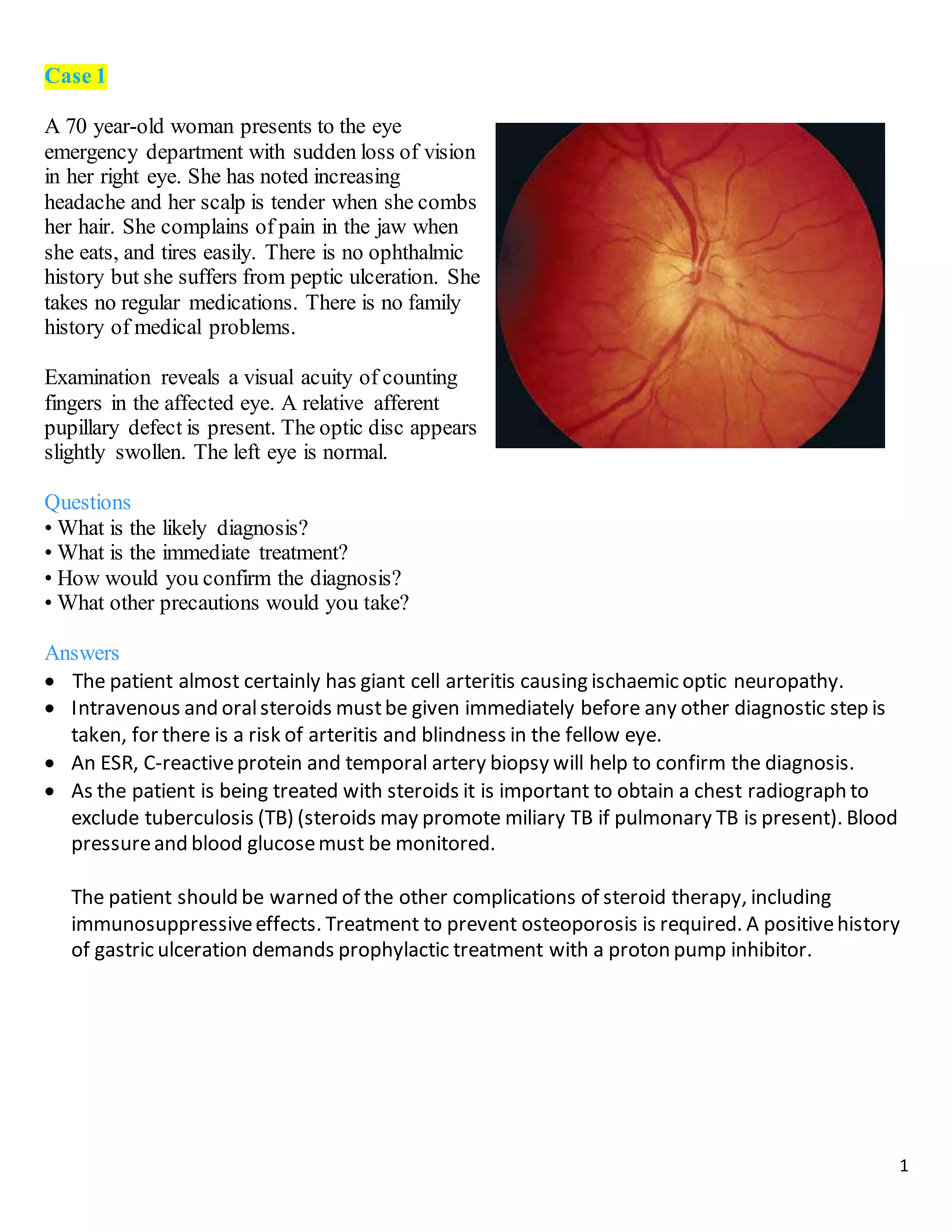
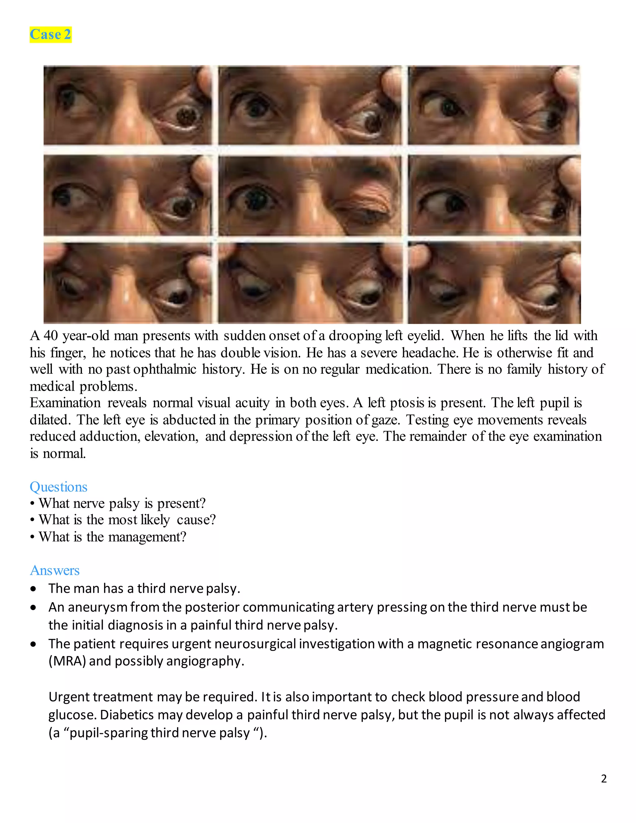
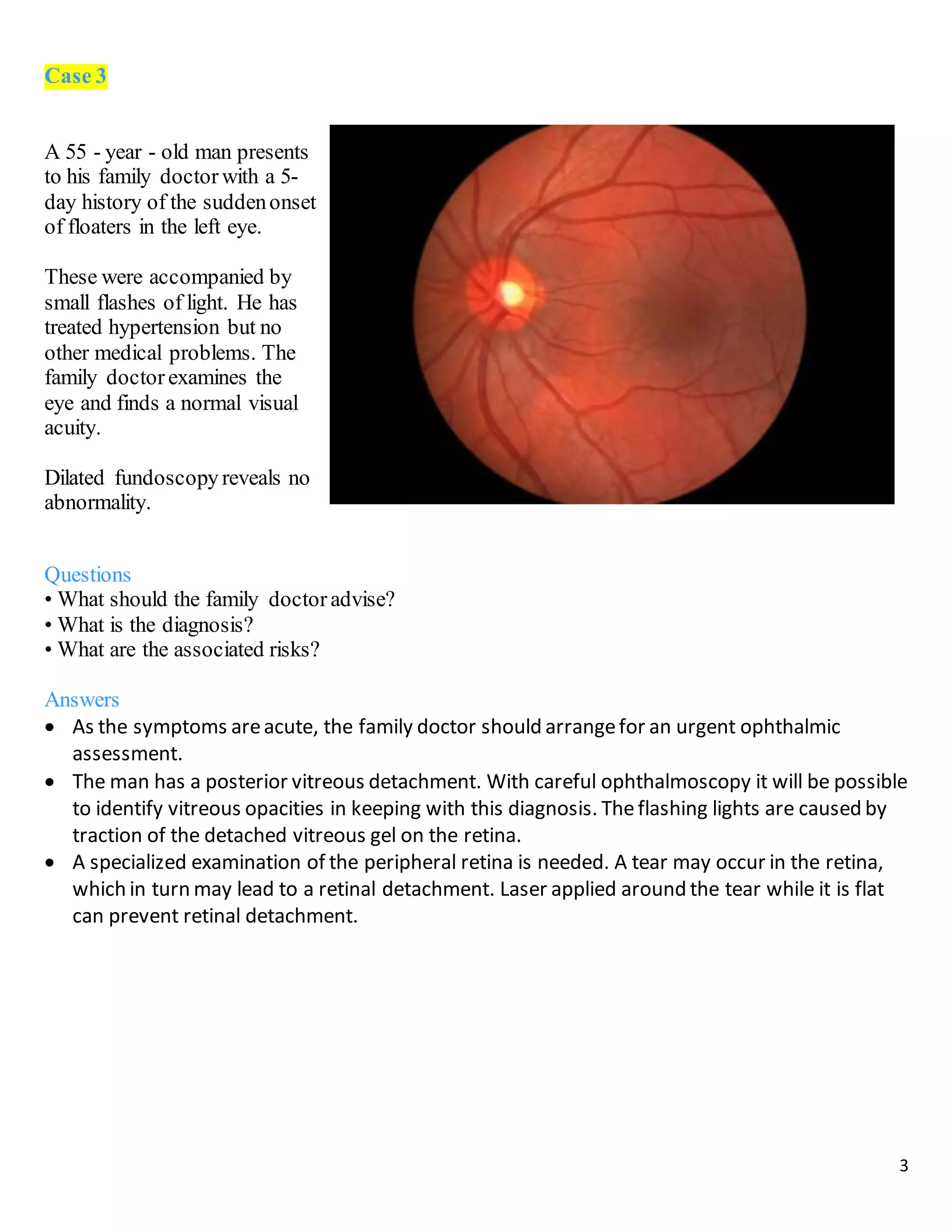
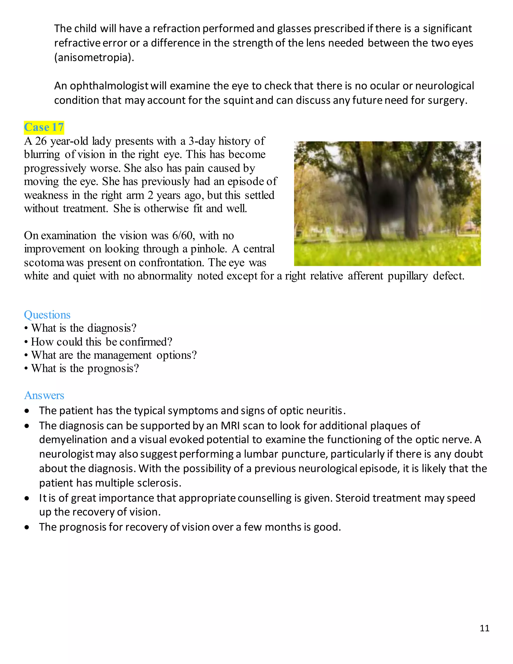
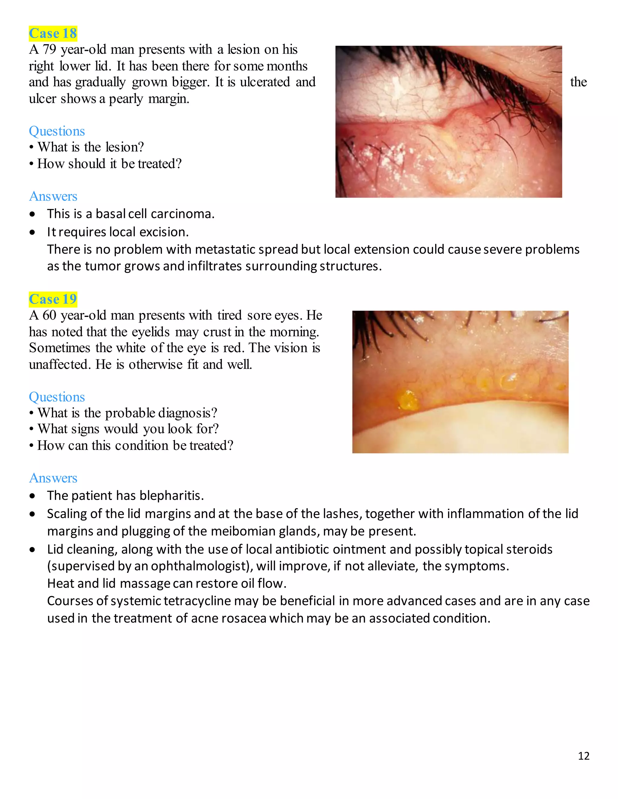
A hordeolum is a common infection of the eyelid margin. Lesions appear as a red, swollen nodule that resembles a pimple in appearance. The infection may be on the external or internal surface of the eyelid and could lead to redness and edema of the lid. Recognition of these lesions requires an understanding of eyelid anatomy. The objective of this case is to review etiology of these lesions and appropriate treatment.
cataract is clouding of the lens inside the eye..
it is very useful topic in medical filed...in this presentation all content is included about cataract like causes, symptoms and treatment...its very useful in your study.
Clinical and dermographics profile of glaucoma patients in Hebron - Palestin...Riyad Banayot
ABSTRACT
BACKGROUND: The purpose of the study was to describe the clinical profile of glaucoma types, treatment modalities, visual outcomes, and intraocular pressure (IOP) control for patients in Palestine.MATERIAL AND METHODS: Data collection was done through the hospital record review, which included basic demographics including file number, age, sex, family history of glaucoma, history of anti-glaucoma and steroid medication, history of ocular trauma or surgery, etiology of secondary glaucoma and history of systemic illness. All the patients had a comprehensive eye examination, including visual acuity, intraocular pressure, vertical cup-disc ratio, and gonioscopy. Data were obtained, tabulated, and organized using Microsoft Excel, and statistical analyses were done using Wizard Version 1.9.49 by Evan Miller.RESULTS: There were 100 females with a mean age of 53 and 101 males with a mean age of 67. Primary open-angle glaucoma and its variants represented 45.3% of all patients, while secondary glaucoma represented 40.3% and primary angle closure glaucoma represented 10.4%. The prevalence of glaucoma increased with age, and the last visual acuity (VA) showed that 39.2% of eyes had Normal/near normal VA. The highest average IOP of 25 mm Hg was recorded among secondary glaucoma patients. Of all glaucoma eyes studied, 64% were on one or two medications, and the most common surgical procedures performed were peripheral iridectomy 18.2% followed by trabeculectomy 15.5%. CONCLUSION: Primary open-angle glaucoma (POAG) was the predominant glaucoma. Glaucoma increased significantly with advancing age. Pseudoexfoliation and neovascular glaucoma comprised the majority of secondary glaucoma.
Acute ocular chemical injury: a descriptive assessment and management review ...Riyad Banayot
Background: The purpose of the study was to assess caregivers’ compliance with the management protocol for
chemical injury at St. John Eye Hospital, Jerusalem.
Materia l and methods: Charts of all new chemical injury patients who presented to St. John Eye Hospital,
Jerusalem, between January and December 2019 were retrospectively reviewed. Data categories collected included:
Presentation, age, sex, injury, irrigation, lids, visual acuity, slit-lamp examination (SLE), management plan, and
medications given. Data were stored and analysed using Excel.
Results: Patients’ presentation date and time, sex, and age were recorded in over 90% of cases. The mechanism of
injury and type of offending chemical were recorded in 65% of cases. The irrigating solution was identified in 50%
of cases. Corrected visual acuity was recorded in both eyes in almost 50% of cases. Limbal ischemia was documented
in 45% of cases, and intraocular pressure (IOP) was recorded in 25%. The management plan and explanation of the
condition to patients were documented in less than 50% of cases. Antibiotics and steroids (drops/ointment) were
prescribed in 92.5% of cases.
Conclusi ons: The results of this study reveal that our documentation needs improvement for several parameters.
Several recommendations were formulated:
1. Emphasize to caregivers that irrigation must be done first.
2. Corrected visual acuity should be attempted for both eyes in all cases, and reasons for not recording it should be
documented.
3. It is important to document and record limbus details, iris details, and IOP in all cases.
open-globe injuries in palestinePalestine: epidemiology and factors associate...Riyad Banayot
Background: The purpose was to describe the epidemiology of open-globe injury (OGI) in Palestine and identify
the prognostic factors associated with profound visual loss.
Materia l and methods: The current study is a retrospective review of hospital files for 83 consecutive patients
with OGI who presented to St. John Eye Hospital, Jerusalem, within 5 years, between 2009 and 2013. Demographic
details included age, gender, wound characteristics, and visual acuity (VA). The Ocular Trauma Classification Group
was used for wound location, classification, and scoring for each case.
Results: We identified 83 OGI that presented to St. John eye hospital. The study group included 62 males and
21 females. The mean age was 16.66 years ± 3.216. The most frequent injuries were playground injuries (59%),
followed by workplace injuries (26.5%). Penetrating injuries represented 45.8% of injuries, and rupture globes
occurred in 39.8% of cases. The most frequent objects causing injury were metal (31.3%) and stone (20.5%). Kinetic
impact projectiles were a statistically significant poor prognostic factor for the visual outcome. Variables that
were statistically significant poor prognostic factors for visual outcome included: retinal detachment, macular scar,
vitreous hemorrhage.
Conclusi on: This study showed that the act of demonstration, street injuries, kinetic impact projectiles, zone III
injuries, globe disruption, retinal detachment, vitreous hemorrhage, and a poor VA at the first visit are poor prognostic
factors for OGI. Recognition of these prognostic factors will help the ophthalmologist evaluate the injury
and its prognosis.
Use of digital retinal camera to detect prevalence and severity of diabetic ...Riyad Banayot
ABStrAct
BAckGround: The purpose of this study was to determine the prevalence of diabetic retinopathy among Palestinian
refugees serviced by the Diabetic Retinopathy Screening Program in the Occupied Palestinian Territories (DRS-
-OPT).
MAterIAl And MethodS: This is a retrospective study of retinal images of 1891 diabetic patients in 15 urban
UNRWA clinics participating in the DRS-OPT program in Palestine over 12 months. A nonmydriatic Canon CR-2
fundus retinal camera was used to capture two 450 non-stereo fundus images for each eye. Qualified graders (nurses)
performed the grading based on the DRS-OPT grading system.
reSultS: Out of the 1891 diabetic patients screened, 1694 had at least one gradable eye. 16% of patients had
diabetic retinopathy (5.7% had mild nonproliferative diabetic retinopathy, 4.3% had moderate nonproliferative
diabetic retinopathy, 1.1% had severe, moderate nonproliferative diabetic retinopathy, and 1.7% had proliferative
diabetic retinopathy. Maculopathy without retinopathy amounted to 3%. Other findings included the identification
of blinding diseases such as age-related macular degeneration and optic disc glaucomatous cupping.
concluSIonS: The retinopathy screening program using a nonmydriatic fundus camera identified diabetic retino-
pathy in 16% of diabetic Palestinian refugees. A total of 72% of these patients were diabetics with nonproliferative
retinopathy. This program can be used to prevent progression by facilitating the education of patients and early
intervention.
Bilateral lens capsule rupture in a patient with previously undiagnosed alpor...Riyad Banayot
Ophthalmologists may be the first to consider the diagnosis of Alport’s Syndrome based on lens changes. Uncontrolled Blood pressure can delay surgery during which time IOP should be monitored closely. Results of lensectomies with foldable IOL implantation are successful. To our knowledge, this is the second report of a case of bilateral lens capsule rupture in a patient with previously undiagnosed Alport’s Syndrome.
Evaluating the optic nerve head in glaucomaRiyad Banayot
The best method readily available to the clinician for performing this examination is high plus lens fundus biomicroscopy. Optimal magnification can be achieved by using a +60D lens which provides 1.5 times the magnification of a 90D lens. During this examination the patient's pupils must be maximally dilated with a combination of mydriatic agents such as 1% Tropicamide and 2.5% Phenylephrine.
Tom Selleck Health: A Comprehensive Look at the Iconic Actor’s Wellness Journeygreendigital
Tom Selleck, an enduring figure in Hollywood. has captivated audiences for decades with his rugged charm, iconic moustache. and memorable roles in television and film. From his breakout role as Thomas Magnum in Magnum P.I. to his current portrayal of Frank Reagan in Blue Bloods. Selleck's career has spanned over 50 years. But beyond his professional achievements. fans have often been curious about Tom Selleck Health. especially as he has aged in the public eye.
Follow us on: Pinterest
Introduction
Many have been interested in Tom Selleck health. not only because of his enduring presence on screen but also because of the challenges. and lifestyle choices he has faced and made over the years. This article delves into the various aspects of Tom Selleck health. exploring his fitness regimen, diet, mental health. and the challenges he has encountered as he ages. We'll look at how he maintains his well-being. the health issues he has faced, and his approach to ageing .
Early Life and Career
Childhood and Athletic Beginnings
Tom Selleck was born on January 29, 1945, in Detroit, Michigan, and grew up in Sherman Oaks, California. From an early age, he was involved in sports, particularly basketball. which played a significant role in his physical development. His athletic pursuits continued into college. where he attended the University of Southern California (USC) on a basketball scholarship. This early involvement in sports laid a strong foundation for his physical health and disciplined lifestyle.
Transition to Acting
Selleck's transition from an athlete to an actor came with its physical demands. His first significant role in "Magnum P.I." required him to perform various stunts and maintain a fit appearance. This role, which he played from 1980 to 1988. necessitated a rigorous fitness routine to meet the show's demands. setting the stage for his long-term commitment to health and wellness.
Fitness Regimen
Workout Routine
Tom Selleck health and fitness regimen has evolved. adapting to his changing roles and age. During his "Magnum, P.I." days. Selleck's workouts were intense and focused on building and maintaining muscle mass. His routine included weightlifting, cardiovascular exercises. and specific training for the stunts he performed on the show.
Selleck adjusted his fitness routine as he aged to suit his body's needs. Today, his workouts focus on maintaining flexibility, strength, and cardiovascular health. He incorporates low-impact exercises such as swimming, walking, and light weightlifting. This balanced approach helps him stay fit without putting undue strain on his joints and muscles.
Importance of Flexibility and Mobility
In recent years, Selleck has emphasized the importance of flexibility and mobility in his fitness regimen. Understanding the natural decline in muscle mass and joint flexibility with age. he includes stretching and yoga in his routine. These practices help prevent injuries, improve posture, and maintain mobilit
Couples presenting to the infertility clinic- Do they really have infertility...Sujoy Dasgupta
Dr Sujoy Dasgupta presented the study on "Couples presenting to the infertility clinic- Do they really have infertility? – The unexplored stories of non-consummation" in the 13th Congress of the Asia Pacific Initiative on Reproduction (ASPIRE 2024) at Manila on 24 May, 2024.
Report Back from SGO 2024: What’s the Latest in Cervical Cancer?bkling
Are you curious about what’s new in cervical cancer research or unsure what the findings mean? Join Dr. Emily Ko, a gynecologic oncologist at Penn Medicine, to learn about the latest updates from the Society of Gynecologic Oncology (SGO) 2024 Annual Meeting on Women’s Cancer. Dr. Ko will discuss what the research presented at the conference means for you and answer your questions about the new developments.
ARTIFICIAL INTELLIGENCE IN HEALTHCARE.pdfAnujkumaranit
Artificial intelligence (AI) refers to the simulation of human intelligence processes by machines, especially computer systems. It encompasses tasks such as learning, reasoning, problem-solving, perception, and language understanding. AI technologies are revolutionizing various fields, from healthcare to finance, by enabling machines to perform tasks that typically require human intelligence.
Title: Sense of Smell
Presenter: Dr. Faiza, Assistant Professor of Physiology
Qualifications:
MBBS (Best Graduate, AIMC Lahore)
FCPS Physiology
ICMT, CHPE, DHPE (STMU)
MPH (GC University, Faisalabad)
MBA (Virtual University of Pakistan)
Learning Objectives:
Describe the primary categories of smells and the concept of odor blindness.
Explain the structure and location of the olfactory membrane and mucosa, including the types and roles of cells involved in olfaction.
Describe the pathway and mechanisms of olfactory signal transmission from the olfactory receptors to the brain.
Illustrate the biochemical cascade triggered by odorant binding to olfactory receptors, including the role of G-proteins and second messengers in generating an action potential.
Identify different types of olfactory disorders such as anosmia, hyposmia, hyperosmia, and dysosmia, including their potential causes.
Key Topics:
Olfactory Genes:
3% of the human genome accounts for olfactory genes.
400 genes for odorant receptors.
Olfactory Membrane:
Located in the superior part of the nasal cavity.
Medially: Folds downward along the superior septum.
Laterally: Folds over the superior turbinate and upper surface of the middle turbinate.
Total surface area: 5-10 square centimeters.
Olfactory Mucosa:
Olfactory Cells: Bipolar nerve cells derived from the CNS (100 million), with 4-25 olfactory cilia per cell.
Sustentacular Cells: Produce mucus and maintain ionic and molecular environment.
Basal Cells: Replace worn-out olfactory cells with an average lifespan of 1-2 months.
Bowman’s Gland: Secretes mucus.
Stimulation of Olfactory Cells:
Odorant dissolves in mucus and attaches to receptors on olfactory cilia.
Involves a cascade effect through G-proteins and second messengers, leading to depolarization and action potential generation in the olfactory nerve.
Quality of a Good Odorant:
Small (3-20 Carbon atoms), volatile, water-soluble, and lipid-soluble.
Facilitated by odorant-binding proteins in mucus.
Membrane Potential and Action Potential:
Resting membrane potential: -55mV.
Action potential frequency in the olfactory nerve increases with odorant strength.
Adaptation Towards the Sense of Smell:
Rapid adaptation within the first second, with further slow adaptation.
Psychological adaptation greater than receptor adaptation, involving feedback inhibition from the central nervous system.
Primary Sensations of Smell:
Camphoraceous, Musky, Floral, Pepperminty, Ethereal, Pungent, Putrid.
Odor Detection Threshold:
Examples: Hydrogen sulfide (0.0005 ppm), Methyl-mercaptan (0.002 ppm).
Some toxic substances are odorless at lethal concentrations.
Characteristics of Smell:
Odor blindness for single substances due to lack of appropriate receptor protein.
Behavioral and emotional influences of smell.
Transmission of Olfactory Signals:
From olfactory cells to glomeruli in the olfactory bulb, involving lateral inhibition.
Primitive, less old, and new olfactory systems with different path
Lung Cancer: Artificial Intelligence, Synergetics, Complex System Analysis, S...Oleg Kshivets
RESULTS: Overall life span (LS) was 2252.1±1742.5 days and cumulative 5-year survival (5YS) reached 73.2%, 10 years – 64.8%, 20 years – 42.5%. 513 LCP lived more than 5 years (LS=3124.6±1525.6 days), 148 LCP – more than 10 years (LS=5054.4±1504.1 days).199 LCP died because of LC (LS=562.7±374.5 days). 5YS of LCP after bi/lobectomies was significantly superior in comparison with LCP after pneumonectomies (78.1% vs.63.7%, P=0.00001 by log-rank test). AT significantly improved 5YS (66.3% vs. 34.8%) (P=0.00000 by log-rank test) only for LCP with N1-2. Cox modeling displayed that 5YS of LCP significantly depended on: phase transition (PT) early-invasive LC in terms of synergetics, PT N0—N12, cell ratio factors (ratio between cancer cells- CC and blood cells subpopulations), G1-3, histology, glucose, AT, blood cell circuit, prothrombin index, heparin tolerance, recalcification time (P=0.000-0.038). Neural networks, genetic algorithm selection and bootstrap simulation revealed relationships between 5YS and PT early-invasive LC (rank=1), PT N0—N12 (rank=2), thrombocytes/CC (3), erythrocytes/CC (4), eosinophils/CC (5), healthy cells/CC (6), lymphocytes/CC (7), segmented neutrophils/CC (8), stick neutrophils/CC (9), monocytes/CC (10); leucocytes/CC (11). Correct prediction of 5YS was 100% by neural networks computing (area under ROC curve=1.0; error=0.0).
CONCLUSIONS: 5YS of LCP after radical procedures significantly depended on: 1) PT early-invasive cancer; 2) PT N0--N12; 3) cell ratio factors; 4) blood cell circuit; 5) biochemical factors; 6) hemostasis system; 7) AT; 8) LC characteristics; 9) LC cell dynamics; 10) surgery type: lobectomy/pneumonectomy; 11) anthropometric data. Optimal diagnosis and treatment strategies for LC are: 1) screening and early detection of LC; 2) availability of experienced thoracic surgeons because of complexity of radical procedures; 3) aggressive en block surgery and adequate lymph node dissection for completeness; 4) precise prediction; 5) adjuvant chemoimmunoradiotherapy for LCP with unfavorable prognosis.
MANAGEMENT OF ATRIOVENTRICULAR CONDUCTION BLOCK.pdfJim Jacob Roy
Cardiac conduction defects can occur due to various causes.
Atrioventricular conduction blocks ( AV blocks ) are classified into 3 types.
This document describes the acute management of AV block.
Flu Vaccine Alert in Bangalore Karnatakaaddon Scans
As flu season approaches, health officials in Bangalore, Karnataka, are urging residents to get their flu vaccinations. The seasonal flu, while common, can lead to severe health complications, particularly for vulnerable populations such as young children, the elderly, and those with underlying health conditions.
Dr. Vidisha Kumari, a leading epidemiologist in Bangalore, emphasizes the importance of getting vaccinated. "The flu vaccine is our best defense against the influenza virus. It not only protects individuals but also helps prevent the spread of the virus in our communities," he says.
This year, the flu season is expected to coincide with a potential increase in other respiratory illnesses. The Karnataka Health Department has launched an awareness campaign highlighting the significance of flu vaccinations. They have set up multiple vaccination centers across Bangalore, making it convenient for residents to receive their shots.
To encourage widespread vaccination, the government is also collaborating with local schools, workplaces, and community centers to facilitate vaccination drives. Special attention is being given to ensuring that the vaccine is accessible to all, including marginalized communities who may have limited access to healthcare.
Residents are reminded that the flu vaccine is safe and effective. Common side effects are mild and may include soreness at the injection site, mild fever, or muscle aches. These side effects are generally short-lived and far less severe than the flu itself.
Healthcare providers are also stressing the importance of continuing COVID-19 precautions. Wearing masks, practicing good hand hygiene, and maintaining social distancing are still crucial, especially in crowded places.
Protect yourself and your loved ones by getting vaccinated. Together, we can help keep Bangalore healthy and safe this flu season. For more information on vaccination centers and schedules, residents can visit the Karnataka Health Department’s official website or follow their social media pages.
Stay informed, stay safe, and get your flu shot today!
Ethanol (CH3CH2OH), or beverage alcohol, is a two-carbon alcohol
that is rapidly distributed in the body and brain. Ethanol alters many
neurochemical systems and has rewarding and addictive properties. It
is the oldest recreational drug and likely contributes to more morbidity,
mortality, and public health costs than all illicit drugs combined. The
5th edition of the Diagnostic and Statistical Manual of Mental Disorders
(DSM-5) integrates alcohol abuse and alcohol dependence into a single
disorder called alcohol use disorder (AUD), with mild, moderate,
and severe subclassifications (American Psychiatric Association, 2013).
In the DSM-5, all types of substance abuse and dependence have been
combined into a single substance use disorder (SUD) on a continuum
from mild to severe. A diagnosis of AUD requires that at least two of
the 11 DSM-5 behaviors be present within a 12-month period (mild
AUD: 2–3 criteria; moderate AUD: 4–5 criteria; severe AUD: 6–11 criteria).
The four main behavioral effects of AUD are impaired control over
drinking, negative social consequences, risky use, and altered physiological
effects (tolerance, withdrawal). This chapter presents an overview
of the prevalence and harmful consequences of AUD in the U.S.,
the systemic nature of the disease, neurocircuitry and stages of AUD,
comorbidities, fetal alcohol spectrum disorders, genetic risk factors, and
pharmacotherapies for AUD.
These simplified slides by Dr. Sidra Arshad present an overview of the non-respiratory functions of the respiratory tract.
Learning objectives:
1. Enlist the non-respiratory functions of the respiratory tract
2. Briefly explain how these functions are carried out
3. Discuss the significance of dead space
4. Differentiate between minute ventilation and alveolar ventilation
5. Describe the cough and sneeze reflexes
Study Resources:
1. Chapter 39, Guyton and Hall Textbook of Medical Physiology, 14th edition
2. Chapter 34, Ganong’s Review of Medical Physiology, 26th edition
3. Chapter 17, Human Physiology by Lauralee Sherwood, 9th edition
4. Non-respiratory functions of the lungs https://academic.oup.com/bjaed/article/13/3/98/278874
1. 1
Case 1
A 70 year-old woman presents to the eye
emergency department with sudden loss of vision
in her right eye. She has noted increasing
headache and her scalp is tender when she combs
her hair. She complains of pain in the jaw when
she eats, and tires easily. There is no ophthalmic
history but she suffers from peptic ulceration. She
takes no regular medications. There is no family
history of medical problems.
Examination reveals a visual acuity of counting
fingers in the affected eye. A relative afferent
pupillary defect is present. The optic disc appears
slightly swollen. The left eye is normal.
Questions
• What is the likely diagnosis?
• What is the immediate treatment?
• How would you confirm the diagnosis?
• What other precautions would you take?
Answers
The patient almost certainly has giant cell arteritis causing ischaemic optic neuropathy.
Intravenous and oralsteroids mustbe given immediately before any other diagnostic step is
taken, for there is a risk of arteritis and blindness in the fellow eye.
An ESR, C-reactiveprotein and temporal artery biopsy will help to confirm the diagnosis.
As the patient is being treated with steroids it is important to obtain a chest radiograph to
exclude tuberculosis (TB) (steroids may promote miliary TB if pulmonary TB is present). Blood
pressureand blood glucosemust be monitored.
The patient should be warned of the other complications of steroid therapy, including
immunosuppressiveeffects. Treatment to prevent osteoporosis is required. A positivehistory
of gastric ulceration demands prophylactic treatment with a proton pump inhibitor.
2. 2
Case 2
A 40 year-old man presents with sudden onset of a drooping left eyelid. When he lifts the lid with
his finger, he notices that he has double vision. He has a severe headache. He is otherwise fit and
well with no past ophthalmic history. He is on no regular medication. There is no family history of
medical problems.
Examination reveals normal visual acuity in both eyes. A left ptosis is present. The left pupil is
dilated. The left eye is abducted in the primary position of gaze. Testing eye movements reveals
reduced adduction, elevation, and depression of the left eye. The remainder of the eye examination
is normal.
Questions
• What nerve palsy is present?
• What is the most likely cause?
• What is the management?
Answers
The man has a third nervepalsy.
An aneurysmfromthe posterior communicating artery pressing on the third nerve mustbe
the initial diagnosis in a painful third nervepalsy.
The patient requires urgent neurosurgical investigation with a magnetic resonanceangiogram
(MRA) and possibly angiography.
Urgent treatment may be required. Itis also important to check blood pressureand blood
glucose. Diabetics may develop a painful third nerve palsy, but the pupil is not always affected
(a “pupil-sparing third nerve palsy “).
3. 3
Case 3
A 55 - year - old man presents
to his family doctor with a 5-
day history of the suddenonset
of floaters in the left eye.
These were accompanied by
small flashes of light. He has
treated hypertension but no
other medical problems. The
family doctor examines the
eye and finds a normal visual
acuity.
Dilated fundoscopy reveals no
abnormality.
Questions
• What should the family doctor advise?
• What is the diagnosis?
• What are the associated risks?
Answers
As the symptoms areacute, the family doctor should arrangefor an urgent ophthalmic
assessment.
The man has a posterior vitreous detachment. With careful ophthalmoscopy it will be possible
to identify vitreous opacities in keeping with this diagnosis. Theflashing lights are caused by
traction of the detached vitreous gel on the retina.
A specialized examination of the peripheral retina is needed. A tear may occur in the retina,
which in turn may lead to a retinal detachment. Laser applied around the tear while it is flat
can prevent retinal detachment.
4. 4
Case 4
A 75 year-old woman attends the emergency
department with nausea and vomiting. She
says that her right eye is painful and red.
Vision is reduced. She is longsighted and
wears glasses for near and distance vision. She
is generally fit. There is no family history of
medical problems.
On examination the emergency doctor finds
the vision to be reduced to counting fingers in
the right eye. The eye is red, the cornea
appears cloudy and edematous, and the pupil
is oval and dilated on the affected side. No view of the fundus is obtained.
Questions
• What is the diagnosis?
• How might it be confirmed?
• What is the treatment?
Answers
The lady is longsighted and has acute angle closureglaucoma.
Tonometry would reveal an intraocular pressureof about 65 mm Hg. If possible, through the
cloudy cornea, gonioscopy would confirm the presenceof a closed angle and a narrow angle
in the fellow eye. This can also be seen using high resolution ultrasound or confocal
microscopy.
The pressuremustbe lowered with intravenous acetazolamide and topical hypotensivedrops,
including pilocarpine, to producemiosis. A peripheral iridotomy is then performed, usually
with a YAG laser, in both eyes, to prevent further attacks.
5. 5
Case 5
A 28 year-old man presents to his ophthalmologist with a
painful, red right eye. The vision has becomeincreasingly
blurred over the last 2 days. He wears softcontact lenses, and
contact lens tolerance has decreased in the preceding days.
The ophthalmologist notes that the vision is reduced to 6/60
in the right eye, the conjunctiva is inflamed, and there is a
central opacity on the cornea. A small hypopyon is present.
Questions
• What is the likely diagnosis?
• What should the optician do?
Answers
The man has a bacterial corneal ulcer secondary to contact lens wear.
The ulcer will be scraped for culture and the contact lens and any lens containers cultured.
Intensive, topical, broad-spectrumantibiotics are administered as an inpatient pending the
result of the microbiological investigation.
Case 6
A mother takes her 8 month-old baby to her
ophthalmologist. He has had a persistently watery
eye since birth. Intermittently there is a yellow
discharge surrounding the eye. The white of the eye
has never been red. The baby is otherwise healthy.
Examination reveals a white, quiet, normal eye.
Slight pressure over the lacrimal sac produces a
yellowish discharge from the normal puncta.
Questions
• What is the diagnosis?
• What advice would you give the mother?
Answers
Itis likely that the child has nasolacrimalobstruction due to an imperforate nasolacrimalduct.
The mother should be reassured thatthis often resolves spontaneously. Thelids should be
kept clean and the region overlying the lacrimal sac massaged gently daily. Antibiotics are
generally not effective.
If the symptoms persistafter the child’s firstbirthday the child can be referred for syringing
and probing of the nasolacrimal duct.
6. 6
Case 7
A 14 year-old complains of intermittent redness and soreness
of the right eye. He has noticed a small lump on the upper
lid. The vision is unaffected.
Examination reveals a small, raised, umbilicated lesion on
the skin of the upper lid, associated with a follicular
conjunctivitis below.
Questions
• What is the likely diagnosis?
• What is the treatment?
Answers
Itis likely that the lid lesion is a molluscumcontagiosum.
Itis treated by excision.
Case 8
A 35 year-old man presents to his family
doctorwith erythematous, swollen right
upper and lower eyelids, worsening over
the previous 2 days. He is unable to open
them. He feels unwell and has a
temperature.
Examination reveals marked lid swelling, a
tender globe and, on manual opening of the
lids, a proptosis with chemotic injected
conjunctiva. Eye movements are limited in all directions. Visual acuity and color vision are
normal, and there is no relative afferent pupillary defect. The optic disc
and retina also appear normal.
Questions
• What is the diagnosis?
• What is the management?
Answers
The man has orbital cellulitis.
Blood cultures and a high nasal swab should be performed, together with an orbital CT scan,
to confirm the diagnosis and delineate any abscess.
7. 7
He requires admission to hospital for intravenous antibiotics and close monitoring of his
vision, color vision and pupillary reflexes, as he is at risk of severeoptic nervedamage.
The ENT surgeons should beinformed, as they may be required to drain an abscess.
The normal acuity and color vision suggestthat the optic nerveis not compromised at
present, but should these signs worsen, urgentsurgicaldrainagewill be required.
Case 9
While working in the laboratory a colleague inadvertently sprays his eyes with
an alkali solution.
Questions
• What is the immediate treatment?
• What should you do next?
Answers
Anesthesia with Benoxinate then irrigate for at least 30 minutes (Normal saline, Lactated
ringer, BSS) until pH returns normal(7).
Evert/double evert lids and removeparticulate matter. If unable to remove particulate
matter, consider general anesthesia.
Debridement of necrotic corneal epithelium, necrotic conjunctival& sub-conjunctivaltissue.
Do not patch the eye.
Take visualacuity for both eyes (with correction, unaided and pinhole)
Start treatment according to grading.
Case 10
A 27 year-old man presents with a 2-day history of a
painful red right eye; the vision is slightly blurred and
he dislikes bright lights. He is otherwise fit and well,
but complains of some backache. He wears no glasses.
Questions
• What is the likely diagnosis?
• What would you expect to find on examination of
the eye?
• What treatment would you give?
• What is the eye condition likely to be associated
with?
Answers
The patient has iritis. Examination would reveal a reduction in visualacuity, redness of the eye
that is worseat the limbus, cells in the anterior chamber and possibly on the cornea (keratic
8. 8
precipitate) or a collection at the bottom of the anterior chamber (hypopyon). Theiris may be
stuck to the lens (posterior synechiae). Theremay be inflammation of the vitreous and retina.
The patient is treated with steroid eye drops to reduce the inflammation and dilating drops to
prevent the formation of posterior synechiae.
The history of backachesuggests that the patient may haveankylosing spondylitis.
Case 11
A 68 year-old lady presents with a mildly painful
red eye and some blurring of vision. One year
previously she had a corneal graft. She is on no
medications and is otherwise well.
Questions
• What is the possible diagnosis?
• What treatment should the patient be given?
Answers
A diagnosis of graft rejection mustbe
considered first.
Intensivetreatment with topical steroids to savethe graft.
Case 12
A 68 year-old hypertensive man noted a fleeting loss of vision in one eye lasting for about a
minute. He described it as a curtain coming down over the vision. Recovery was complete. There
was no pain. Examination reveals no abnormality.
Questions
• What is the diagnosis?
• What treatment would you advise?
Answers
The patient has had an episode of amaurosis fugax, mostlikely caused by the
passageof a fi brin-platelet embolus through the retinal circulation.
The patient requires treatment with antiplatelet drugs and a cardiovascular work - up.
The most likely abnormality is a plaque on the carotid artery, which may require surgery.
9. 9
Case 13
A 60 year-old lady presented to her family doctor with
gradual loss of vision over some months. She noticed that
the problem was particularly bad in bright sunshine. The
eye was not painful or red. She was otherwise well.
Questions
• What is the probable diagnosis?
• How can the diagnosis be confirmed?
• What treatment may be advised?
Answers
Itis likely that the lady has a cataract.
These can be readily seen with a slit lamp, but are also well visualized with the direct
ophthalmoscopein the red reflex.
The advantages and possiblecomplications of cataractsurgery should be discussed with her
once the diagnosis has been confirmed.
Case 14
An 80 year-old lady who has already lost the vision in one
eye develops distortion and reduction of vision over a few
days in her good eye.
Examination reveals an acuity of 6/12, an early cataract, and
an abnormality at the macula.
Questions
• What is the likely diagnosis?
• What treatment may be helpful?
Answers
The rapid onset suggests thatthe cataract has little to do
with the new visual disturbance. Itis most likely due to age-related macular degeneration
(AMD).
In somepatients, with wet AMD, where a fibrovascular membrane grows beneath the macula
that can be shown on a fluorescein angiogram, anti-VEGF injections into the vitreous may be
helpful in preventing further progression and in somecases improving vision.
10. 10
Case 15
A 30 year-old builder was using a hammer to hit a steel chisel. He felt
something hit his eye and the vision became blurred. He is fit and well
and there is no history of medical problems.
On examination, vision was reduced to 6/12. A fluorescein staining
lesion was seen on the cornea but this appeared Seidel-negative. A
small hyphema was seen in the anterior chamber, and in the red reflex
observed with a direct ophthalmoscopea well delineated lens opacity
was seen. The retina appeared normal.
Questions
• What is the cause of the reduced acuity?
• What is the likely origin of the lens opacity?
• What is the possible management of the patient?
Answers
The relatively good acuity suggests that there has been no damage to the macula.
Itis likely that a piece of steel travelling at high velocity has penetrated the cornea, traversed
the iris (resulting in the hyphema) and passed into or through the lens (causing the opacity).
The corneal wound, if self-sealing, will probably not require suturing.
The exact location of the foreign body must be determined. Although it is unlikely to causean
infection (heat generated by the impact of the hammer on the metal may effectively sterilize
the fragment) it may causeretinal toxicity if it has entered the vitreous cavity or retina. If it is
enclosed in the lens there is less chance of retinal toxicity developing but the patient is at high
risk of developing a subsequentcataractthat may require an operation.
A foreign body that impacts on the retina or the vitreous body requires a vitrectomy to
remove it, with careful examination of the retina for tears.
Case 16
A 2 year-old child was thought to have a squint by her parents. The finding was confirmed by her
family doctor and she was referred to hospital.
Question
• What examination must be conducted in hospital?
Answer
Having taken a full history, an orthoptistwill measurethe visualacuity of the child,
examine the range of eye movements, determine the presence type of squintwith a cover
test, trying to assess thedegree of binocular vision present.
11. 11
The child will have a refraction performed and glasses prescribed if there is a significant
refractiveerror or a difference in the strength of the lens needed between the two eyes
(anisometropia).
An ophthalmologistwill examine the eye to check that there is no ocular or neurological
condition that may account for the squintand can discuss any futureneed for surgery.
Case 17
A 26 year-old lady presents with a 3-day history of
blurring of vision in the right eye. This has become
progressively worse. She also has pain caused by
moving the eye. She has previously had an episode of
weakness in the right arm 2 years ago, but this settled
without treatment. She is otherwise fit and well.
On examination the vision was 6/60, with no
improvement on looking through a pinhole. A central
scotomawas present on confrontation. The eye was
white and quiet with no abnormality noted except for a right relative afferent pupillary defect.
Questions
• What is the diagnosis?
• How could this be confirmed?
• What are the management options?
• What is the prognosis?
Answers
The patient has the typical symptoms and signs of optic neuritis.
The diagnosis can be supported by an MRI scan to look for additional plaques of
demyelination and a visual evoked potential to examine the functioning of the optic nerve. A
neurologistmay also suggestperforming a lumbar puncture, particularly if there is any doubt
about the diagnosis. With the possibility of a previous neurological episode, it is likely that the
patient has multiple sclerosis.
Itis of great importance that appropriatecounselling is given. Steroid treatment may speed
up the recovery of vision.
The prognosis for recovery of vision over a few months is good.
12. 12
Case 18
A 79 year-old man presents with a lesion on his
right lower lid. It has been there for some months
and has gradually grown bigger. It is ulcerated and the
ulcer shows a pearly margin.
Questions
• What is the lesion?
• How should it be treated?
Answers
This is a basalcell carcinoma.
Itrequires local excision.
There is no problem with metastatic spread but local extension could causesevere problems
as the tumor grows and infiltrates surrounding structures.
Case 19
A 60 year-old man presents with tired sore eyes. He
has noted that the eyelids may crust in the morning.
Sometimes the white of the eye is red. The vision is
unaffected. He is otherwise fit and well.
Questions
• What is the probable diagnosis?
• What signs would you look for?
• How can this condition be treated?
Answers
The patient has blepharitis.
Scaling of the lid margins and at the base of the lashes, together with inflammation of the lid
margins and plugging of the meibomian glands, may be present.
Lid cleaning, along with the useof local antibiotic ointment and possibly topical steroids
(supervised by an ophthalmologist), will improve, if not alleviate, the symptoms.
Heat and lid massagecan restore oil flow.
Courses of systemic tetracycline may be beneficial in more advanced cases and are in any case
used in the treatment of acne rosacea which may be an associated condition.
13. 13
Case 20
A 30 year-old man developed an acute red eye first
on the right and then in the left eye, associated with
a watery discharge. Vision is unaffected but the eye
irritates. He is otherwise fit and well.
Questions
• What is the diagnosis?
• What confirmatory signs would you look for on
examination?
• What precautions would you take following your
examination?
Answers
The patient has viral conjunctivitis – probably adenovirus.
Examination for a preauricular lymph node and conjunctivalfollicles on the lower tarsus
would confirm the diagnosis.
This formof conjunctivitis is highly contagious; it is important to ensurethat hands and
equipment arethoroughly cleaned following the examination, and that the importance of
good hygieneis emphasized to the patient.