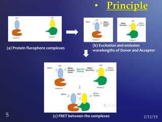
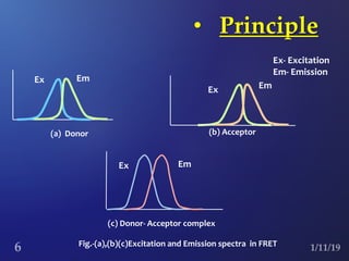
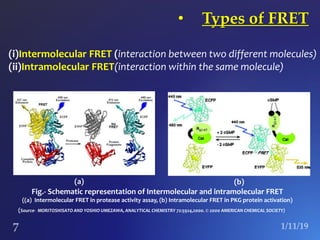
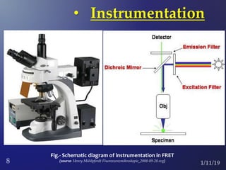
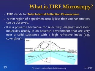
The document discusses three fluorescence microscopy techniques: FRET, FRAP, and TIRF microscopy. It explains the principles, instrumentation, sample preparation, applications, and future aspects of each technique. FRET involves energy transfer between fluorophores and is used to study molecular interactions and structure. FRAP examines fluorophore diffusion by photobleaching a region and monitoring recovery. It provides information about molecular mobility. TIRF microscopy uses evanescent waves to selectively excite fluorophores very near a surface and is applied to studies of cellular processes at membranes. All three techniques provide insights into biological phenomena at the molecular level.
wo-dimensional gel electrophoresis, abbreviated as 2-DE or 2-D electrophoresis, is a form of gel electrophoresis commonly used to analyze proteins. Mixtures of proteins are separated by two properties in two dimensions on 2D gels. 2-DE was first independently introduced by O'Farrell and Klose in 1975.
Gel electrophoresis native, denaturing&reducingLovnish Thakur
Electrophoresis is a technique used to separate and sometimes purify macromolecules - especially proteins and nucleic acids - that differ in size, charge or conformation.
It is a subtype of the gel electrophoresis whereby the normal gel is replaced with polyacrylamide gels used as support media.
Gels are made by free radical-induced polymerization of acrylamide and N,N’-Methylenebisacrylamide.
It is the most widely used technique of electrophoresis.
this is about chromatofocusing. technique useful for the final purification of proteins..this technique is based on isoelectric point of the proteins..
What is the Surface characterization techniques of Fourier-transform.pdfarishmarketing21
What is the Surface characterization techniques of Fourier-transform infrared spectroscopy
(FTIR) and Optical Imaging and Spectroscopy (microscopy, TIRF)
explain what are they measuring, their uniqueness, working principle briefly
Solution
Fourier transform infrared spectroscopy (FTIR) is a powerful analysis tool for characterizing and
identifying organic molecules. It is the spectroscopic technique that is the most widely used for
determining the characteristics of new membranes. In attenuated total reflectance mode, this type
of spectroscopy enables functional groups present over a depth of about 1 m to be identified.
During ATR analysis, the sample is kept in contact with a crystal allowing total internal
reflection. An infrared ray arrives at the crystal where the material under study has been placed.
The internal reflection of the ray in the crystal gives rise to an evanescent wave which, at each
reflection, continues beyond the surface of the crystal and penetrates the sample over about 1 m.
The penetration depth depends on the wavelength, the angle of incidence of the beam on the
crystal, and the nature of the crystal.
Spectra are thus obtained (curves of absorbance vs. wavelength) that have absorption peaks
characteristic of the functions present at the membrane surface.
FTIR-ATR is a sensitive, nondestructive method that can be used qualitatively and
quantitatively. However, it requires prior drying of the membrane sample.
One of the applications of FTIR-ATR is the characterization of modified surfaces. In this case,
the spectra show bands characteristic of the basic membrane with, in most cases, bands
characteristic of the new functional groups related to the modification. FTIR-ATR also enables
the efficiency of membrane cleaning to be assessed .
This method can also be used to analyze the adsorption of macromolecules at the membrane
surface and to check whether the conformation of the adsorbed compounds (e.g., proteins) has
been modified by comparing the spectra of the adsorbed product with those of the same product
in solution . When the deposits are very small, however, it is difficult to determine the presence
of fouling agents; this necessitates the use of elaborate data-processing methods that eliminate
the contributions of the membrane and water from the raw spectra.
UNIQUENESS:
There are three principal advantages for an FT spectrometer compared to a scanning (dispersive)
spectrometer.
TIRF
Total internal reflection fluorescence (TIRF) microscopy (TIRFM) is an elegant optical
technique that provides for the excitation of fluorophores in an extremely thin axial region
(‘optical section’). The method is based on the principle that when excitation light is totally
internally reflected in a transparent solid (e.g., coverglass) at its interface with liquid an
electromagnetic field, called the evanescent wave, is generated in the liquid at the solid-liquid
interface and is the same frequency as the excitation light. Since the inte.
Lightoptical nanoscopy for the use in biomedical applications and material sciences, detection in attomolar concentrations
* Use of standard fluorophores like GFP, RFP, YFP, Alexa, Fluorescein (no photoswitch necessary)
2CLM Two Color Localisation microscopy in the nanoscale
* Optical resolution 10 nm in 2D, 40 nm in 3D
* Very fast in processing, complete picture (2000 images) with processing in 3 minutes
In this lab report, I have presented the ATR- FT-IR of as polyvinylprrolidone . I have also analyzed the Proton FT NMR, The differential scanning calorimeter (DSC) of Polyvinylpyrrolidone and lastly ,the FT-Raman- of polyvinylprrolidone. Raman and FTIR spectroscopy are the perfect complement to one another for a wide variety of analyses. Where FTIR is strong at identifying functional groups, Raman spectroscopy is well-suited to giving information about molecular backbones
The term biophotonics denotes a combination of biology and photonics, with photonics being the science and technology of generation, manipulation, and detection of photons, quantum units of light. Photonics is related to electronics and photons. Photons play a central role in information technologies such as fiber optics the way electrons do in electronics.
Biophotonics can also be described as the "development and application of optical techniques, particularly imaging, to the study of biological molecules, cells and tissue". One of the main benefits of using optical techniques which make up biophotonics is that they preserve the integrity of the biological cells being examined.
IR SPECTROSCOPY-INTRODUCTION, PRINCIPLE, TYPE OF VIBRATIONS, INSTRUMENTATION, APPLICATION{ FOR the m.pharm 1st year 2019
Presented by DIPSANKAR BERA(M.PHARM STUDENT)
Slide 1: Title Slide
Extrachromosomal Inheritance
Slide 2: Introduction to Extrachromosomal Inheritance
Definition: Extrachromosomal inheritance refers to the transmission of genetic material that is not found within the nucleus.
Key Components: Involves genes located in mitochondria, chloroplasts, and plasmids.
Slide 3: Mitochondrial Inheritance
Mitochondria: Organelles responsible for energy production.
Mitochondrial DNA (mtDNA): Circular DNA molecule found in mitochondria.
Inheritance Pattern: Maternally inherited, meaning it is passed from mothers to all their offspring.
Diseases: Examples include Leber’s hereditary optic neuropathy (LHON) and mitochondrial myopathy.
Slide 4: Chloroplast Inheritance
Chloroplasts: Organelles responsible for photosynthesis in plants.
Chloroplast DNA (cpDNA): Circular DNA molecule found in chloroplasts.
Inheritance Pattern: Often maternally inherited in most plants, but can vary in some species.
Examples: Variegation in plants, where leaf color patterns are determined by chloroplast DNA.
Slide 5: Plasmid Inheritance
Plasmids: Small, circular DNA molecules found in bacteria and some eukaryotes.
Features: Can carry antibiotic resistance genes and can be transferred between cells through processes like conjugation.
Significance: Important in biotechnology for gene cloning and genetic engineering.
Slide 6: Mechanisms of Extrachromosomal Inheritance
Non-Mendelian Patterns: Do not follow Mendel’s laws of inheritance.
Cytoplasmic Segregation: During cell division, organelles like mitochondria and chloroplasts are randomly distributed to daughter cells.
Heteroplasmy: Presence of more than one type of organellar genome within a cell, leading to variation in expression.
Slide 7: Examples of Extrachromosomal Inheritance
Four O’clock Plant (Mirabilis jalapa): Shows variegated leaves due to different cpDNA in leaf cells.
Petite Mutants in Yeast: Result from mutations in mitochondrial DNA affecting respiration.
Slide 8: Importance of Extrachromosomal Inheritance
Evolution: Provides insight into the evolution of eukaryotic cells.
Medicine: Understanding mitochondrial inheritance helps in diagnosing and treating mitochondrial diseases.
Agriculture: Chloroplast inheritance can be used in plant breeding and genetic modification.
Slide 9: Recent Research and Advances
Gene Editing: Techniques like CRISPR-Cas9 are being used to edit mitochondrial and chloroplast DNA.
Therapies: Development of mitochondrial replacement therapy (MRT) for preventing mitochondrial diseases.
Slide 10: Conclusion
Summary: Extrachromosomal inheritance involves the transmission of genetic material outside the nucleus and plays a crucial role in genetics, medicine, and biotechnology.
Future Directions: Continued research and technological advancements hold promise for new treatments and applications.
Slide 11: Questions and Discussion
Invite Audience: Open the floor for any questions or further discussion on the topic.
A brief information about the SCOP protein database used in bioinformatics.
The Structural Classification of Proteins (SCOP) database is a comprehensive and authoritative resource for the structural and evolutionary relationships of proteins. It provides a detailed and curated classification of protein structures, grouping them into families, superfamilies, and folds based on their structural and sequence similarities.
Cancer cell metabolism: special Reference to Lactate PathwayAADYARAJPANDEY1
Normal Cell Metabolism:
Cellular respiration describes the series of steps that cells use to break down sugar and other chemicals to get the energy we need to function.
Energy is stored in the bonds of glucose and when glucose is broken down, much of that energy is released.
Cell utilize energy in the form of ATP.
The first step of respiration is called glycolysis. In a series of steps, glycolysis breaks glucose into two smaller molecules - a chemical called pyruvate. A small amount of ATP is formed during this process.
Most healthy cells continue the breakdown in a second process, called the Kreb's cycle. The Kreb's cycle allows cells to “burn” the pyruvates made in glycolysis to get more ATP.
The last step in the breakdown of glucose is called oxidative phosphorylation (Ox-Phos).
It takes place in specialized cell structures called mitochondria. This process produces a large amount of ATP. Importantly, cells need oxygen to complete oxidative phosphorylation.
If a cell completes only glycolysis, only 2 molecules of ATP are made per glucose. However, if the cell completes the entire respiration process (glycolysis - Kreb's - oxidative phosphorylation), about 36 molecules of ATP are created, giving it much more energy to use.
IN CANCER CELL:
Unlike healthy cells that "burn" the entire molecule of sugar to capture a large amount of energy as ATP, cancer cells are wasteful.
Cancer cells only partially break down sugar molecules. They overuse the first step of respiration, glycolysis. They frequently do not complete the second step, oxidative phosphorylation.
This results in only 2 molecules of ATP per each glucose molecule instead of the 36 or so ATPs healthy cells gain. As a result, cancer cells need to use a lot more sugar molecules to get enough energy to survive.
introduction to WARBERG PHENOMENA:
WARBURG EFFECT Usually, cancer cells are highly glycolytic (glucose addiction) and take up more glucose than do normal cells from outside.
Otto Heinrich Warburg (; 8 October 1883 – 1 August 1970) In 1931 was awarded the Nobel Prize in Physiology for his "discovery of the nature and mode of action of the respiratory enzyme.
WARNBURG EFFECT : cancer cells under aerobic (well-oxygenated) conditions to metabolize glucose to lactate (aerobic glycolysis) is known as the Warburg effect. Warburg made the observation that tumor slices consume glucose and secrete lactate at a higher rate than normal tissues.
Nutraceutical market, scope and growth: Herbal drug technologyLokesh Patil
As consumer awareness of health and wellness rises, the nutraceutical market—which includes goods like functional meals, drinks, and dietary supplements that provide health advantages beyond basic nutrition—is growing significantly. As healthcare expenses rise, the population ages, and people want natural and preventative health solutions more and more, this industry is increasing quickly. Further driving market expansion are product formulation innovations and the use of cutting-edge technology for customized nutrition. With its worldwide reach, the nutraceutical industry is expected to keep growing and provide significant chances for research and investment in a number of categories, including vitamins, minerals, probiotics, and herbal supplements.
This pdf is about the Schizophrenia.
For more details visit on YouTube; @SELF-EXPLANATORY;
https://www.youtube.com/channel/UCAiarMZDNhe1A3Rnpr_WkzA/videos
Thanks...!
Mammalian Pineal Body Structure and Also Functions
FRET, FRAP, TIFR MICROSCOPY
1. -presented by
Baishali Tamuli (BBI17008)
Jyotishman Sarma (BBI17009)
FRET, FRAP and TIRF
Microscopy
Principles and Applications of
TEZPUR UNIVERSITY
2. FRET stands for Förster resonance energy transfer or
fluorescence resonance energy transfer, one of the modern
advancements in microscopic techniques crucial for
understanding various biological processes.
• What is FRET?
3. FRET is named after German physicist
Theodor Förster who was pioneer in
discovering the Förster distance or
radius and interaction of molecules
which are in close proximity. His theory
of FRET was first published in 1946.
• Discovery of FRET
4. FRET involves the transfer of energy from an excited molecular
fluorophore (donor) to another fluorophore (acceptor) non
radiatively whenever the distance between the donor and the
acceptor is smaller than Förster radius.
The efficiency of FRET is dependent on the inverse sixth power
of intermolecular separation making it a sensitive technique for
investigating a variety of biological phenomena that produce
changes in molecular proximity.
• Principle
5. • Principle
(a) Protein flurophore complexes
(b) Excitation and emission
wavelengths of Donor and Acceptor
(c) FRET between the complexes
6. • Principle
(a) Donor (b) Acceptor
(c) Donor- Acceptor complex
Fig.-(a),(b)(c)Excitation and Emission spectra in FRET
Em
EmEx
Ex
Ex- Excitation
Em- Emission
EmEx
8. • Instrumentation
Fig.- Schematic diagram of instrumentation in FRET
(source- Henry Mühlpfordt Fluoreszenzmikroskopie_2008-09-28.svg)
9. • Sample preparation
In 1960s Osamu Shimomura discovered that a certain species of jellyfish
(Aequorea victoria) owes its luminescent character to the presence of
fluorescent proteins, such as aequorin and the green fluorescent protein
(GFP).
In GFP, the light-absorbing/emitting chromophore is formed by self
modification (i.e., by an autocatalytic reaction) of three of the amino acids
that make up the primary structure of the GFP polypeptide
Live-cell imaging studies can often be made more informative by the
simultaneous use of GFP variants that exhibit different spectral properties.
Variants of GFP that fluoresce in shades of blue (BFP), yellow (YFP), and
cyan (CFP) were generated by Roger Tsien of the University of California,
San Diego, through directed mutagenesis of the GFP gene.
10. • How FRET helps?
Major applications of FRET:
Molecular interactions (eg.- Protein-protein interactions)
Structure elucidation of biomolecules
Ligand receptor binding
Molecular colocalization (eg.- Studying lipid rafts)
Developing biosensors and chemosensors
11. • Future aspects
Better understanding of signalling pathways
Cellular interaction with the environment
Hormone receptor binding
Trsansport of biomolecules within a system
Drug designing
12. FRAP stands for Fluorescence recovery after
photobleaching, is a method for determining
the kinetics of diffusion through living tissues,
cells or a system.
• What is FRAP?
13. • Principle
The general method is to label a specific cell component with
a fluorescent molecule, image that cell, photobleach a small
portion of the cell, then image the recovery of fluorescence
over time.
Diffusion or active movement of molecules within the cell
replace bleached fluorophore with unbleached molecules
that were located in a different part of the cell.
Over time fluorescence in the bleached region recovers.
14. Instrumentation and sample preparation
The basic apparatus comprises an optical microscope, a light source and
some fluorescent probe.
Fluorescent emission is contingent upon absorption of a specific optical
wavelength or color.
The technique begins by saving a background image of the sample before
photobleaching.
Next, the light source is focused onto a small patch of the viewable area
either by switching to a higher magnification microscope objective or with
laser light of the appropriate wavelength.
15. Instrumentation and sample preparation
The fluorophores in this region receives high intensity illumination which
causes their fluorescence lifetime to quickly elapse (limited to roughly 105
photons before extinction). Now the image in the microscope is that of a
uniformly fluorescent field with a noticeable dark spot.
As Brownian motion proceeds, the still-fluorescing probes will diffuse
throughout the sample and replace the non-fluorescent probes in the
bleached region.
The initial and final photograph gives the extent of mobility which can be
calculated by the equation,
where, D is diffusion constant , ω is the radius of the beam and tD is the
characteristic diffusion time
17. • Applications
Major applications of FRAP are,
To study brownian motion which is dependent on molecular
size, local environment and binding interactions.
Understandingmoleculartrafficking.
Intracellular transport
Continuity of compartments
Stability of molecular complexes
18. • Drawbacks of FRAP
FRAP can only follow the average movement of a relatively
large number of labeled molecules (hundreds to thousands)
when they diffuse over a relatively large distance (e.g. 1 µm).
As a result, researchers using FRAP cannot distinguish
between proteins that are truly immobile and ones that can
only diffuse over a limited distance in the time allowed.
19. What is TIRF Microscopy?
TIRF stands for Total Internal Reflection Fluorescence.
A thin region of a specimen, usually less than 200 nanometers
can be observed.
It is a powerful technique for selectively imaging fluorescent
molecules usually in an aqueous environment that are very
near a solid substance with a high refractive index (e.g.
coverglass).
Fig source: onlinephysicstuition.com.my
20. The idea of using total internal reflection in
microscope was first described by E.J. Ambrose
in 1956.
This idea further extended by Daniel Axelrod at
the University of Michigan.
Ann Arbor in the early 1980s introduced
TIRFM.
THE STORY BEHIND
21. • What is the principle involved?
Based on the principle of total internal reflection of light.
By Snell’s Law, θcritical = sin-1 (n1/n2)
where, n1= refractive index of air(less dense)
n2= refractive index of glass (more dense)
Fig source: onlinephysicstuition.com.my
(c)(b)(a)
22. The evanescent field intensity decays exponentially with increasing distance
from the interface into the low-index medium as
where I0 is the intensity at the interface, z is the
perpendicular distance from the
interface ,and d is the penetration depth
The penetration depth (d) is determined by:
d = λ /4π(n1
2sin2θ – n2
2)1/2
fig source: ncbi.nlm.nih.gov
Iz = I0e(-z/d)
23. (Components- 1. Specimen 2. Evanescent wave range
3. Cover slip 4. Immersion oil 5. Objective 6.
Emission beam (signal) 7. Excitation beam)
(Components- 1. Objective 2. Emission beam (signal)
3. Immersion oil 4. Cover slip 5. Specimen 6.
Evanescent wave range 7. Excitation beam 8. Quartz
prism)
Based on the type of objective used, two types of TIRFM are available.
(i) Objective based (ii) Prism based
Basic Instrumental Approaches
Fig source: microscopy.com
ncbi.nlm.nih/pmc/articles
24. Epifluorescence versus TIRF Microscopy
Epifluorescence TIRF microscopy
Fig A: Hela cells recorded by standard
epifluorescence microscopy
Fig B: Hela cells recorded by TIRF
micoscopy
Fig source: ncbi.nlm.nih.gov/pmc/articles
25. Applications
Excellent technique for combining kinetic studies with
spatial information in live samples or even in vitro.
localization of single molecules is achievable with a
precision of 1 nm.
Examination of membrane-fusion processes such as vesicle
trafficking.
Useful in studying cellular signaling at the level of plasma
membrane.
Source: leica-microsystems.com
26. Prospects for future development
Other source of light can also be used if modifications
are done to block light in the central region.
Acquisition of image data at multiple wavelengths is an
area of great promise for TIRFM.
Improvement of Single molecule studies.
Refinement of genetic and molecular manipulation
techniques combined with optical detection.
27. FRET
What is FRET?
What is the principle underlying FRET technology?
What are the different types of FRET?
What is the basic Instrumentation involved?
How is the sample prepared for FRAP?
Mention some of the applications and future aspects of FRET
Questions
28. FRAP
what is FRAP?
What is the principle involved?
What is the basic instrumentation and sample preparation
of FRAP?
Describe the study of lipid membrane movement using
FRAP.
What are the drawbacks of FRAP?
Mention some applications of FRAP.
Questions
29. TIRF
What is TIRF?
What is the basic principle underlying the TIRF technology?
What are the conditions for total internal reflection?
What is evanescent wave?
What is critical angle?
On what factors do the penetration depth of evanescent wave
depend?
Mention two basic instrumental approaches involved in TIFR
microscopy.
Write the advantages of TIRFM over standard fluorescence
microscope.
Mention some applications and future prospects of TIRFM.
Questions
The efficiency of FRET is dependent on the inverse sixth power of intermolecular separation making it a sensitive technique for investigating a variety of biological phenomena that produce changes in molecular proximity