Download as PDF, PPTX

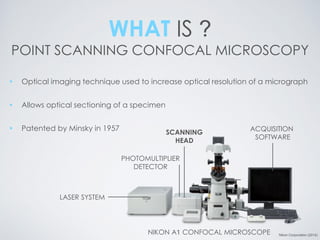
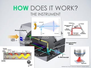
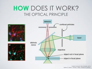
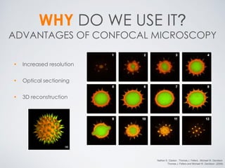
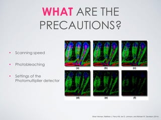

Point scanning confocal microscopy is an optical imaging technique that enhances resolution and allows for optical sectioning of specimens. Key advantages include improved resolution, 3D reconstruction capabilities, while precautions involve managing scanning speed and photobleaching. The document references various sources for further reading on the topic.


![Polymer [ बहुलक ] Chemistry Notes PDF - Irfanullah Mehar - JJ Sir Chemistry.pdf](https://cdn.slidesharecdn.com/ss_thumbnails/polymerchemistrynotespdf-irfanullahmehar-jjsirchemistry-260210172118-3f9b37f7-thumbnail.jpg?width=640&height=640&fit=bounds)