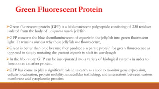
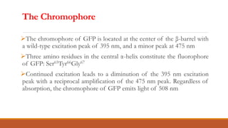
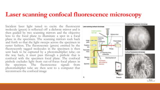
Green Fluorescent Protein
Green fluorescent protein (GFP) is a bioluminescent polypeptide consisting of 238 residues
isolated from the body of Aequorea victoria jellyfish
GFP converts the blue chemiluminescent of aequorin in the jellyfish into green fluorescent
light. It remains unclear why these jellyfish use fluorescence,
Green is better than blue because they produce a separate protein for green fluorescence as
opposed to simply mutating the present aequorin to shift its wavelength
In the laboratory, GFP can be incorporated into a variety of biological systems in order to
function as a marker protein.
GFP has come to play a significant role in research as a tool to monitor gene expression,
cellular localization, protein mobility, intracellular trafficking, and interactions between various
membrane and cytoplasmic proteins
Primary & Secondary Structure
A 21 kDa protein consisting of 238 residues
strung together to form a secondary structure of
five α-helices and one eleven-stranded β-pleated
sheet, where each strand contains nine to thirteen
residues each.
11-strand β-barrel, with an α-helical segment
threaded up through the interior of the barrel
Structurally, the barrel α-helical segment is near
the center of the β-barrel cavity.
The Chromophore
The chromophore of GFP is located at the center of the β-barrel with
a wild-type excitation peak of 395 nm, and a minor peak at 475 nm
Three amino residues in the central α-helix constitute the fluorophore
of GFP: Ser65Tyr66Gly67
Continued excitation leads to a diminution of the 395 nm excitation
peak with a reciprocal amplification of the 475 nm peak. Regardless of
absorption, the chromophore of GFP emits light of 508 nm
Using GFP as a Research Tool
1. Fluorescence resonance energy transfer (FRET).
2. Microscopy:
Fluorescence microscopy
Laser scanning confocal fluorescence microscopy
Other tools include
Centrifugation: density gradient and rate zonal centrifugation
Electrophoresis: SDS-PAGE
Fluorescence resonance energy transfer (FRET).
(A) The two proteins of interest are expressed in cells as
fusion proteins with either blue fluorescent protein
(BFP) (protein X) or GFP (protein Y). Excitation of BFP
with violet light results in the emission of blue
fluorescent light by BFP; excitation of GFP with blue
light yields green fluorescence.
(B) If the two proteins do not bind each other inside the
cell, excitation of the BFP molecule with violet light
results simply in blue fluorescence. If, however, (C) the
two proteins do bind each other, they will be close
enough to permit resonant energy transfer between
the excited BFP molecule and the GFP protein, resulting
in green fluorescence after violet excitation.
A. Optical layout of a fluorescence
microscope. Incident light tuned to excite
the fluorescent molecule is reflected by a
dichroic mirror, and then focused on the
sample; fluorescent light (longer
wavelength than excitation light) emitted
by the sample passes through the dichroic
mirror for viewing.
B. Immunofluorescent micrograph of a human
skin fibroblast, stained with fluorescent
antiactin antibody. Cells were fixed,
permeabilized, and then incubated with
fluorescein-coupled antibody. Unbound
antibody was washed away before viewing
FLUORESCENCE MICROSCOPE
Laser scanning confocal fluorescence microscopy
Incident laser light tuned to excite the fluorescent
molecule (green) is reflected off a dichroic mirror and is
then guided by two scanning mirrors and the objective
lens to the focal plane to illuminate a spot in a focal
plane in the specimen. The scanning mirrors rock back
and forth so that the light sweeps across the specimen in
raster fashion. The fluorescence (green) emitted by the
fluorescently tagged molecules in the specimen is then
sent back to be captured by a photomultiplier tube; on
the way back, it must pass through a pinhole that is
confocal with the specimen focal plane. The confocal
pinhole excludes light from out-of-focus focal planes in
the specimen. The fluorescence signals from
photomultiplier tube are then sent to a computer that
reconstructs the confocal image
Application of GFP
GFP in cell biology and biotechnology: protein fusions, imagining whole organism,
and transcriptional reporters.
GFP in the study of bacterial protein localization
GFP in host–pathogen interaction research; Salmonella typhimurium, Yersinia
pseudotuberculosis, and Mycobacterium marinum
Use of GFP as reporter gene i.e monitor gene expression in different kinds of cells
mapping (mec-7 gene in nematodes)
GFP as active indicator in case of sturding phosphorylation sites
GFP as fusion tag gene encoding the protein of interest and expressed in the cell

Green Fluorescent Protein.ppt

  • 1.
  • 2.
    Green Fluorescent Protein Greenfluorescent protein (GFP) is a bioluminescent polypeptide consisting of 238 residues isolated from the body of Aequorea victoria jellyfish GFP converts the blue chemiluminescent of aequorin in the jellyfish into green fluorescent light. It remains unclear why these jellyfish use fluorescence, Green is better than blue because they produce a separate protein for green fluorescence as opposed to simply mutating the present aequorin to shift its wavelength In the laboratory, GFP can be incorporated into a variety of biological systems in order to function as a marker protein. GFP has come to play a significant role in research as a tool to monitor gene expression, cellular localization, protein mobility, intracellular trafficking, and interactions between various membrane and cytoplasmic proteins
  • 3.
    Primary & SecondaryStructure A 21 kDa protein consisting of 238 residues strung together to form a secondary structure of five α-helices and one eleven-stranded β-pleated sheet, where each strand contains nine to thirteen residues each. 11-strand β-barrel, with an α-helical segment threaded up through the interior of the barrel Structurally, the barrel α-helical segment is near the center of the β-barrel cavity.
  • 4.
    The Chromophore The chromophoreof GFP is located at the center of the β-barrel with a wild-type excitation peak of 395 nm, and a minor peak at 475 nm Three amino residues in the central α-helix constitute the fluorophore of GFP: Ser65Tyr66Gly67 Continued excitation leads to a diminution of the 395 nm excitation peak with a reciprocal amplification of the 475 nm peak. Regardless of absorption, the chromophore of GFP emits light of 508 nm
  • 5.
    Using GFP asa Research Tool 1. Fluorescence resonance energy transfer (FRET). 2. Microscopy: Fluorescence microscopy Laser scanning confocal fluorescence microscopy Other tools include Centrifugation: density gradient and rate zonal centrifugation Electrophoresis: SDS-PAGE
  • 6.
    Fluorescence resonance energytransfer (FRET). (A) The two proteins of interest are expressed in cells as fusion proteins with either blue fluorescent protein (BFP) (protein X) or GFP (protein Y). Excitation of BFP with violet light results in the emission of blue fluorescent light by BFP; excitation of GFP with blue light yields green fluorescence. (B) If the two proteins do not bind each other inside the cell, excitation of the BFP molecule with violet light results simply in blue fluorescence. If, however, (C) the two proteins do bind each other, they will be close enough to permit resonant energy transfer between the excited BFP molecule and the GFP protein, resulting in green fluorescence after violet excitation.
  • 7.
    A. Optical layoutof a fluorescence microscope. Incident light tuned to excite the fluorescent molecule is reflected by a dichroic mirror, and then focused on the sample; fluorescent light (longer wavelength than excitation light) emitted by the sample passes through the dichroic mirror for viewing. B. Immunofluorescent micrograph of a human skin fibroblast, stained with fluorescent antiactin antibody. Cells were fixed, permeabilized, and then incubated with fluorescein-coupled antibody. Unbound antibody was washed away before viewing FLUORESCENCE MICROSCOPE
  • 8.
    Laser scanning confocalfluorescence microscopy Incident laser light tuned to excite the fluorescent molecule (green) is reflected off a dichroic mirror and is then guided by two scanning mirrors and the objective lens to the focal plane to illuminate a spot in a focal plane in the specimen. The scanning mirrors rock back and forth so that the light sweeps across the specimen in raster fashion. The fluorescence (green) emitted by the fluorescently tagged molecules in the specimen is then sent back to be captured by a photomultiplier tube; on the way back, it must pass through a pinhole that is confocal with the specimen focal plane. The confocal pinhole excludes light from out-of-focus focal planes in the specimen. The fluorescence signals from photomultiplier tube are then sent to a computer that reconstructs the confocal image
  • 9.
    Application of GFP GFPin cell biology and biotechnology: protein fusions, imagining whole organism, and transcriptional reporters. GFP in the study of bacterial protein localization GFP in host–pathogen interaction research; Salmonella typhimurium, Yersinia pseudotuberculosis, and Mycobacterium marinum Use of GFP as reporter gene i.e monitor gene expression in different kinds of cells mapping (mec-7 gene in nematodes) GFP as active indicator in case of sturding phosphorylation sites GFP as fusion tag gene encoding the protein of interest and expressed in the cell