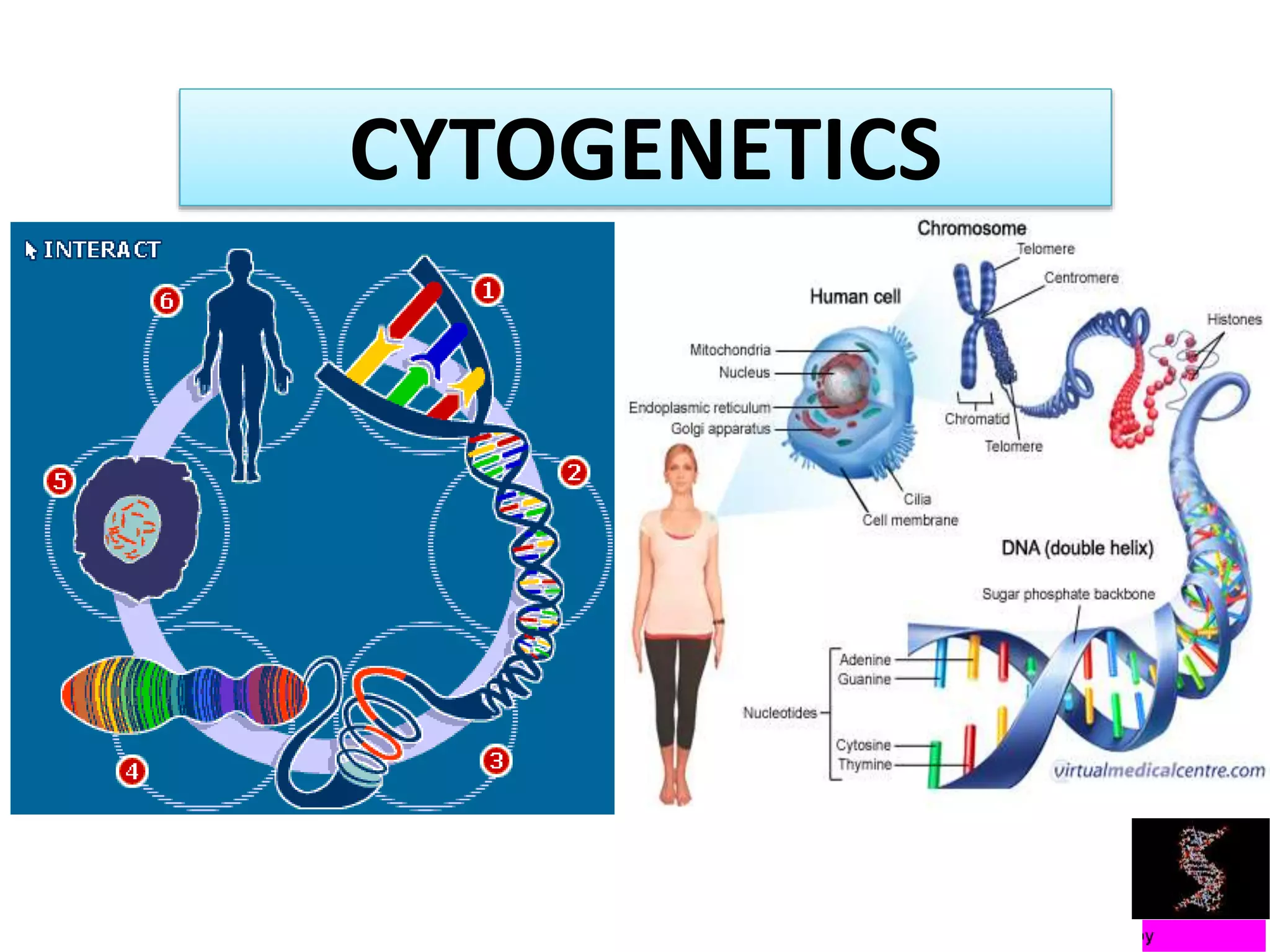
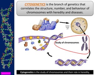
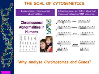
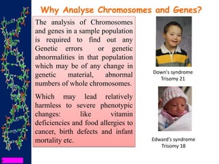
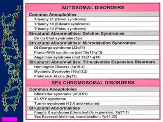
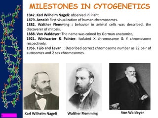
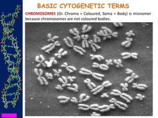
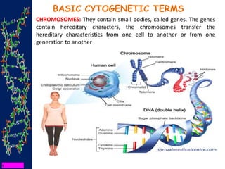
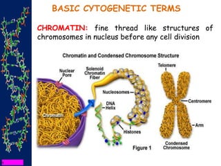
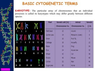
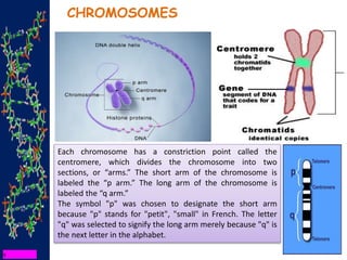
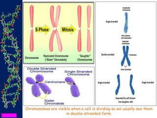
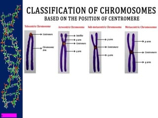
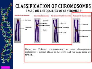
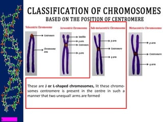
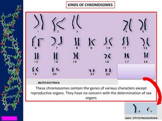
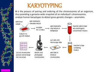
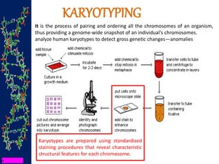
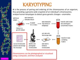
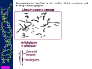
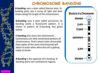
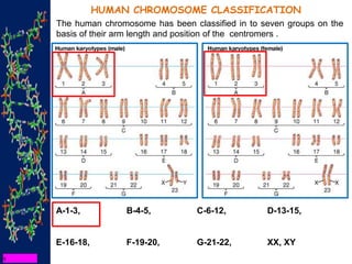
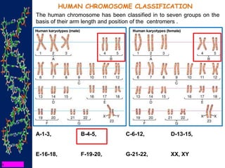
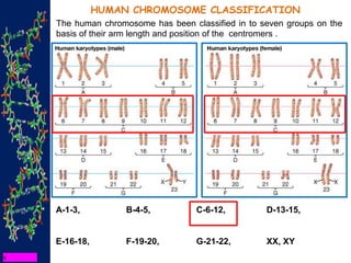
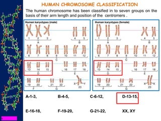
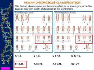
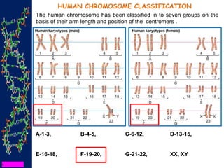
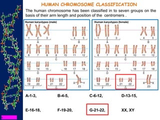
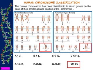
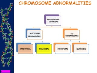
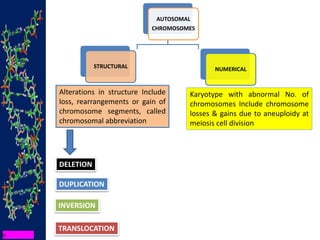
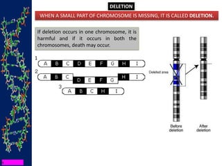
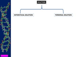
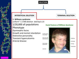
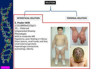
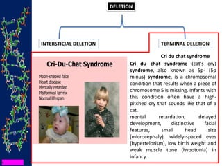
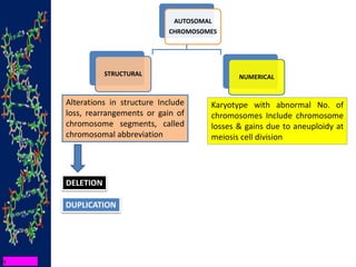
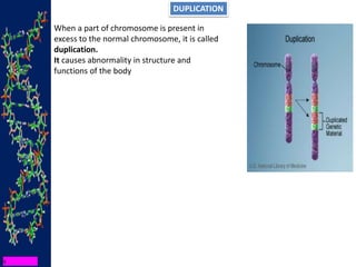
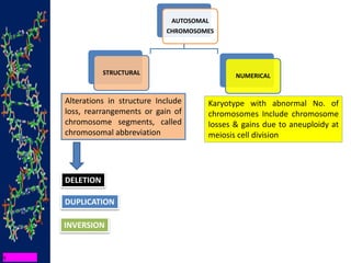
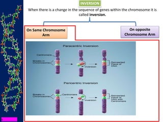
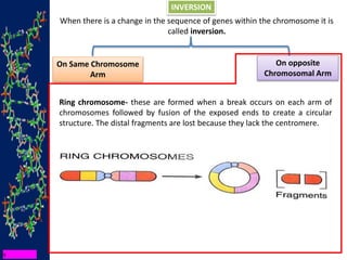
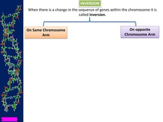
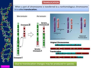
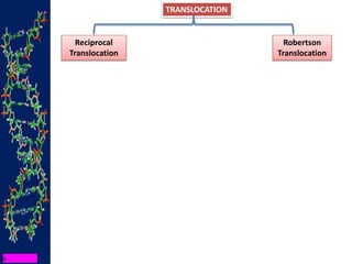
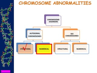
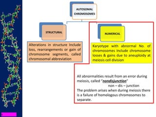
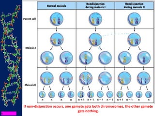
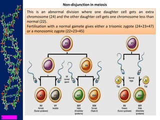
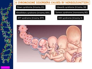
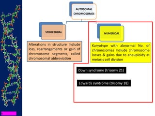
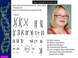
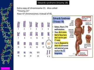
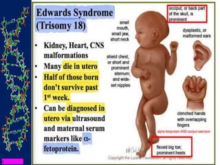
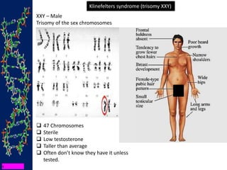
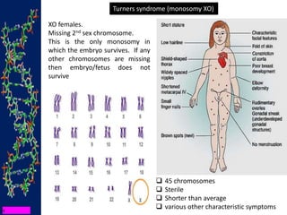
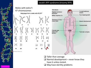
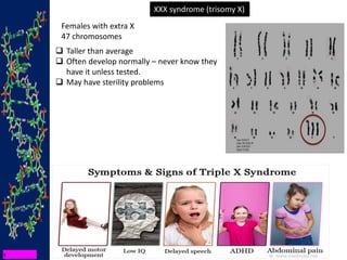
3. ORGANIZATION OF THE LECTURE
Definition and
principles
•Tools
•Applications of
cytogenetic analyses
–Diagnostic
–Prognostic
–Pathogenetic and
Therapeutic
4. Cytogenetics is the study of chromosomes and their role in heredity.
CYTOGENETICS is the branch of genetics that
correlates the structure, number, and behaviour of
chromosomes with heredity and diseases.
Study of chromosomes
5. THE GOAL OF CYTOGENETICS:
1. diagnosis of chromosomal
abnormalities.
2. localisation of any (often abnormal)
chromosomal region/DNA sequence.
Why Analyse Chromosomes and Genes?
6. The analysis of Chromosomes
and genes in a sample population
is required to find out any
Genetic errors or genetic
abnormalities in that population
which may be of any change in
genetic material, abnormal
numbers of whole chromosomes.
Which may lead relatively
harmless to severe phenotypic
changes: like vitamin
deficiencies and food allergies to
cancer, birth defects and infant
mortality etc.
Why Analyse Chromosomes and Genes?
Down's syndrome
Trisomy 21
Edward's syndrome
Trisomy 18
7.
8. MILESTONES IN CYTOGENETICS
1842. Karl Wilhelm Nageli: observed in Plant
1879. Arnold: First visualization of human chromosomes.
1882. Walther Flemming : behavior in animal cells was described, the
discoverer of mitosis,
1888. Von Waldeyer: The name was coined by German anatomist,
1921. Winiwarter & Painter: Isolated X chromosome & Y chromosome
respectively.
1956. Tijio and Levan: : Described correct chromosome number as 22 pair of
autosomes and 2 sex chromosomes.
Karl Wilhelm Nageli Walther Flemming Von Waldeyer
11. BASIC CYTOGENETIC TERMS
CHROMOSOMES: They contain small bodies, called genes. The genes
contain hereditary characters, the chromosomes transfer the
hereditary characteristics from one cell to another or from one
generation to another
12. CHROMATIN: fine thread like structures of
chromosomes in nucleus before any cell division
BASIC CYTOGENETIC TERMS
13. KARYOTYPE: The particular array of chromosomes that an individual
possesses is called its karyotypes which may differ greatly between different
species
BASIC CYTOGENETIC TERMS
14. Ideograms are a schematic representation of chromosomes. They
show the relative size of the chromosomes and their banding patterns.
15. CHROMOSOMES
Each chromosome has a constriction point called the
centromere, which divides the chromosome into two
sections, or “arms.” The short arm of the chromosome is
labeled the “p arm.” The long arm of the chromosome is
labeled the “q arm.”
The symbol "p" was chosen to designate the short arm
because "p" stands for "petit", "small" in French. The letter
"q" was selected to signify the long arm merely because "q" is
the next letter in the alphabet.
16. Chromosomes are visible when a cell is dividing so we usually see them
in double-stranded form.
17.
18. These are V-shaped chromosomes. In these chromosomes
centromere is present almost in the centre and two equal arms are
formed.
19. These are J or L-shaped chromosomes, lit these chromo-
somes centromere is present in the centre in such a
manner that two unequal! arms are formed
20. These are also rod-shaped chromosomes, but the
centromere is between two chromatids in such a
manner that one arm is short and another is long.
22. KINDS OF CHROMOSOMES
HOMOLOGOUS CHROMOSOMES:
Chromosomes are found in pairs, one set comes from Father and
another from Mother. These paired chromosomes are identical in all
respects and termed as homologous chromosomes.
23. KINDS OF CHROMOSOMES
These chromosomes contain the genes of reproductive organs. They
determine the sex. These chromosomes are of two types, X-chromosome
and Y-chromosome. X chromosomes determine female and V chromosomes
determine male.
24. KINDS OF CHROMOSOMES
These chromosomes contain the genes of various characters except
reproductive organs. They have no concern with the determination of sex
organs
25. KARYOTYPING
It is the process of pairing and ordering all the chromosomes of an organism,
thus providing a genome-wide snapshot of an individual's chromosomes.
analyze human karyotypes to detect gross genetic changes—anomalies
26. KARYOTYPING
It is the process of pairing and ordering all the chromosomes of an organism,
thus providing a genome-wide snapshot of an individual's chromosomes.
analyze human karyotypes to detect gross genetic changes—anomalies
Karyotypes are prepared using standardized
staining procedures that reveal characteristic
structural features for each chromosome.
27. KARYOTYPING
It is the process of pairing and ordering all the chromosomes of an organism,
thus providing a genome-wide snapshot of an individual's chromosomes.
analyze human karyotypes to detect gross genetic changes—anomalies
Chromosomes can be photographed or visualized
using a computer, and then analyzed
29. Chromosomes are identified by size, position of the centromere, and
banding and staining regions
G - Banding
Q - Banding
C - Banding
R - Banding
T - Banding
NOR - Banding
High Resolution Banding
Restriction Endonuclease Banding
30. CHROMOSOME BANDING
G-banding uses a stain called Giemsa stain. G-
banding gives you a series of light and dark
stripes along the length of the chromosome.
Q-banding uses a stain called quinacrine. Q-
banding yields a fluorescent pattern. It is
similar in pattern to G-banding, but glows
yellow.
C-banding only stains the centromeres.
Centromeres are little constricted portions of
chromosomes. That's where sister chromatids
(two copies of the same chromosome) will
attach to each other when the cell is getting
ready to divide.
R-banding is the opposite of C-banding. R-
banding stains non-centromeric regions.
31. HUMAN CHROMOSOME CLASSIFICATION
The human chromosome has been classified in to seven groups on the
basis of their arm length and position of the centromers .
A-1-3, B-4-5, C-6-12, D-13-15,
E-16-18, F-19-20, G-21-22, XX, XY
32. HUMAN CHROMOSOME CLASSIFICATION
The human chromosome has been classified in to seven groups on the
basis of their arm length and position of the centromers .
A-1-3, B-4-5, C-6-12, D-13-15,
E-16-18, F-19-20, G-21-22, XX, XY
33. HUMAN CHROMOSOME CLASSIFICATION
The human chromosome has been classified in to seven groups on the
basis of their arm length and position of the centromers .
A-1-3, B-4-5, C-6-12, D-13-15,
E-16-18, F-19-20, G-21-22, XX, XY
34. HUMAN CHROMOSOME CLASSIFICATION
The human chromosome has been classified in to seven groups on the
basis of their arm length and position of the centromers .
A-1-3, B-4-5, C-6-12, D-13-15,
E-16-18, F-19-20, G-21-22, XX, XY
35. HUMAN CHROMOSOME CLASSIFICATION
The human chromosome has been classified in to seven groups on the
basis of their arm length and position of the centromers .
A-1-3, B-4-5, C-6-12, D-13-15,
E-16-18, F-19-20, G-21-22, XX, XY
36. HUMAN CHROMOSOME CLASSIFICATION
The human chromosome has been classified in to seven groups on the
basis of their arm length and position of the centromers .
A-1-3, B-4-5, C-6-12, D-13-15,
E-16-18, F-19-20, G-21-22, XX, XY
37. HUMAN CHROMOSOME CLASSIFICATION
The human chromosome has been classified in to seven groups on the
basis of their arm length and position of the centromers .
A-1-3, B-4-5, C-6-12, D-13-15,
E-16-18, F-19-20, G-21-22, XX, XY
38. HUMAN CHROMOSOME CLASSIFICATION
The human chromosome has been classified in to seven groups on the
basis of their arm length and position of the centromers .
A-1-3, B-4-5, C-6-12, D-13-15,
E-16-18, F-19-20, G-21-22, XX, XY
40. AUTOSOMAL
CHROMOSOMES
STRUCTURAL NUMERICAL
Alterations in structure Include
loss, rearrangements or gain of
chromosome segments, called
chromosomal abbreviation
Karyotype with abnormal No. of
chromosomes Include chromosome
losses & gains due to aneuploidy at
meiosis cell division
DELETION
DUPLICATION
TRANSLOCATION
INVERSION
41. DELETION
WHEN A SMALL PART OF CHROMOSOME IS MISSING, IT IS CALLED DELETION.
If deletion occurs in one chromosome, it is
harmful and if it occurs in both the
chromosomes, death may occur.
43. DELETION
INTERSTICIAL DELETION TERMINAL DELETION
1. Williams syndrome
uniform” 1.5 MB deletion del(7)q11.23
1/20,000 all populations
Phenotype
Dysmorphic facies
Growth and mental retardation
Distinctive personality
Transient hypercalcemia
Arterial disease
44. DELETION
INTERSTICIAL DELETION TERMINAL DELETION
2. Prader-Willi
1/10,000Del(15)q11-
13…..Paternal
Uniparental Disomy
Phenotype:
Mild to moderate MR
Hypotonia, poor feeding in infancy
Short stature, small hands and feet,
small external genitalia
Hyperphagia (compulsive
overeating), obesity
45. DELETION
INTERSTICIAL DELETION TERMINAL DELETION
Cri du chat syndrome
Cri du chat syndrome (cat's cry)
syndrome, also known as 5p- (5p
minus) syndrome, is a chromosomal
condition that results when a piece of
chromosome 5 is missing. Infants with
this condition often have a high-
pitched cry that sounds like that of a
cat.
mental retardation, delayed
development, distinctive facial
features, small head size
(microcephaly), widely-spaced eyes
(hypertelorism), low birth weight and
weak muscle tone (hypotonia) in
infancy.
46. AUTOSOMAL
CHROMOSOMES
STRUCTURAL NUMERICAL
Alterations in structure Include
loss, rearrangements or gain of
chromosome segments, called
chromosomal abbreviation
Karyotype with abnormal No. of
chromosomes Include chromosome
losses & gains due to aneuploidy at
meiosis cell division
DELETION
DUPLICATION
47. DUPLICATION
When a part of chromosome is present in
excess to the normal chromosome, it is called
duplication.
It causes abnormality in structure and
functions of the body
48. AUTOSOMAL
CHROMOSOMES
STRUCTURAL NUMERICAL
Alterations in structure Include
loss, rearrangements or gain of
chromosome segments, called
chromosomal abbreviation
Karyotype with abnormal No. of
chromosomes Include chromosome
losses & gains due to aneuploidy at
meiosis cell division
DELETION
DUPLICATION
INVERSION
49. INVERSION
When there is a change in the sequence of genes within the chromosome it is
called inversion.
On Same Chromosome
Arm
On opposite
Chromosome Arm
50. INVERSION
When there is a change in the sequence of genes within the chromosome it is
called inversion.
On Same Chromosome
Arm
On opposite
Chromosome Arm
51. INVERSION
When there is a change in the sequence of genes within the chromosome it is
called inversion.
On Same Chromosome
Arm
On opposite
Chromosomal Arm
Ring chromosome- these are formed when a break occurs on each arm of
chromosomes followed by fusion of the exposed ends to create a circular
structure. The distal fragments are lost because they lack the centromere.
52. INVERSION
When there is a change in the sequence of genes within the chromosome it is
called inversion.
On Same Chromosome
Arm
On opposite
Chromosome Arm
53. AUTOSOMAL
CHROMOSOMES
STRUCTURAL NUMERICAL
Alterations in structure Include
loss, rearrangements or gain of
chromosome segments, called
chromosomal abbreviation
Karyotype with abnormal No. of
chromosomes Include chromosome
losses & gains due to aneuploidy at
meiosis cell division
DELETION
DUPLICATION
TRANSLOCATION
INVERSION
54. TRANSLOCATION
Due to translocation changes may be produced in species.
When a part of chromosome is transferred to a nonhomologous chromosome
it is called translocation.
57. AUTOSOMAL
CHROMOSOMES
STRUCTURAL NUMERICAL
Alterations in structure Include
loss, rearrangements or gain of
chromosome segments, called
chromosomal abbreviation
Karyotype with abnormal No. of
chromosomes Include chromosome
losses & gains due to aneuploidy at
meiosis cell division
All abnormalities result from an error during
meiosis, called “nondisjunction”
non – dis – junction
The problem arises when during meiosis there
is a failure of homologous chromosomes to
separate.
59. Non-disjunction in meiosis
This is an abnormal division where one daughter cell gets an extra
chromosome (24) and the other daughter cell gets one chromosome less than
normal (22).
Fertilisation with a normal gamete gives either a trisomic zygote (24+23=47)
or a monosomic zygote (22+23=45)
60. 2% of sperms have Chromosomal
abnormalities
20% of ova have Chromosomal
abnormalities
So among 100 conceptions, there
are 25% chromosome
abnormalities
In every 100 pregnancies, there
occurs 15 spontaneous
miscarriages,
50% of which have chromosome
abnormalities
Among 160 births, one baby is
born
with a chromosome abnormality
Frequencies of chromosome Abnormalities
62. AUTOSOMAL
CHROMOSOMES
STRUCTURAL NUMERICAL
Alterations in structure Include
loss, rearrangements or gain of
chromosome segments, called
chromosomal abbreviation
Karyotype with abnormal No. of
chromosomes Include chromosome
losses & gains due to aneuploidy at
meiosis cell division
Down syndrome (trisomy 21)
Edwards syndrome (trisomy 18)
63. Extra copy of chromosome 21. Also called
“Trisomy 21”
Have 47 chromosomes instead of 46,
Down syndrome (trisomy 21)
Short stature
Mental retardation
Reduced life span
Characteristic features
John Langdon Down (the British
physician) who described the
syndrome in 1866.
64. Edwards syndrome (trisomy 18)
Extra copy of chromosome 21. Also called
“Trisomy 21”
Have 47 chromosomes instead of 46,
65.
66. Klinefelters syndrome (trisomy XXY)
XXY – Male
Trisomy of the sex chromosomes
47 Chromosomes
Sterile
Low testosterone
Taller than average
Often don’t know they have it unless
tested.
67. Turners syndrome (monosomy XO)
45 chromosomes
Sterile
Shorter than average
various other characteristic symptoms
XO females.
Missing 2nd sex chromosome.
This is the only monosomy in
which the embryo survives. If any
other chromosomes are missing
then embryo/fetus does not
survive
68. Jacob’s XYY syndrome (trisomy XYY)
Taller than average
Normal development – never know they
have it unless tested.
May have sterility problems
Males with extra Y.
47 chromosomes
69. XXX syndrome (trisomy X)
Taller than average
Often develop normally – never know they
have it unless tested.
May have sterility problems
Females with extra X
47 chromosomes