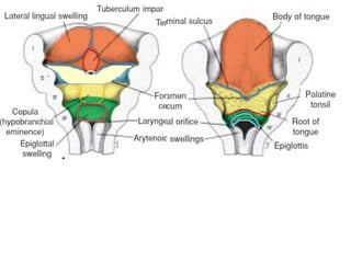
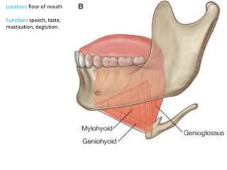
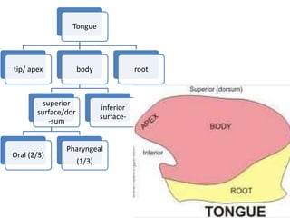
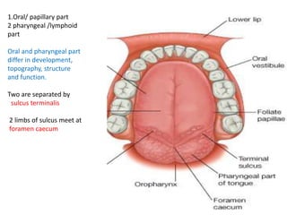
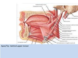
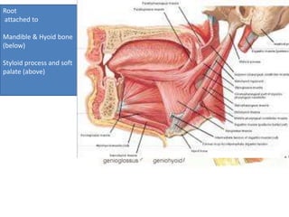
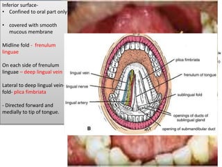
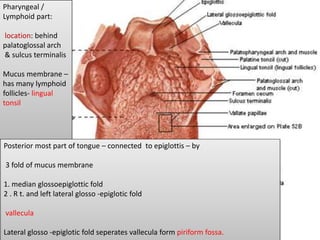
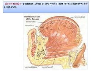
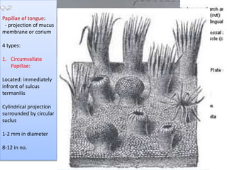
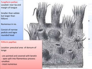
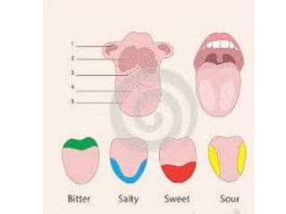
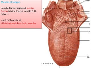
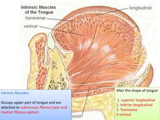
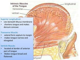
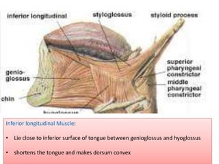
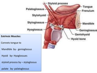
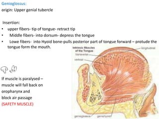
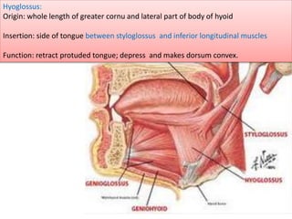
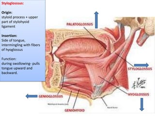
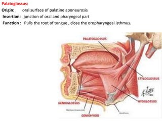
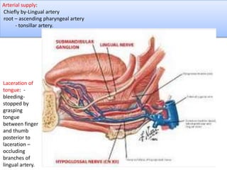
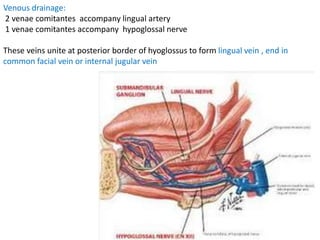
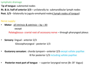
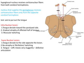
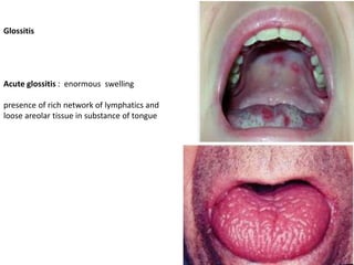
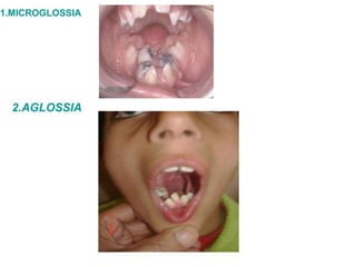
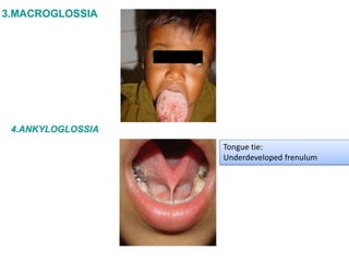
TONGUE
Dr. Prabhakar Yadav
Associate Professor
(MBBS, MS)
Department of Human Anatomy
B.P. Koirala Institute of Health Sciences
Location: floor of mouth
Function: speech, taste,
mastication, deglution.
Tongue
tip/ apex body
superior
surface/dor
-sum
Oral (2/3)
Pharyngeal
(1/3)
inferior
surface-
root
1.Oral/ papillary part
2 pharyngeal /lymphoid
part
Oral and pharyngeal part
differ in development,
topography, structure
and function.
Two are separated by
sulcus terminalis
2 limbs of sulcus meet at
foramen caecum
Apex/Tip –behind upper incisor
Root
attached to
Mandible & Hyoid bone
(below)
Styloid process and soft
palate (above)
Oral Part:
in front of palatoglossus
arch-
foliate papillae
Superior surface-
median furrow/ groove-
covered with papillae
Inferior surface-
• Confined to oral part only
• covered with smooth
mucous membrane
Midline fold - frenulum
linguae
On each side of frenulum
linguae – deep lingual vein
Lateral to deep lingual vein-
fold- plica fimbriata
- Directed forward and
medially to tip of tongue.
Pharyngeal /
Lymphoid part:
location: behind
palatoglossal arch
& sulcus terminalis
Mucus membrane –
has many lymphoid
follicles- lingual
tonsil
Posterior most part of tongue – connected to epiglottis – by
3 fold of mucus membrane
1. median glossoepiglottic fold
2 . R t. and left lateral glosso -epiglotic fold
vallecula
Lateral glosso -epiglotic fold seperates vallecula form piriform fossa.
base of tongue – posterior surface of pharyngeal part -forms anterior wall of
oropharynx
Papillae of tongue:
- projection of mucus
membrane or corium
4 types:
1. Circumvallate
Papillae:
Located: immediately
infront of sulcus
termanilis
Cylindrical projection
surrounded by circular
suclus
1-2 mm in diameter
8-12 in no.
Fungiform paillae:
Located: near tip and
margin of tongue
Smaller than vallate
but larger than
filiform
Numerous in no.
Consist of narrow
pedicle and lagre
rounded head
Filiform papillae:
Location: presulcal area of dorsum of
tonge
- are pointed and covered with keratin
- apex split into filamentous process
-smallest
- most numerous
Muscles of tongue:
middle fibrous septum ( median
furrow) divide tongue into Rt. & Lt.
halves .
each half consist of
4 Intrinsic and 4 extrinsic muscles.
Intrinsic Muscles:
Occupy upper part of tongue and are
attached to submucous fibrous layer and
median fibrous septum
Alter the shape of tongue
1. superior longitudinal
2. Inferior longitudinal
3. Transverse
4 vertical
Superior Longitudinal:
• Lies beneath Mucus membrane
• shortens tongue and makes
dorsum concave
Transverse Muscle:
• extend form septum to margin
• makes tongue narrow and
elongated
Verticle Muscle:
• located at border of anterior
part of tongue
• Makes tongue broad and
flattened.
Inferior longitudinal Muscle:
• Lie close to inferior surface of tongue between genioglossus and hyoglossus
• shortens the tongue and makes dorsum convex
Extrinsic Muscles:
Connets tongue to
Mandible by- genioglossus
Hyoid by- Hyoglossues
styloid process by – styloglossus
palate by- palatoglossus
Genioglossus:
origin: Upper genial tubercle
Insertion:
• upper fibers- tip of tongue- retract tip
• Middle fibers- into dorsum- depress the tongue
• Lowe fibers- into Hyoid bone-pulls posterior part of tongue forward – protude the
tongue form the mouth.
If muscle is paralysed –
muscle will fall back on
oropharynx and
block air passage
(SAFETY MUSCLE)
Hyoglossus:
Origin: whole length of greater cornu and lateral part of body of hyoid
Insertion: side of tongue between styloglossus and inferior longitudinal muscles
Function: retract protuded tongue; depress and makes dorsum convex.
Superficial relation:
1 styloglossus,
2 lingual nerve,
3.submandibular
ganglion,
4 deep part of
submandibular gland
5submandibular duct ,
6. hypoglossal nerve and
veins accompanying it
Deep relation: 1.Inferior longitudinal muscle,
2. Genioglossus,
3. middle constrictor of pharynx
,
4.glosssopharyngeal nerve
5. stylohyoid ligament,
6. lingual artery
Strucutue passing deep to posterior border of hyoglossus :
form above downward :
1 Glossopharyngeal nerve,
2 stylohyoid ligament,
3. lingual artery.
Styloglosssus:
Origin:
styloid process + upper
part of stylohyoid
ligament
Insertion:
Side of tongue,
intermingling with fibers
of hyoglossus
Function:
during swollowing- pulls
tongue upward and
backward.
Palatoglossus:
Origin: oral surface of palatine aponeurosis
Insertion: junction of oral and pharyngeal part
Function : Pulls the root of tongue , close the oropharyngeal isthmus.
Arterial supply:
Chiefly by-Lingual artery
root – ascending pharyngeal artery
- tonsillar artery.
Laceration of
tongue: -
bleeding-
stopped by
grasping
tongue
between finger
and thumb
posterior to
laceration –
occluding
branches of
lingual artery.
Venous drainage:
2 venae comitantes accompany lingual artery
1 venae comitantes accompany hypoglossal nerve
These veins unite at posterior border of hyoglossus to form lingual vein , end in
common facial vein or internal jugular vein
Lymphatic drainage
Tip of tongue- submental nodes
Rt. & Lt. half of anterior 2/3 – unilaterally to submandibular lymph nodes
Post. 1/3 – bilaterally to jugulo-omohyoid nodes( lymph nodes of tongue)
Nerve supply:
• Motor- all intrinsic & extrinsic – by – XII
except
Palatoglossus- cranial root of accessory nerve – through pharyngeal plexus
• Sensory- lingual - anterior 2/3
Glossopharyngeal - posterior 1/3
• Gustarory sensaton: chorda tympani---anterior 2/3 except vallate papillae
IX for posterior 1/3 including vallate papillae
• Posterior most part of tongue – superior laryngeal nerve (Br. Of Vagus)
hypoglossal nucleus receives corticonuclear fibers
from both cerebral hemispheres.
nucleus that supplies the genioglossus receives
corticonuclear fibers only from the opposite
cerebral hemisphere
test :ask to put out the tongue
Infra Nuclear lesion:
1.Tongue deviate toward the paralyzed side.
2. Gradual atrophy of affected half of tongue
3. Muscular twitching
Supra Nuclear Lesion:
1.Tongue deviate to the side opposite the lesion.
2.No atrophy or fibrillation/ twitching
3. Tongue – stiff, moves very sluggishly – defective
articulation
Glossitis
Acute glossitis : enormous swelling
presence of rich network of lymphatics and
loose areolar tissue in substance of tongue
Tongue tie:
Underdeveloped frenulum
Non fusion of 2 lingual swelling
Tongue

Tongue

  • 1.
    TONGUE Dr. Prabhakar Yadav AssociateProfessor (MBBS, MS) Department of Human Anatomy B.P. Koirala Institute of Health Sciences
  • 3.
    Location: floor ofmouth Function: speech, taste, mastication, deglution.
  • 4.
    Tongue tip/ apex body superior surface/dor -sum Oral(2/3) Pharyngeal (1/3) inferior surface- root
  • 5.
    1.Oral/ papillary part 2pharyngeal /lymphoid part Oral and pharyngeal part differ in development, topography, structure and function. Two are separated by sulcus terminalis 2 limbs of sulcus meet at foramen caecum
  • 6.
  • 7.
    Root attached to Mandible &Hyoid bone (below) Styloid process and soft palate (above)
  • 8.
    Oral Part: in frontof palatoglossus arch- foliate papillae Superior surface- median furrow/ groove- covered with papillae Inferior surface- • Confined to oral part only • covered with smooth mucous membrane Midline fold - frenulum linguae On each side of frenulum linguae – deep lingual vein Lateral to deep lingual vein- fold- plica fimbriata - Directed forward and medially to tip of tongue.
  • 9.
    Pharyngeal / Lymphoid part: location:behind palatoglossal arch & sulcus terminalis Mucus membrane – has many lymphoid follicles- lingual tonsil Posterior most part of tongue – connected to epiglottis – by 3 fold of mucus membrane 1. median glossoepiglottic fold 2 . R t. and left lateral glosso -epiglotic fold vallecula Lateral glosso -epiglotic fold seperates vallecula form piriform fossa.
  • 10.
    base of tongue– posterior surface of pharyngeal part -forms anterior wall of oropharynx
  • 11.
    Papillae of tongue: -projection of mucus membrane or corium 4 types: 1. Circumvallate Papillae: Located: immediately infront of sulcus termanilis Cylindrical projection surrounded by circular suclus 1-2 mm in diameter 8-12 in no.
  • 12.
    Fungiform paillae: Located: neartip and margin of tongue Smaller than vallate but larger than filiform Numerous in no. Consist of narrow pedicle and lagre rounded head Filiform papillae: Location: presulcal area of dorsum of tonge - are pointed and covered with keratin - apex split into filamentous process -smallest - most numerous
  • 14.
    Muscles of tongue: middlefibrous septum ( median furrow) divide tongue into Rt. & Lt. halves . each half consist of 4 Intrinsic and 4 extrinsic muscles.
  • 15.
    Intrinsic Muscles: Occupy upperpart of tongue and are attached to submucous fibrous layer and median fibrous septum Alter the shape of tongue 1. superior longitudinal 2. Inferior longitudinal 3. Transverse 4 vertical
  • 16.
    Superior Longitudinal: • Liesbeneath Mucus membrane • shortens tongue and makes dorsum concave Transverse Muscle: • extend form septum to margin • makes tongue narrow and elongated Verticle Muscle: • located at border of anterior part of tongue • Makes tongue broad and flattened.
  • 17.
    Inferior longitudinal Muscle: •Lie close to inferior surface of tongue between genioglossus and hyoglossus • shortens the tongue and makes dorsum convex
  • 18.
    Extrinsic Muscles: Connets tongueto Mandible by- genioglossus Hyoid by- Hyoglossues styloid process by – styloglossus palate by- palatoglossus
  • 19.
    Genioglossus: origin: Upper genialtubercle Insertion: • upper fibers- tip of tongue- retract tip • Middle fibers- into dorsum- depress the tongue • Lowe fibers- into Hyoid bone-pulls posterior part of tongue forward – protude the tongue form the mouth. If muscle is paralysed – muscle will fall back on oropharynx and block air passage (SAFETY MUSCLE)
  • 20.
    Hyoglossus: Origin: whole lengthof greater cornu and lateral part of body of hyoid Insertion: side of tongue between styloglossus and inferior longitudinal muscles Function: retract protuded tongue; depress and makes dorsum convex.
  • 21.
    Superficial relation: 1 styloglossus, 2lingual nerve, 3.submandibular ganglion, 4 deep part of submandibular gland 5submandibular duct , 6. hypoglossal nerve and veins accompanying it
  • 22.
    Deep relation: 1.Inferiorlongitudinal muscle, 2. Genioglossus, 3. middle constrictor of pharynx , 4.glosssopharyngeal nerve 5. stylohyoid ligament, 6. lingual artery
  • 23.
    Strucutue passing deepto posterior border of hyoglossus : form above downward : 1 Glossopharyngeal nerve, 2 stylohyoid ligament, 3. lingual artery.
  • 24.
    Styloglosssus: Origin: styloid process +upper part of stylohyoid ligament Insertion: Side of tongue, intermingling with fibers of hyoglossus Function: during swollowing- pulls tongue upward and backward.
  • 25.
    Palatoglossus: Origin: oral surfaceof palatine aponeurosis Insertion: junction of oral and pharyngeal part Function : Pulls the root of tongue , close the oropharyngeal isthmus.
  • 26.
    Arterial supply: Chiefly by-Lingualartery root – ascending pharyngeal artery - tonsillar artery. Laceration of tongue: - bleeding- stopped by grasping tongue between finger and thumb posterior to laceration – occluding branches of lingual artery.
  • 27.
    Venous drainage: 2 venaecomitantes accompany lingual artery 1 venae comitantes accompany hypoglossal nerve These veins unite at posterior border of hyoglossus to form lingual vein , end in common facial vein or internal jugular vein
  • 28.
    Lymphatic drainage Tip oftongue- submental nodes Rt. & Lt. half of anterior 2/3 – unilaterally to submandibular lymph nodes Post. 1/3 – bilaterally to jugulo-omohyoid nodes( lymph nodes of tongue) Nerve supply: • Motor- all intrinsic & extrinsic – by – XII except Palatoglossus- cranial root of accessory nerve – through pharyngeal plexus • Sensory- lingual - anterior 2/3 Glossopharyngeal - posterior 1/3 • Gustarory sensaton: chorda tympani---anterior 2/3 except vallate papillae IX for posterior 1/3 including vallate papillae • Posterior most part of tongue – superior laryngeal nerve (Br. Of Vagus)
  • 29.
    hypoglossal nucleus receivescorticonuclear fibers from both cerebral hemispheres. nucleus that supplies the genioglossus receives corticonuclear fibers only from the opposite cerebral hemisphere test :ask to put out the tongue Infra Nuclear lesion: 1.Tongue deviate toward the paralyzed side. 2. Gradual atrophy of affected half of tongue 3. Muscular twitching Supra Nuclear Lesion: 1.Tongue deviate to the side opposite the lesion. 2.No atrophy or fibrillation/ twitching 3. Tongue – stiff, moves very sluggishly – defective articulation
  • 30.
    Glossitis Acute glossitis :enormous swelling presence of rich network of lymphatics and loose areolar tissue in substance of tongue
  • 32.
  • 33.
    Non fusion of2 lingual swelling