SlideShare a Scribd company logo
Development of Pharyngeal Arches & Pouches
Dr. Prabhakar Yadav
Associate Professor
Department of Human Anatomy
B.P. Koirala Institute of Health Sciences
After establishment of head fold,
foregut is bounded ventrally by
pericardium & dorsally by developing
brain.
Foregut is initially separated from
stomatodeum by buccopharyngeal
membrane, when it breaks down,
foregut opens to exterior.
Developing brain & pericardium are
seperated by stomatodeum -
………………………
Elongation of region between stomatodaeum &
pericardium forms- Neck.
Elongation is due to series of mesodermal
thickening in the wall of cranial most part of foregut
- pharyngeal / branchial arches - provide support to
lateral & ventral of wall of primitive pharynx.
•Before appearance of pharyngeal arches endoderma of foregut is separated form ectoderm by
mesoderm.
•Mesoderm form thickening in the form of six bars that runs dorsoventrally in the floor of developing
pharynx and fuse with corrosponding bar of opposite side to form pharyngeal arch.
•Initially there are six arches. The fifth arch is small and rudimentary, and soon disappears. Pharyngeal
arches are numbered craniocaudally as 1, 2, 3, 4, and 6.
Between two adjoining arches,
endoderm extend outward in the form
of –endodermal or branchial or
pharyngeal pouch.
Between two adjoining arches,
ectoderm invaginate inward in the
form of – ectodermal cleft.
Between two adjoining arches where
pharyngeal cleft and pouches are
opposed to each other forms-
Pharyngeal membrane
Structures formed in mesoderm of each arch
1. Skeletal element: Is cartilaginous, may remain
crtilaginous, may develop into bone or may
disappear
2. Striated Muscle: is supplied by nerve of the arch.
• Later muscle may or may not retain its
attachment to skeletal element of the arch.
• Muscle may subdivide to form number of distinct
muscles that may migrate away form pharyngeal
region but still carry their nerve with them.
Structures formed in mesoderm of each arch
3. Arteries of Pharyngeal Arches: Each pharyngeal
arch has its own artery that connects aortic sac
with the dorsal aorta.
Nerves of the Pharyngeal Arches:
Each arch is supplied by a nerve that Supplies-Skeletal Muscles
Its sensory branch supplies- overlying ectoderm &
endoderm(skin and mucous membrane).
•Morphologically each pharyngeal arch is supplied by 2 nerves.
Nerve that runs along cranial border of the arch- post-trematic
Nerve that runs along its caudal border is - pretrematic nerve.
In human beings, pretrematic nerves disappear from all arches
except first arch where it persists as chorda tympani nerve.
Derivatives of skeletal elements:
Meckel’s cartilage: cartilage of 1st arch
• From dorsal end- Incus & malleus
•Ventral part- is surrounded by
developing mandible & is absorbed.
•Part extending from region of middle
ear to mandible disappears but its
perichondrium forms:
 anteriror ligament of malleus &
 sphenomandibular ligament.
Mesenchyme of the first arch is
responsible for formation of bones of
face including mandible, maxilla,
zygomatic , palatine & part of temporal
bone
Mandibulofacial dysostosis, Treacher collins
syndrome or first arch syndrome:
Entire first arch may remain underdeveloped on one
or both sides, affecting maxilla, mandible external
ear & lower eyelid ( coloboma type defect) , palate
• prominence of cheek is absent,
• ear may be displaced ventrally and caudally,
•presence of cleft palate, faulty dentition
Reichert’s cartilage : Cartilage of 2nd arch
Derivatives
• Stapes
•Styloid process
•Stylohyoid ligament
•Smaller ( lesser) cornu of hyoid bone
•Superior (upper) part of body of hyoid
bone
Cartilage of the third arch:
Derivatives
•Greater cournu of hyoid bone
•Lower part of body of hyoid bone
Cartilage of fourth & sixth arch:
Derivatives
cartilage of larynx viz., thyroid,
cricoid, arytenoid, corniculate, and cuneiform
except epiglottis, which develops from caudal part
of hypobranchial eminence
Muscles derived from pharyngeal arch
1st arch: Muscles of mastication
(temporalis, masseter, medial &lateral
pterygoids), mylohyoid, anterior belly of
digastric, tensor veli palatini & tensor
tympani
2nd arch: Muscles of facial expression
(buccinator, occipitofrontalis, platysma,
orbicularis oris, orbicularis oculi),
posterior belly of digastric
stylohyoid, stapedius & Auricular muscles
3rd arch: stylopharyngeus
4th & 6th arch: Muscle of larynx & pharynx
(Cricothyroid, levator palati,constrictors of
pharynx, and intrinsic muscles of the larynx)
Nerve of the arch & muscle supplied by them:
Arch Nerve of Arch Muscle of Arch
First arch mandibular
nerve
Muscles of mastication
(temporalis, masseter, medial
and lateral pterygoids), mylohyoid, anterior
belly of
digastric, tensor veli palatini,
and tensor tympani
Second arch Facial Muscles of facial expression
(buccinator,occipitofrontalis,
platysma,orbicularis oris, orbicularis oculi),
posterior belly of digastric
stylohyoid, stapedius & Auricular muscles
Third arch Glossophryngeal stylopharyngeus
Fourth and
sixth arches
Superior laryngeal
Recurrent laryngeal
Muscle of larynx & pharynx
(Cricothyroid, levator palati,
constrictors of pharynx, and
intrinsic muscles of the larynx)
Fate of endodermal pouches:
First pharyngeal pouch: elongates to form a
diverticulum called tubotympanic recess.
Distal part- of tubotympanic recess expands-
forms middle ear cavity & mastoid antrum.
proximal part- remains tubular-forms
pharyngotympanic/ (eustachian / auditory) tube,-
communication between nasopharynx and
tympanic cavity (middle ear).
Fate of endodermal pouches:
Second pharyngeal pouch: Epithelium of ventral
part of this pouch contribute to forms tonsil.
(Dorsal part take part in formation of tubotymanic
recess.)
Third pharyngeal pouch: give rise to inferior
parathyroid & thymus
Fourth pharngeal pouch: gives rise to superior
parathyroid gland & may contribute to thyroid
gland
Fifth or ultimobranchial pouch: gets incorporated
with fourth pouch, & forms caudal pharyngeal
complex.
Superior parathyroid gland & parafollicular cells
of thyroid arise from complex.
Pharyngeal Clefts (Grooves)
There are four pharyngeal clefts or grooves
First pharyngeal cleft gives rise to -epithelial lining
of external auditory meatus.
Pinna / auricle is formed by series of swellings
( hillocks) that arise on first & second arch
whereas
2, 3 & 4th cleft are obliterated.
Pharyngeal Clefts (Grooves)
Second arch grows faster than succeding arch &
comes to overhang them.
Space between overhanging second arch & the
third arch, fourth arch & sixth arch is called cervical
sinus.
Cavity of cervical sinus is lined by ectoderm-
normally gets oblitereted
Part of cervical sinus may persist & give rise to
swelling that lie in the neck along anterior border of
sternocleidomastoid – called branchial cyst .
When branchial cyst ruptures a branchial fistula is
formed- found on lateral aspect of neck along the
anterior border of sternocleidomastoid.
THANK YOU

More Related Content

What's hot

Pterygopalatine ganglion 1
Pterygopalatine ganglion 1Pterygopalatine ganglion 1
Pterygopalatine ganglion 1Omar Eraky
 
Anterior triangle of the neck
Anterior triangle of the neckAnterior triangle of the neck
Anterior triangle of the neck
Moamer Gabsa
 
PHARYNGEAL ARCHES,POUCHES
PHARYNGEAL ARCHES,POUCHESPHARYNGEAL ARCHES,POUCHES
PHARYNGEAL ARCHES,POUCHES
Jamil Kifayatullah
 
Anatomy of Submandibular Gland
Anatomy of Submandibular GlandAnatomy of Submandibular Gland
Anatomy of Submandibular GlandFuad Ridha Mahabot
 
Development of pharyngeal apparatus
Development of pharyngeal apparatusDevelopment of pharyngeal apparatus
Development of pharyngeal apparatus
Mohamed Autifi
 
Temporal & Infratemporal Region -I
Temporal &  Infratemporal  Region -ITemporal &  Infratemporal  Region -I
Temporal & Infratemporal Region -I
Prabhakar Yadav
 
Pharyngeal arches pouches and clefts
Pharyngeal arches pouches and cleftsPharyngeal arches pouches and clefts
Pharyngeal arches pouches and clefts
Muhammad Ramzan Ul Rehman
 
Otic Ganglia
Otic GangliaOtic Ganglia
Otic Ganglia
Usama Rana
 
Suboccipital triangle
Suboccipital triangleSuboccipital triangle
Suboccipital triangle
mgmcri1234
 
Blood supply of face
Blood supply of faceBlood supply of face
Blood supply of face1423262214
 
Facial artery dr gosai
Facial artery dr gosaiFacial artery dr gosai
Facial artery dr gosai
Dr.B.B. Gosai
 
Development of Face, Nose and Palate (Special Embryology)
Development of Face, Nose and Palate (Special Embryology)Development of Face, Nose and Palate (Special Embryology)
Development of Face, Nose and Palate (Special Embryology)
Dr. Sherif Fahmy
 
blood supply nerve supply of face
blood supply nerve supply of faceblood supply nerve supply of face
blood supply nerve supply of face
spmc bikaner
 
Infra temporal fossa
Infra temporal fossaInfra temporal fossa
Infra temporal fossa
mgmcri1234
 
The hyoglossal muscle
The hyoglossal muscleThe hyoglossal muscle
The hyoglossal muscle
Idris Siddiqui
 
Development of face
Development of faceDevelopment of face
Development of face
Dr Sudeep Madhusudan Chaudhari
 
Development of head and neck
Development of head and neckDevelopment of head and neck
Development of head and neck
Mohamed El Fiky
 
Parotid gland
 Parotid gland  Parotid gland
Parotid gland
Mohamed El Fiky
 
Pharyngeal arches and pouches
Pharyngeal arches and pouchesPharyngeal arches and pouches
Pharyngeal arches and pouches
Sajeesh Raj
 
Development of the tongue
Development of the tongueDevelopment of the tongue
Development of the tongueLheanne Tesoro
 

What's hot (20)

Pterygopalatine ganglion 1
Pterygopalatine ganglion 1Pterygopalatine ganglion 1
Pterygopalatine ganglion 1
 
Anterior triangle of the neck
Anterior triangle of the neckAnterior triangle of the neck
Anterior triangle of the neck
 
PHARYNGEAL ARCHES,POUCHES
PHARYNGEAL ARCHES,POUCHESPHARYNGEAL ARCHES,POUCHES
PHARYNGEAL ARCHES,POUCHES
 
Anatomy of Submandibular Gland
Anatomy of Submandibular GlandAnatomy of Submandibular Gland
Anatomy of Submandibular Gland
 
Development of pharyngeal apparatus
Development of pharyngeal apparatusDevelopment of pharyngeal apparatus
Development of pharyngeal apparatus
 
Temporal & Infratemporal Region -I
Temporal &  Infratemporal  Region -ITemporal &  Infratemporal  Region -I
Temporal & Infratemporal Region -I
 
Pharyngeal arches pouches and clefts
Pharyngeal arches pouches and cleftsPharyngeal arches pouches and clefts
Pharyngeal arches pouches and clefts
 
Otic Ganglia
Otic GangliaOtic Ganglia
Otic Ganglia
 
Suboccipital triangle
Suboccipital triangleSuboccipital triangle
Suboccipital triangle
 
Blood supply of face
Blood supply of faceBlood supply of face
Blood supply of face
 
Facial artery dr gosai
Facial artery dr gosaiFacial artery dr gosai
Facial artery dr gosai
 
Development of Face, Nose and Palate (Special Embryology)
Development of Face, Nose and Palate (Special Embryology)Development of Face, Nose and Palate (Special Embryology)
Development of Face, Nose and Palate (Special Embryology)
 
blood supply nerve supply of face
blood supply nerve supply of faceblood supply nerve supply of face
blood supply nerve supply of face
 
Infra temporal fossa
Infra temporal fossaInfra temporal fossa
Infra temporal fossa
 
The hyoglossal muscle
The hyoglossal muscleThe hyoglossal muscle
The hyoglossal muscle
 
Development of face
Development of faceDevelopment of face
Development of face
 
Development of head and neck
Development of head and neckDevelopment of head and neck
Development of head and neck
 
Parotid gland
 Parotid gland  Parotid gland
Parotid gland
 
Pharyngeal arches and pouches
Pharyngeal arches and pouchesPharyngeal arches and pouches
Pharyngeal arches and pouches
 
Development of the tongue
Development of the tongueDevelopment of the tongue
Development of the tongue
 

Similar to Pharyngeal arches & pouches

phayrangeal apparatus By Dr.Aisha Sadaf IBMS -KMU-peshawar
phayrangeal apparatus By Dr.Aisha Sadaf IBMS -KMU-peshawarphayrangeal apparatus By Dr.Aisha Sadaf IBMS -KMU-peshawar
phayrangeal apparatus By Dr.Aisha Sadaf IBMS -KMU-peshawarAisha Sadaf
 
Branchial arches 2020
Branchial arches 2020Branchial arches 2020
Branchial arches 2020
sallamahmed1
 
Branchial arches
Branchial archesBranchial arches
Branchial arches
DrpriyonaJohn
 
Embryology of Pharyngeal Arches by Dr Niazur Rahman
Embryology of Pharyngeal Arches by Dr Niazur RahmanEmbryology of Pharyngeal Arches by Dr Niazur Rahman
Embryology of Pharyngeal Arches by Dr Niazur Rahman
S M Niazur Rahman
 
fetal head development(week 12)
fetal head development(week 12)fetal head development(week 12)
fetal head development(week 12)
warkaa ali
 
Embryology of head&neck es - copy
Embryology of head&neck es - copyEmbryology of head&neck es - copy
Embryology of head&neck es - copy
sallamahmed1
 
Orofacial development
Orofacial developmentOrofacial development
Orofacial development
Rano Ya Naso
 
Development of ear
Development of ear  Development of ear
Development of ear
Prabhakar Yadav
 
Lecture 11 Pharyngeal Aches Chapter 9 Pdf
Lecture 11 Pharyngeal Aches Chapter 9 PdfLecture 11 Pharyngeal Aches Chapter 9 Pdf
Lecture 11 Pharyngeal Aches Chapter 9 Pdfmsu
 
Overview of pharyngeal arch derivatives beba
Overview of pharyngeal arch derivatives bebaOverview of pharyngeal arch derivatives beba
Overview of pharyngeal arch derivatives beba
BISRATGETACHEWMD
 
Pharyngeal Apparatus .pptx
Pharyngeal Apparatus .pptxPharyngeal Apparatus .pptx
Pharyngeal Apparatus .pptx
AbiolaNerd
 
Embryology of branchial arches applied anatomy by osama elhamzawy
Embryology of branchial arches applied anatomy by osama elhamzawyEmbryology of branchial arches applied anatomy by osama elhamzawy
Embryology of branchial arches applied anatomy by osama elhamzawy
Osama Elhamzawy
 
Pharyngeal Arches, Pouches and clefts-1.pptx
Pharyngeal Arches, Pouches and clefts-1.pptxPharyngeal Arches, Pouches and clefts-1.pptx
Pharyngeal Arches, Pouches and clefts-1.pptx
EmmanuelIshioma
 
PHARYNGEAL APPARATUS BY JITENDRA GURJAR.pptx
PHARYNGEAL APPARATUS BY JITENDRA GURJAR.pptxPHARYNGEAL APPARATUS BY JITENDRA GURJAR.pptx
PHARYNGEAL APPARATUS BY JITENDRA GURJAR.pptx
64Narendra
 
Lec 5 skull
Lec 5 skullLec 5 skull
Lec 5 skull
Meinah Razak
 
Embryology of Pharyngeal arches and it's clinical aspect.ppt
Embryology of Pharyngeal arches and it's clinical aspect.pptEmbryology of Pharyngeal arches and it's clinical aspect.ppt
Embryology of Pharyngeal arches and it's clinical aspect.ppt
druttamnepal
 
Development of face
Development of faceDevelopment of face
Development of face
dr. supratim tripathi
 
Skull base anatomy by Dr. Aditya Tiwari
Skull base anatomy by Dr. Aditya TiwariSkull base anatomy by Dr. Aditya Tiwari
Skull base anatomy by Dr. Aditya Tiwari
Aditya Tiwari
 
Embryology and anatomy of external ear
Embryology and anatomy of external earEmbryology and anatomy of external ear
Embryology and anatomy of external ear
FalaqFaiyaz
 
Pharyngeal apparatus.pptx
Pharyngeal apparatus.pptxPharyngeal apparatus.pptx
Pharyngeal apparatus.pptx
Sundip Charmode
 

Similar to Pharyngeal arches & pouches (20)

phayrangeal apparatus By Dr.Aisha Sadaf IBMS -KMU-peshawar
phayrangeal apparatus By Dr.Aisha Sadaf IBMS -KMU-peshawarphayrangeal apparatus By Dr.Aisha Sadaf IBMS -KMU-peshawar
phayrangeal apparatus By Dr.Aisha Sadaf IBMS -KMU-peshawar
 
Branchial arches 2020
Branchial arches 2020Branchial arches 2020
Branchial arches 2020
 
Branchial arches
Branchial archesBranchial arches
Branchial arches
 
Embryology of Pharyngeal Arches by Dr Niazur Rahman
Embryology of Pharyngeal Arches by Dr Niazur RahmanEmbryology of Pharyngeal Arches by Dr Niazur Rahman
Embryology of Pharyngeal Arches by Dr Niazur Rahman
 
fetal head development(week 12)
fetal head development(week 12)fetal head development(week 12)
fetal head development(week 12)
 
Embryology of head&neck es - copy
Embryology of head&neck es - copyEmbryology of head&neck es - copy
Embryology of head&neck es - copy
 
Orofacial development
Orofacial developmentOrofacial development
Orofacial development
 
Development of ear
Development of ear  Development of ear
Development of ear
 
Lecture 11 Pharyngeal Aches Chapter 9 Pdf
Lecture 11 Pharyngeal Aches Chapter 9 PdfLecture 11 Pharyngeal Aches Chapter 9 Pdf
Lecture 11 Pharyngeal Aches Chapter 9 Pdf
 
Overview of pharyngeal arch derivatives beba
Overview of pharyngeal arch derivatives bebaOverview of pharyngeal arch derivatives beba
Overview of pharyngeal arch derivatives beba
 
Pharyngeal Apparatus .pptx
Pharyngeal Apparatus .pptxPharyngeal Apparatus .pptx
Pharyngeal Apparatus .pptx
 
Embryology of branchial arches applied anatomy by osama elhamzawy
Embryology of branchial arches applied anatomy by osama elhamzawyEmbryology of branchial arches applied anatomy by osama elhamzawy
Embryology of branchial arches applied anatomy by osama elhamzawy
 
Pharyngeal Arches, Pouches and clefts-1.pptx
Pharyngeal Arches, Pouches and clefts-1.pptxPharyngeal Arches, Pouches and clefts-1.pptx
Pharyngeal Arches, Pouches and clefts-1.pptx
 
PHARYNGEAL APPARATUS BY JITENDRA GURJAR.pptx
PHARYNGEAL APPARATUS BY JITENDRA GURJAR.pptxPHARYNGEAL APPARATUS BY JITENDRA GURJAR.pptx
PHARYNGEAL APPARATUS BY JITENDRA GURJAR.pptx
 
Lec 5 skull
Lec 5 skullLec 5 skull
Lec 5 skull
 
Embryology of Pharyngeal arches and it's clinical aspect.ppt
Embryology of Pharyngeal arches and it's clinical aspect.pptEmbryology of Pharyngeal arches and it's clinical aspect.ppt
Embryology of Pharyngeal arches and it's clinical aspect.ppt
 
Development of face
Development of faceDevelopment of face
Development of face
 
Skull base anatomy by Dr. Aditya Tiwari
Skull base anatomy by Dr. Aditya TiwariSkull base anatomy by Dr. Aditya Tiwari
Skull base anatomy by Dr. Aditya Tiwari
 
Embryology and anatomy of external ear
Embryology and anatomy of external earEmbryology and anatomy of external ear
Embryology and anatomy of external ear
 
Pharyngeal apparatus.pptx
Pharyngeal apparatus.pptxPharyngeal apparatus.pptx
Pharyngeal apparatus.pptx
 

More from Prabhakar Yadav

Larynx
Larynx  Larynx
Diaphragm
Diaphragm   Diaphragm
Diaphragm
Prabhakar Yadav
 
Lungs,Pleura & Tracheo-bronchial Tree
Lungs,Pleura & Tracheo-bronchial TreeLungs,Pleura & Tracheo-bronchial Tree
Lungs,Pleura & Tracheo-bronchial Tree
Prabhakar Yadav
 
Medulla oblongata
Medulla oblongata Medulla oblongata
Medulla oblongata
Prabhakar Yadav
 
Nasal cavity and Paranasal Air Sinuses
Nasal cavity and Paranasal Air Sinuses Nasal cavity and Paranasal Air Sinuses
Nasal cavity and Paranasal Air Sinuses
Prabhakar Yadav
 
Tongue
TongueTongue
Lacrimal apparatus & nose
Lacrimal apparatus & noseLacrimal apparatus & nose
Lacrimal apparatus & nose
Prabhakar Yadav
 
Functional cortical area & white matter of cerebrum
Functional cortical area & white matter of cerebrumFunctional cortical area & white matter of cerebrum
Functional cortical area & white matter of cerebrum
Prabhakar Yadav
 
Cerebrum external features
Cerebrum external featuresCerebrum external features
Cerebrum external features
Prabhakar Yadav
 
Pharynx & Palate
Pharynx & PalatePharynx & Palate
Pharynx & Palate
Prabhakar Yadav
 
Knee joint
Knee joint   Knee joint
Knee joint
Prabhakar Yadav
 
Popliteal fossa & back of thigh
Popliteal fossa & back of thigh  Popliteal fossa & back of thigh
Popliteal fossa & back of thigh
Prabhakar Yadav
 
Bilaminar & trilaminar embryonic germ disc
Bilaminar & trilaminar embryonic germ discBilaminar & trilaminar embryonic germ disc
Bilaminar & trilaminar embryonic germ disc
Prabhakar Yadav
 
Perineum & external genitalia
Perineum & external genitaliaPerineum & external genitalia
Perineum & external genitalia
Prabhakar Yadav
 
Boundaries & contents of Axilla
Boundaries & contents of Axilla   Boundaries & contents of Axilla
Boundaries & contents of Axilla
Prabhakar Yadav
 
Temperomandibular joint
Temperomandibular  jointTemperomandibular  joint
Temperomandibular joint
Prabhakar Yadav
 
Temporal & infratemporal regions II
Temporal & infratemporal regions IITemporal & infratemporal regions II
Temporal & infratemporal regions II
Prabhakar Yadav
 
Duodenum & pancreas
Duodenum & pancreas   Duodenum & pancreas
Duodenum & pancreas
Prabhakar Yadav
 
Development of Hepatobiliary System
Development of Hepatobiliary System Development of Hepatobiliary System
Development of Hepatobiliary System
Prabhakar Yadav
 
Development of eye
Development of eye  Development of eye
Development of eye
Prabhakar Yadav
 

More from Prabhakar Yadav (20)

Larynx
Larynx  Larynx
Larynx
 
Diaphragm
Diaphragm   Diaphragm
Diaphragm
 
Lungs,Pleura & Tracheo-bronchial Tree
Lungs,Pleura & Tracheo-bronchial TreeLungs,Pleura & Tracheo-bronchial Tree
Lungs,Pleura & Tracheo-bronchial Tree
 
Medulla oblongata
Medulla oblongata Medulla oblongata
Medulla oblongata
 
Nasal cavity and Paranasal Air Sinuses
Nasal cavity and Paranasal Air Sinuses Nasal cavity and Paranasal Air Sinuses
Nasal cavity and Paranasal Air Sinuses
 
Tongue
TongueTongue
Tongue
 
Lacrimal apparatus & nose
Lacrimal apparatus & noseLacrimal apparatus & nose
Lacrimal apparatus & nose
 
Functional cortical area & white matter of cerebrum
Functional cortical area & white matter of cerebrumFunctional cortical area & white matter of cerebrum
Functional cortical area & white matter of cerebrum
 
Cerebrum external features
Cerebrum external featuresCerebrum external features
Cerebrum external features
 
Pharynx & Palate
Pharynx & PalatePharynx & Palate
Pharynx & Palate
 
Knee joint
Knee joint   Knee joint
Knee joint
 
Popliteal fossa & back of thigh
Popliteal fossa & back of thigh  Popliteal fossa & back of thigh
Popliteal fossa & back of thigh
 
Bilaminar & trilaminar embryonic germ disc
Bilaminar & trilaminar embryonic germ discBilaminar & trilaminar embryonic germ disc
Bilaminar & trilaminar embryonic germ disc
 
Perineum & external genitalia
Perineum & external genitaliaPerineum & external genitalia
Perineum & external genitalia
 
Boundaries & contents of Axilla
Boundaries & contents of Axilla   Boundaries & contents of Axilla
Boundaries & contents of Axilla
 
Temperomandibular joint
Temperomandibular  jointTemperomandibular  joint
Temperomandibular joint
 
Temporal & infratemporal regions II
Temporal & infratemporal regions IITemporal & infratemporal regions II
Temporal & infratemporal regions II
 
Duodenum & pancreas
Duodenum & pancreas   Duodenum & pancreas
Duodenum & pancreas
 
Development of Hepatobiliary System
Development of Hepatobiliary System Development of Hepatobiliary System
Development of Hepatobiliary System
 
Development of eye
Development of eye  Development of eye
Development of eye
 

Recently uploaded

Honest Reviews of Tim Han LMA Course Program.pptx
Honest Reviews of Tim Han LMA Course Program.pptxHonest Reviews of Tim Han LMA Course Program.pptx
Honest Reviews of Tim Han LMA Course Program.pptx
timhan337
 
Biological Screening of Herbal Drugs in detailed.
Biological Screening of Herbal Drugs in detailed.Biological Screening of Herbal Drugs in detailed.
Biological Screening of Herbal Drugs in detailed.
Ashokrao Mane college of Pharmacy Peth-Vadgaon
 
The Roman Empire A Historical Colossus.pdf
The Roman Empire A Historical Colossus.pdfThe Roman Empire A Historical Colossus.pdf
The Roman Empire A Historical Colossus.pdf
kaushalkr1407
 
aaaaaaaaaaaaaaaaaaaaaaaaaaaaaaaaaaaaaaaaa
aaaaaaaaaaaaaaaaaaaaaaaaaaaaaaaaaaaaaaaaaaaaaaaaaaaaaaaaaaaaaaaaaaaaaaaaaaaaaaaaaa
aaaaaaaaaaaaaaaaaaaaaaaaaaaaaaaaaaaaaaaaa
siemaillard
 
Embracing GenAI - A Strategic Imperative
Embracing GenAI - A Strategic ImperativeEmbracing GenAI - A Strategic Imperative
Embracing GenAI - A Strategic Imperative
Peter Windle
 
Polish students' mobility in the Czech Republic
Polish students' mobility in the Czech RepublicPolish students' mobility in the Czech Republic
Polish students' mobility in the Czech Republic
Anna Sz.
 
Operation Blue Star - Saka Neela Tara
Operation Blue Star   -  Saka Neela TaraOperation Blue Star   -  Saka Neela Tara
Operation Blue Star - Saka Neela Tara
Balvir Singh
 
June 3, 2024 Anti-Semitism Letter Sent to MIT President Kornbluth and MIT Cor...
June 3, 2024 Anti-Semitism Letter Sent to MIT President Kornbluth and MIT Cor...June 3, 2024 Anti-Semitism Letter Sent to MIT President Kornbluth and MIT Cor...
June 3, 2024 Anti-Semitism Letter Sent to MIT President Kornbluth and MIT Cor...
Levi Shapiro
 
Unit 2- Research Aptitude (UGC NET Paper I).pdf
Unit 2- Research Aptitude (UGC NET Paper I).pdfUnit 2- Research Aptitude (UGC NET Paper I).pdf
Unit 2- Research Aptitude (UGC NET Paper I).pdf
Thiyagu K
 
Acetabularia Information For Class 9 .docx
Acetabularia Information For Class 9  .docxAcetabularia Information For Class 9  .docx
Acetabularia Information For Class 9 .docx
vaibhavrinwa19
 
TESDA TM1 REVIEWER FOR NATIONAL ASSESSMENT WRITTEN AND ORAL QUESTIONS WITH A...
TESDA TM1 REVIEWER  FOR NATIONAL ASSESSMENT WRITTEN AND ORAL QUESTIONS WITH A...TESDA TM1 REVIEWER  FOR NATIONAL ASSESSMENT WRITTEN AND ORAL QUESTIONS WITH A...
TESDA TM1 REVIEWER FOR NATIONAL ASSESSMENT WRITTEN AND ORAL QUESTIONS WITH A...
EugeneSaldivar
 
Chapter 3 - Islamic Banking Products and Services.pptx
Chapter 3 - Islamic Banking Products and Services.pptxChapter 3 - Islamic Banking Products and Services.pptx
Chapter 3 - Islamic Banking Products and Services.pptx
Mohd Adib Abd Muin, Senior Lecturer at Universiti Utara Malaysia
 
Mule 4.6 & Java 17 Upgrade | MuleSoft Mysore Meetup #46
Mule 4.6 & Java 17 Upgrade | MuleSoft Mysore Meetup #46Mule 4.6 & Java 17 Upgrade | MuleSoft Mysore Meetup #46
Mule 4.6 & Java 17 Upgrade | MuleSoft Mysore Meetup #46
MysoreMuleSoftMeetup
 
CACJapan - GROUP Presentation 1- Wk 4.pdf
CACJapan - GROUP Presentation 1- Wk 4.pdfCACJapan - GROUP Presentation 1- Wk 4.pdf
CACJapan - GROUP Presentation 1- Wk 4.pdf
camakaiclarkmusic
 
How libraries can support authors with open access requirements for UKRI fund...
How libraries can support authors with open access requirements for UKRI fund...How libraries can support authors with open access requirements for UKRI fund...
How libraries can support authors with open access requirements for UKRI fund...
Jisc
 
Model Attribute Check Company Auto Property
Model Attribute  Check Company Auto PropertyModel Attribute  Check Company Auto Property
Model Attribute Check Company Auto Property
Celine George
 
A Strategic Approach: GenAI in Education
A Strategic Approach: GenAI in EducationA Strategic Approach: GenAI in Education
A Strategic Approach: GenAI in Education
Peter Windle
 
The Challenger.pdf DNHS Official Publication
The Challenger.pdf DNHS Official PublicationThe Challenger.pdf DNHS Official Publication
The Challenger.pdf DNHS Official Publication
Delapenabediema
 
Instructions for Submissions thorugh G- Classroom.pptx
Instructions for Submissions thorugh G- Classroom.pptxInstructions for Submissions thorugh G- Classroom.pptx
Instructions for Submissions thorugh G- Classroom.pptx
Jheel Barad
 
678020731-Sumas-y-Restas-Para-Colorear.pdf
678020731-Sumas-y-Restas-Para-Colorear.pdf678020731-Sumas-y-Restas-Para-Colorear.pdf
678020731-Sumas-y-Restas-Para-Colorear.pdf
CarlosHernanMontoyab2
 

Recently uploaded (20)

Honest Reviews of Tim Han LMA Course Program.pptx
Honest Reviews of Tim Han LMA Course Program.pptxHonest Reviews of Tim Han LMA Course Program.pptx
Honest Reviews of Tim Han LMA Course Program.pptx
 
Biological Screening of Herbal Drugs in detailed.
Biological Screening of Herbal Drugs in detailed.Biological Screening of Herbal Drugs in detailed.
Biological Screening of Herbal Drugs in detailed.
 
The Roman Empire A Historical Colossus.pdf
The Roman Empire A Historical Colossus.pdfThe Roman Empire A Historical Colossus.pdf
The Roman Empire A Historical Colossus.pdf
 
aaaaaaaaaaaaaaaaaaaaaaaaaaaaaaaaaaaaaaaaa
aaaaaaaaaaaaaaaaaaaaaaaaaaaaaaaaaaaaaaaaaaaaaaaaaaaaaaaaaaaaaaaaaaaaaaaaaaaaaaaaaa
aaaaaaaaaaaaaaaaaaaaaaaaaaaaaaaaaaaaaaaaa
 
Embracing GenAI - A Strategic Imperative
Embracing GenAI - A Strategic ImperativeEmbracing GenAI - A Strategic Imperative
Embracing GenAI - A Strategic Imperative
 
Polish students' mobility in the Czech Republic
Polish students' mobility in the Czech RepublicPolish students' mobility in the Czech Republic
Polish students' mobility in the Czech Republic
 
Operation Blue Star - Saka Neela Tara
Operation Blue Star   -  Saka Neela TaraOperation Blue Star   -  Saka Neela Tara
Operation Blue Star - Saka Neela Tara
 
June 3, 2024 Anti-Semitism Letter Sent to MIT President Kornbluth and MIT Cor...
June 3, 2024 Anti-Semitism Letter Sent to MIT President Kornbluth and MIT Cor...June 3, 2024 Anti-Semitism Letter Sent to MIT President Kornbluth and MIT Cor...
June 3, 2024 Anti-Semitism Letter Sent to MIT President Kornbluth and MIT Cor...
 
Unit 2- Research Aptitude (UGC NET Paper I).pdf
Unit 2- Research Aptitude (UGC NET Paper I).pdfUnit 2- Research Aptitude (UGC NET Paper I).pdf
Unit 2- Research Aptitude (UGC NET Paper I).pdf
 
Acetabularia Information For Class 9 .docx
Acetabularia Information For Class 9  .docxAcetabularia Information For Class 9  .docx
Acetabularia Information For Class 9 .docx
 
TESDA TM1 REVIEWER FOR NATIONAL ASSESSMENT WRITTEN AND ORAL QUESTIONS WITH A...
TESDA TM1 REVIEWER  FOR NATIONAL ASSESSMENT WRITTEN AND ORAL QUESTIONS WITH A...TESDA TM1 REVIEWER  FOR NATIONAL ASSESSMENT WRITTEN AND ORAL QUESTIONS WITH A...
TESDA TM1 REVIEWER FOR NATIONAL ASSESSMENT WRITTEN AND ORAL QUESTIONS WITH A...
 
Chapter 3 - Islamic Banking Products and Services.pptx
Chapter 3 - Islamic Banking Products and Services.pptxChapter 3 - Islamic Banking Products and Services.pptx
Chapter 3 - Islamic Banking Products and Services.pptx
 
Mule 4.6 & Java 17 Upgrade | MuleSoft Mysore Meetup #46
Mule 4.6 & Java 17 Upgrade | MuleSoft Mysore Meetup #46Mule 4.6 & Java 17 Upgrade | MuleSoft Mysore Meetup #46
Mule 4.6 & Java 17 Upgrade | MuleSoft Mysore Meetup #46
 
CACJapan - GROUP Presentation 1- Wk 4.pdf
CACJapan - GROUP Presentation 1- Wk 4.pdfCACJapan - GROUP Presentation 1- Wk 4.pdf
CACJapan - GROUP Presentation 1- Wk 4.pdf
 
How libraries can support authors with open access requirements for UKRI fund...
How libraries can support authors with open access requirements for UKRI fund...How libraries can support authors with open access requirements for UKRI fund...
How libraries can support authors with open access requirements for UKRI fund...
 
Model Attribute Check Company Auto Property
Model Attribute  Check Company Auto PropertyModel Attribute  Check Company Auto Property
Model Attribute Check Company Auto Property
 
A Strategic Approach: GenAI in Education
A Strategic Approach: GenAI in EducationA Strategic Approach: GenAI in Education
A Strategic Approach: GenAI in Education
 
The Challenger.pdf DNHS Official Publication
The Challenger.pdf DNHS Official PublicationThe Challenger.pdf DNHS Official Publication
The Challenger.pdf DNHS Official Publication
 
Instructions for Submissions thorugh G- Classroom.pptx
Instructions for Submissions thorugh G- Classroom.pptxInstructions for Submissions thorugh G- Classroom.pptx
Instructions for Submissions thorugh G- Classroom.pptx
 
678020731-Sumas-y-Restas-Para-Colorear.pdf
678020731-Sumas-y-Restas-Para-Colorear.pdf678020731-Sumas-y-Restas-Para-Colorear.pdf
678020731-Sumas-y-Restas-Para-Colorear.pdf
 

Pharyngeal arches & pouches

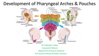
  • 1. Development of Pharyngeal Arches & Pouches Dr. Prabhakar Yadav Associate Professor Department of Human Anatomy B.P. Koirala Institute of Health Sciences
  • 2. After establishment of head fold, foregut is bounded ventrally by pericardium & dorsally by developing brain. Foregut is initially separated from stomatodeum by buccopharyngeal membrane, when it breaks down, foregut opens to exterior. Developing brain & pericardium are seperated by stomatodeum - ………………………
  • 3. Elongation of region between stomatodaeum & pericardium forms- Neck. Elongation is due to series of mesodermal thickening in the wall of cranial most part of foregut - pharyngeal / branchial arches - provide support to lateral & ventral of wall of primitive pharynx.
  • 4. •Before appearance of pharyngeal arches endoderma of foregut is separated form ectoderm by mesoderm. •Mesoderm form thickening in the form of six bars that runs dorsoventrally in the floor of developing pharynx and fuse with corrosponding bar of opposite side to form pharyngeal arch. •Initially there are six arches. The fifth arch is small and rudimentary, and soon disappears. Pharyngeal arches are numbered craniocaudally as 1, 2, 3, 4, and 6.
  • 5. Between two adjoining arches, endoderm extend outward in the form of –endodermal or branchial or pharyngeal pouch. Between two adjoining arches, ectoderm invaginate inward in the form of – ectodermal cleft. Between two adjoining arches where pharyngeal cleft and pouches are opposed to each other forms- Pharyngeal membrane
  • 6. Structures formed in mesoderm of each arch 1. Skeletal element: Is cartilaginous, may remain crtilaginous, may develop into bone or may disappear 2. Striated Muscle: is supplied by nerve of the arch. • Later muscle may or may not retain its attachment to skeletal element of the arch. • Muscle may subdivide to form number of distinct muscles that may migrate away form pharyngeal region but still carry their nerve with them.
  • 7. Structures formed in mesoderm of each arch 3. Arteries of Pharyngeal Arches: Each pharyngeal arch has its own artery that connects aortic sac with the dorsal aorta.
  • 8. Nerves of the Pharyngeal Arches: Each arch is supplied by a nerve that Supplies-Skeletal Muscles Its sensory branch supplies- overlying ectoderm & endoderm(skin and mucous membrane). •Morphologically each pharyngeal arch is supplied by 2 nerves. Nerve that runs along cranial border of the arch- post-trematic Nerve that runs along its caudal border is - pretrematic nerve. In human beings, pretrematic nerves disappear from all arches except first arch where it persists as chorda tympani nerve.
  • 9.
  • 10. Derivatives of skeletal elements: Meckel’s cartilage: cartilage of 1st arch • From dorsal end- Incus & malleus •Ventral part- is surrounded by developing mandible & is absorbed. •Part extending from region of middle ear to mandible disappears but its perichondrium forms:  anteriror ligament of malleus &  sphenomandibular ligament. Mesenchyme of the first arch is responsible for formation of bones of face including mandible, maxilla, zygomatic , palatine & part of temporal bone
  • 11. Mandibulofacial dysostosis, Treacher collins syndrome or first arch syndrome: Entire first arch may remain underdeveloped on one or both sides, affecting maxilla, mandible external ear & lower eyelid ( coloboma type defect) , palate • prominence of cheek is absent, • ear may be displaced ventrally and caudally, •presence of cleft palate, faulty dentition
  • 12. Reichert’s cartilage : Cartilage of 2nd arch Derivatives • Stapes •Styloid process •Stylohyoid ligament •Smaller ( lesser) cornu of hyoid bone •Superior (upper) part of body of hyoid bone Cartilage of the third arch: Derivatives •Greater cournu of hyoid bone •Lower part of body of hyoid bone
  • 13. Cartilage of fourth & sixth arch: Derivatives cartilage of larynx viz., thyroid, cricoid, arytenoid, corniculate, and cuneiform except epiglottis, which develops from caudal part of hypobranchial eminence
  • 14. Muscles derived from pharyngeal arch 1st arch: Muscles of mastication (temporalis, masseter, medial &lateral pterygoids), mylohyoid, anterior belly of digastric, tensor veli palatini & tensor tympani 2nd arch: Muscles of facial expression (buccinator, occipitofrontalis, platysma, orbicularis oris, orbicularis oculi), posterior belly of digastric stylohyoid, stapedius & Auricular muscles 3rd arch: stylopharyngeus 4th & 6th arch: Muscle of larynx & pharynx (Cricothyroid, levator palati,constrictors of pharynx, and intrinsic muscles of the larynx)
  • 15. Nerve of the arch & muscle supplied by them: Arch Nerve of Arch Muscle of Arch First arch mandibular nerve Muscles of mastication (temporalis, masseter, medial and lateral pterygoids), mylohyoid, anterior belly of digastric, tensor veli palatini, and tensor tympani Second arch Facial Muscles of facial expression (buccinator,occipitofrontalis, platysma,orbicularis oris, orbicularis oculi), posterior belly of digastric stylohyoid, stapedius & Auricular muscles Third arch Glossophryngeal stylopharyngeus Fourth and sixth arches Superior laryngeal Recurrent laryngeal Muscle of larynx & pharynx (Cricothyroid, levator palati, constrictors of pharynx, and intrinsic muscles of the larynx)
  • 16. Fate of endodermal pouches: First pharyngeal pouch: elongates to form a diverticulum called tubotympanic recess. Distal part- of tubotympanic recess expands- forms middle ear cavity & mastoid antrum. proximal part- remains tubular-forms pharyngotympanic/ (eustachian / auditory) tube,- communication between nasopharynx and tympanic cavity (middle ear).
  • 17. Fate of endodermal pouches: Second pharyngeal pouch: Epithelium of ventral part of this pouch contribute to forms tonsil. (Dorsal part take part in formation of tubotymanic recess.) Third pharyngeal pouch: give rise to inferior parathyroid & thymus Fourth pharngeal pouch: gives rise to superior parathyroid gland & may contribute to thyroid gland Fifth or ultimobranchial pouch: gets incorporated with fourth pouch, & forms caudal pharyngeal complex. Superior parathyroid gland & parafollicular cells of thyroid arise from complex.
  • 18. Pharyngeal Clefts (Grooves) There are four pharyngeal clefts or grooves First pharyngeal cleft gives rise to -epithelial lining of external auditory meatus. Pinna / auricle is formed by series of swellings ( hillocks) that arise on first & second arch whereas 2, 3 & 4th cleft are obliterated.
  • 19. Pharyngeal Clefts (Grooves) Second arch grows faster than succeding arch & comes to overhang them. Space between overhanging second arch & the third arch, fourth arch & sixth arch is called cervical sinus. Cavity of cervical sinus is lined by ectoderm- normally gets oblitereted
  • 20. Part of cervical sinus may persist & give rise to swelling that lie in the neck along anterior border of sternocleidomastoid – called branchial cyst . When branchial cyst ruptures a branchial fistula is formed- found on lateral aspect of neck along the anterior border of sternocleidomastoid.