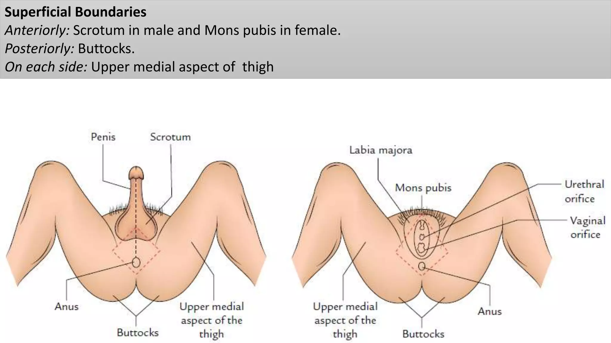
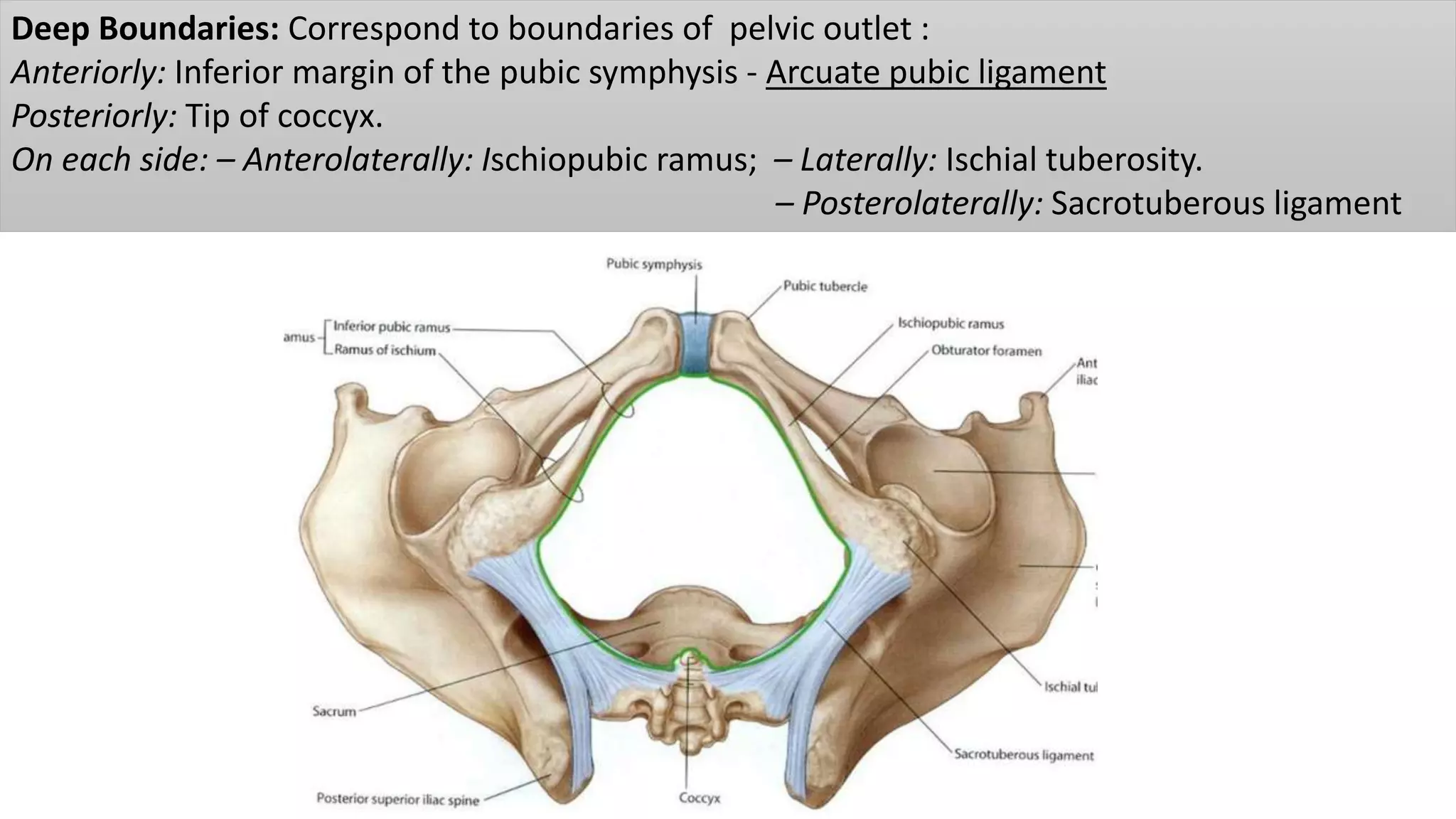
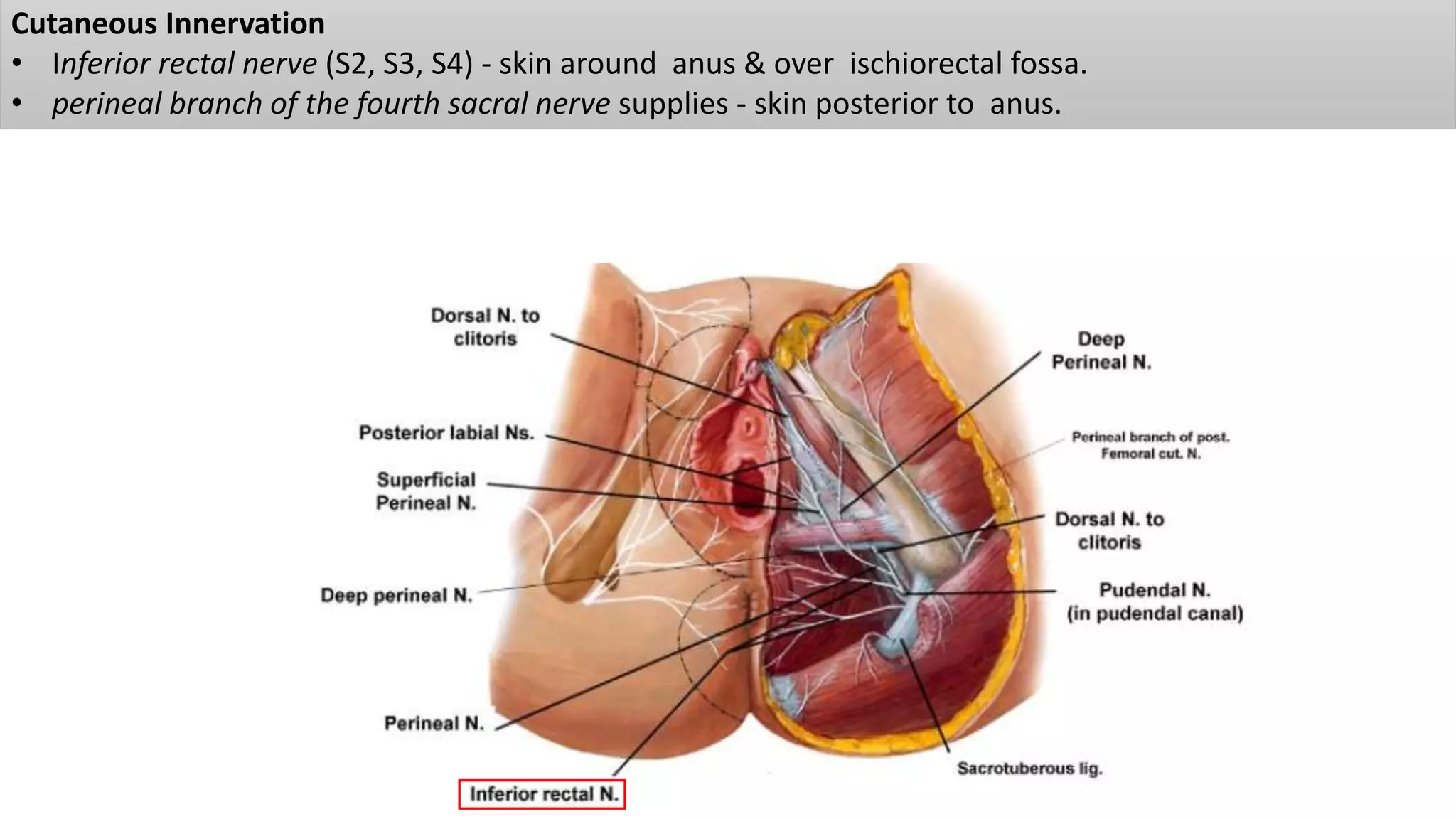
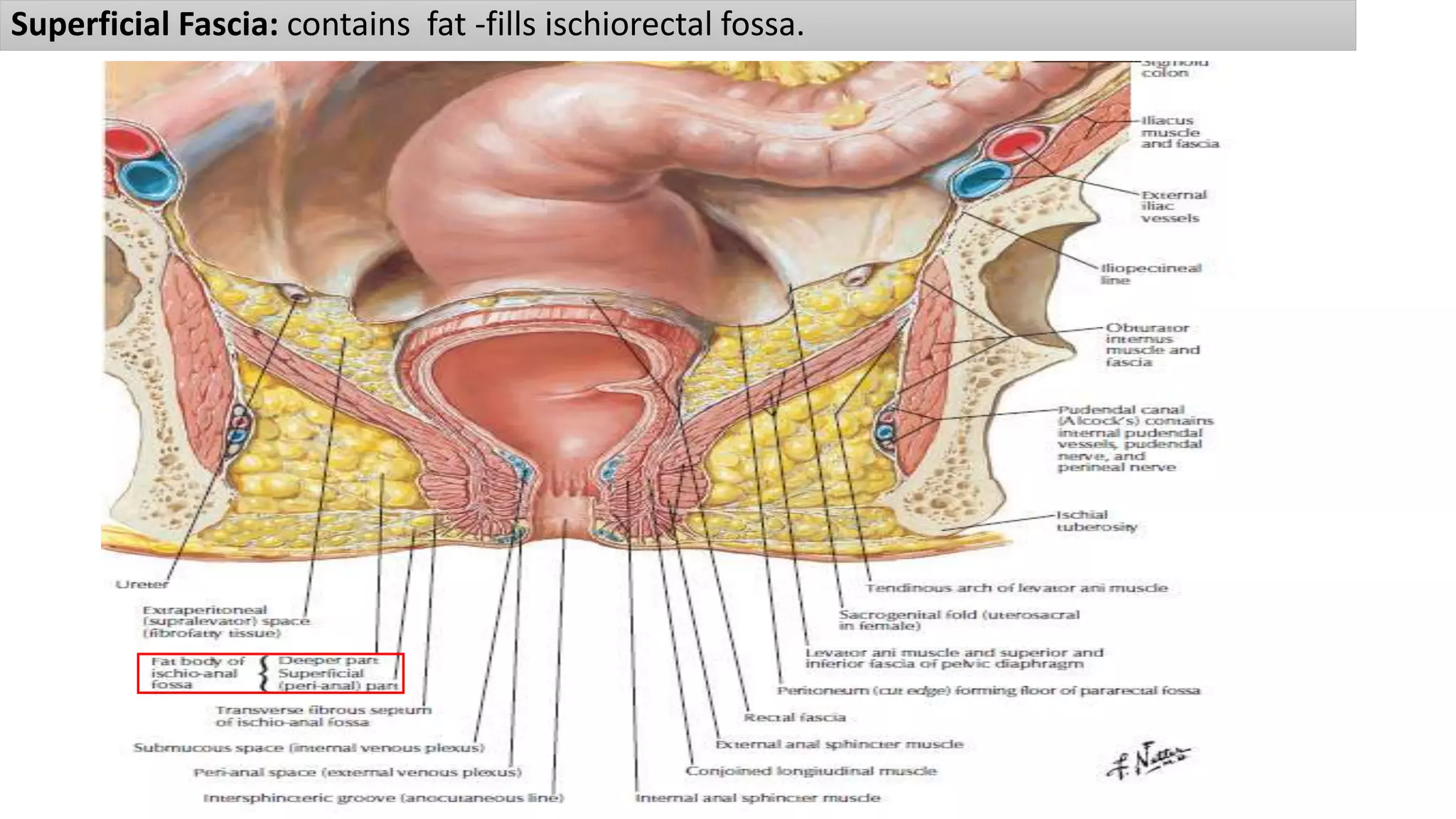
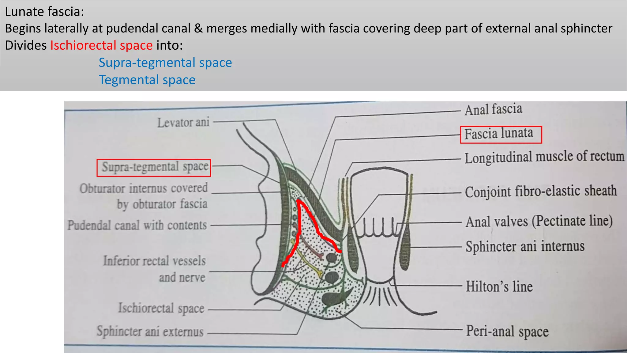
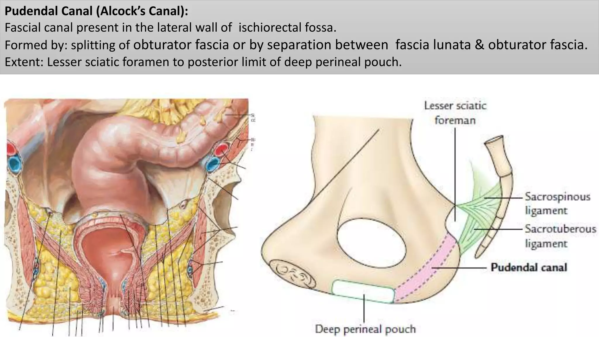
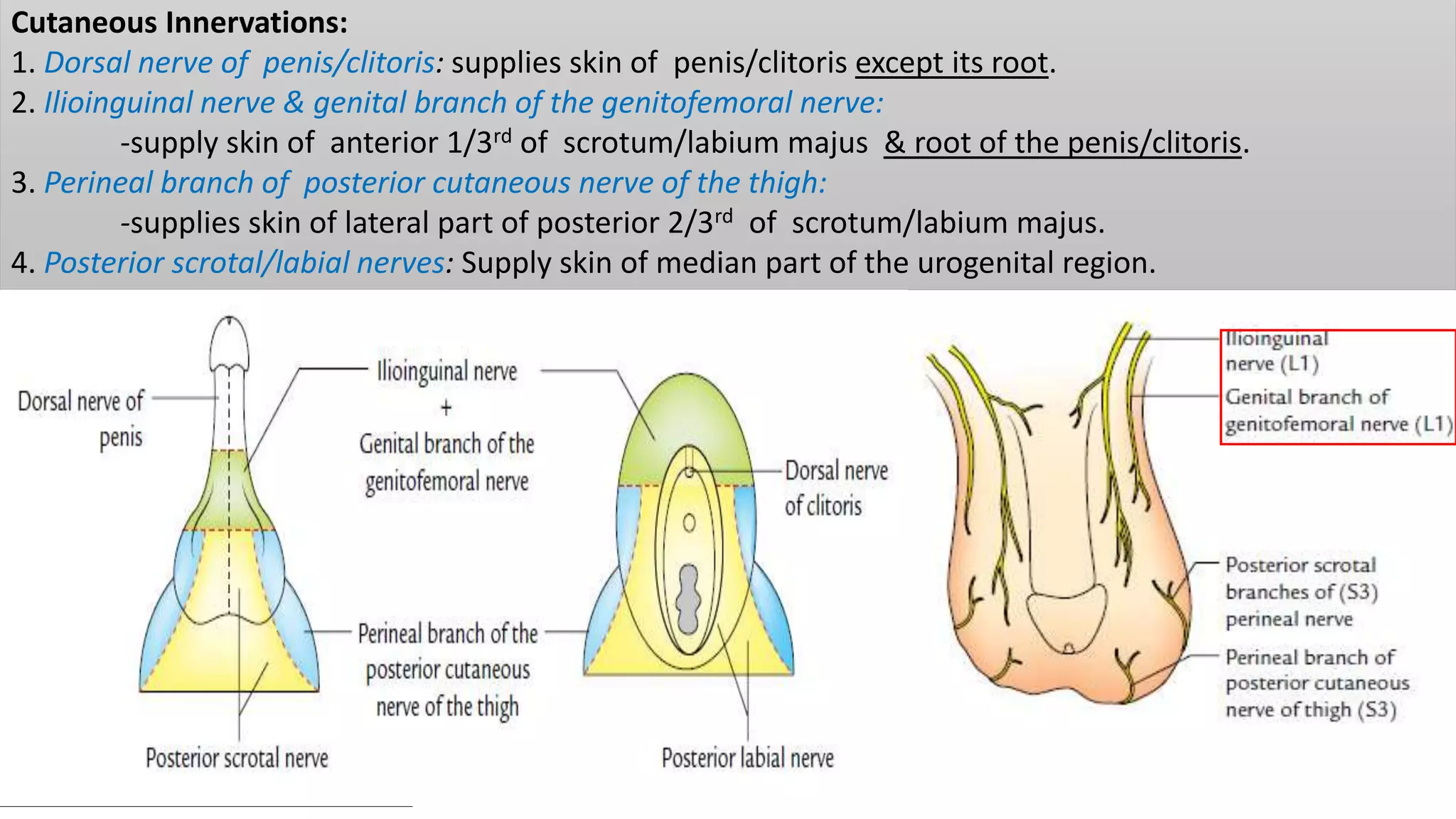
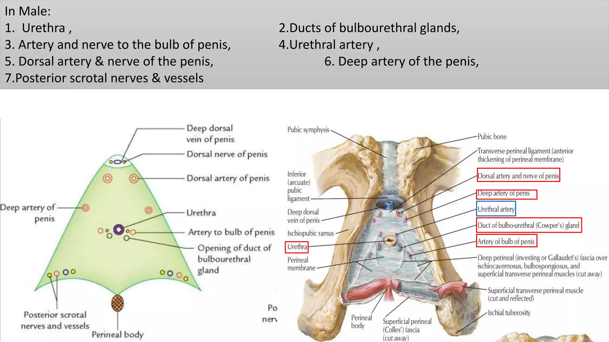
Perineum & External Genitalia(I)
Female Reproductive System
Dr. Prabhakar Yadav
Associate Professor
Department of Human Anatomy
B.P. Koirala Institute of Health Sciences
Perineum: Diamond shaped region in the lowest region of trunk below the pelvic diaphragm, in
the interval between two thighs.
Ceiling : diaphragm
Lateral walls: formed by walls of pelvic cavity below the attachment of levator ani muscle .
Superficial Boundaries
Anteriorly: Scrotum in male and Mons pubis in female.
Posteriorly: Buttocks.
On each side: Upper medial aspect of thigh
Deep Boundaries: Correspond to boundaries of pelvic outlet :
Anteriorly: Inferior margin of the pubic symphysis - Arcuate pubic ligament
Posteriorly: Tip of coccyx.
On each side: – Anterolaterally: Ischiopubic ramus; – Laterally: Ischial tuberosity.
– Posterolaterally: Sacrotuberous ligament
DIVISIONS
(a)Urogenital triangle (anterior perineum): traversed by urethra in male & urethra & vagina in female
(b) Anal triangle (posterior perineum): Traversed by anal canal in both male & female and
contains Ischioanal fossa on each side
ANAL TRIANGLE
In front: Horizontal line joining anterior ends of ischial tuberosities.
Laterally: Ischial tuberosities
Inferolaterally: Sacrotuberous ligaments
Posteriorly: coccyx.
Cutaneous Innervation
• Inferior rectal nerve (S2, S3, S4) - skin around anus & over ischiorectal fossa.
• perineal branch of the fourth sacral nerve supplies - skin posterior to anus.
Superficial Fascia: contains fat -fills ischiorectal fossa.
Deep Fascia : Formed by inferior fascia of pelvic diaphragm &
fascia covering obturator internus below the attachment of the levator ani
Anococcygeal Ligament:
• Fibrofatty mass permeated with muscle fibres derived from levator ani & external anal sphincter.
• Extends from anus to tip of the coccyx
• Supports the rectum
Perineal Body:
Fibromuscular node situated in median plane; 1.25 cm in front of anal margin & close to bulb of penis. 2
2 Unpaired muscles:
I.External anal sphincter , II. Fibres of longitudional muscle coat of anal canal
4 Paired Muscles :
I. Bulbospongiosus
II/III.superficial & deep transversus perenei
IV.Levator ani
In female:
sphincter urethrovaginalis -may be
Damaged during parturition.
Episiotomy(perineotomy)
Internal & External Anal Sphincter:
internal anal spincter- surrounded in its upper 3/4th of Anal canal & end at the level of white line of Hilton
-is supplied by the autonomic nerve fibres -involuntary.
External anal sphincter- surrounds whole length of the anal canal.
is supplied by inferior rectal nerve & perineal branch of fourth sacral nerve.
is under voluntary control and keeps the anus and anal canal closed
Subcutaneous Part:
Part below the internal anal sphincter in the perianal space;
surrounds the lower part of the anal canal.
has no bony attachment.
Is traversed by fibroelastic septa derived from the conjoint longitudinal coat.
Superficial Part
is elliptical in shape
arises from the tip of coccyx & anococcygeal raphe
surrounds lower part of internal sphincter then gets inserted into perineal body in front.
Deep Part:
• surrounds upper part of the internal anal sphincter.
• It has no bony attachments.
• Puborectalis blends with deep part of external sphincter behind & forms a sling around anorectal
junction, which is attached anteriorly to the back of the pubis.
• In the resting state, anorectal tube is angled forward at this level and contraction of puborectalis sling
will increase this angle, an important factor in the continence mechanism.
Ischiorectal Fossa (Ischioanal Fossa):
is a wedge-shaped, fat-filled space situated on each side of anal canal below the pelvic diaphragm.
Two fossae communicate with each other behind the anal canal.
Help in dilatation of anal canal, during defecation
Boundaries
Lateral: Fascia covering
obturator internus muscle &
ischial tuberosity.
Medial: Fascia covering
levator ani muscle & external
anal sphincter.
Floor: skin.
Roof: Meeting point of the
fascia covering obturator
internus & inferior fascia of
pelvic diaphragm.
Posterior: Sacrotuberous
ligament
Anterior: Posterior border of
perineal membrane.
Recesses : are extensions of fossa beyond its boundaries
(1) Anterior recess extends forwards above the Perineal Membrane, reaching up to the posterior surface
of body of pubis. It is closely related to the prostate or the vagina.
(2) Posterior recess extends deep to sacrotuberous ligament; recess is smaller than the anterior recess
(3) Horseshoe recess
connects two ischiorectal fossae
behind the anal canal
Contents: 1. Ischiorectal pad of fat.
2. Inferior rectal nerves and vessels.
3. Perineal branch of fourth sacral nerve enters posterior angle of the fossa running over levator ani to
external anal sphincter which it innervates.
4. Posterior scrotal (or labial) nerves and vessels cross the anterolateral part of the fossa to enter the
urogenital triangle
Contents:.
5. Pudendal canal with its contents , lie along lateral wall.
6. Perforating cutaneous branches of nerve S2,S3,S4 that appear at lower border of gluteus maximus.
Pudendal canal
or
Alcock’s canal
Spaces & canals in the Region of Ischiorectal Fossa:
Perianal Space: Subcutaneous space on each side of anal orifice.
Boundary:
Above: perianal fascia - extends from white line of Hilton medially to pudendal canal laterally.
Below: perianal skin.
Perianal space is subdivided into numerous compartments by fibroelastic septa.
Infection in this space causing perianal abscess is therefore very painful.
Ischiorectal/Ischioanal space:
Fat in this space is loosely arranged in large loculi formed by incomplete delicate septa.
Lunate fascia:
Begins laterally at pudendal canal & merges medially with fascia covering deep part of external anal sphincter
Divides Ischiorectal space into:
Supra-tegmental space
Tegmental space
Perianal and Ischiorectal spaces are common sites of abscesses
Anorectal fistula or fistula in ano:
abscess may burst into the anal canal or rectum internally, and on to the surface of perineum externally.
If abscess bursts only externally &healing does not follow- External sinus
Pudendal Canal (Alcock’s Canal):
Fascial canal present in the lateral wall of ischiorectal fossa.
Formed by: splitting of obturator fascia or by separation between fascia lunata & obturator fascia.
Extent: Lesser sciatic foramen to posterior limit of deep perineal pouch.
Contents
1. Pudendal nerve which divides within the canal into the dorsal nerve of penis and perineal nerve.
2. Internal pudendal vessels.
UROGENITAL TRIANGLE :Area between ischiopubic rami, in front of a horizontal line joining the anterior
ends of two ischial tuberosities.
On the surface, urogenital triangle presents:
• Penis and scrotum in male
• External genitalia & orifices of urethra and vagina in female.
Cutaneous Innervations:
1. Dorsal nerve of penis/clitoris: supplies skin of penis/clitoris except its root.
2. Ilioinguinal nerve & genital branch of the genitofemoral nerve:
-supply skin of anterior 1/3rd of scrotum/labium majus & root of the penis/clitoris.
3. Perineal branch of posterior cutaneous nerve of the thigh:
-supplies skin of lateral part of posterior 2/3rd of scrotum/labium majus.
4. Posterior scrotal/labial nerves: Supply skin of median part of the urogenital region.
Suuperficial fascia: fatty layer is continuous with that of the surrounding region
Membranous layer ( Colles fascia) is attached posteriorly to posterior border of
perineal membrane & on each side to pubic arch below the crus penis.
Anteriorly, it is continuous with the dartos fascia of scrotum & penis, and with fascia of Scarpa of anterior
abdominal wall
Rupture of urethra:
Superficial extravasation of urine :
Deep extravasation of urine;
Perineal membrane (Deep fascia) :
• Triangular fascial sheet, across urogenital triangle -attached to ischiopubic rami .
• Its Posterior margin of is fused centrally to the perineal body & is continuous with fascia over deep
transversus perinei
• Its Anterior margin thickens to form a transverse perineal ligament.
• Gap between transverse perineal ligament & arcuate pubic ligament provides passage to deep dorsal vein
of penis
In Male:
1. Urethra , 2.Ducts of bulbourethral glands,
3. Artery and nerve to the bulb of penis, 4.Urethral artery ,
5. Dorsal artery & nerve of the penis, 6. Deep artery of the penis,
7.Posterior scrotal nerves & vessels
In Female:
1. Urethra, 2. Vagina,
3. Artery and nerve to the bulb of the vestibule, 4. Dorsal artery and nerve of clitoris.
5. Deep artery of clitoris 6. Posterior labial vessels and nerves
Urogenital region is divide into two parts by perineal membrane
Below perineal membrane : Superficial perineal space /pouch
Above perineal membrane : Deep perineal space/ pouch
Above the perineal membrane, urethra is surrounded by sphincter urethrae muscle(superficial urethral
sphincter).
Deep aspect of superficial urethral sphincter is covered by - Endopelvic fascia, continuous with posterior
edge of perineal membrane and is attached to the sides of ischiopubic rami.
Deep perineal Space: space situated deep to perineal membrane & below endopelvic fascia
Boundary:
Superficial: perineal membrane, Deep: endopelvic fascia, Sides: ischiopubic rami,
Anteriorly: gap between two ligaments:
Contents
Male: Female:
1. urethra Urethra and vagina
2. Deep transverse perineal muscles Deep transverse perineal muscles
3. Sphincter urethra muscle Shincter urethra muscle
4. Dorsal nerve of penis Dorsal nerve of Clitoris
5. Artery of penis Artery of Clitoris
6. Bulbourethral gland No glannds
Superficial perinela space:
Boundary:
• Superficial: Colles’fascia
• Deep: Perineal membrane
• Posterior: Closed by fusion of perineal membrane with colles’ fascia
• Anterior: Open & continuous with the space of scortum, penis & anterior abdominal wall
• On each side: Ischopubic rami
Contents: Erectile tissue , Muscles , Vessels and nerves & Glands
Erectile tissue & glads:
Male Female
• Root of penis Body of clitoris
• No any glands Greater vestibular gland with duct.
Vessels
Male
Branches of the internal
pudendal artery
• Perineal artery
• Dorsal artery of penis
• Deep artery of penis
Female
• Perineal artery
• Dorsal artery of clitoris
• Deep artery of clitoris
Nerves:
Male:
• Perineal nerve
• Dorsal nerve of penis
• Posterior scrotal nerve
Female:
Perineal nerve
Dorsal nerve of clitoris
Posterior labial nerve
Pudendal nerve
Branches:
Internal pudendal artery
Transverse perineal artery
Female External Genitalia/ Pudendum/ Vulva
1. Vestibule of vagina
2. Labia minora
3. Clitoris
4. Labia majora
5. Mons Pubis
6. Bulbs of vestibule
7. Greater vestibular glands
Vestibule of Vagina
•Elliptical space between labia minora.
•Contains :
-Urethral orifice anteriorly and vaginal orifice posteriorly- covered by hymen (incompletely).
-Openings of greater vestibular glands and paraurethral glands (of Skene).
Labia minora:
•2 thin folds of hairless skin on either side of midline.
•Anteriorly, it bifurcate, forming medial & lateral fold.
•Medial folds unite to form frenulum of clitoris- joins glans
clitoris.
•Lateral folds unite with glans clitoris & body of clitoris to
form prepuce of clitoris (hood).
•Posterior to the vestibule, Labia minora unite to form the
frenulum of labia minora (fourchette).
Clitoris: erectile structure is composed of-
two corpora cavernosa and glans clitoris
• Divided into: root & body:
Root of clitoris consists only of the two crura
Body of clitoris- formed by free distal ends of
two corpora cavernosa,
Glans clitoris: is attached to the distal end of
body and is connected to bulbs of vestibule by
small bands of erectile tissue.
Labia Majora:
-are two broad folds of hairy skin with underlying
pad of fat, lateral to labia minora.
-unites anteriorly to form the mons pubis.
-posteriorly Labia majora are separated by a
depression- posterior commissure- overlies perineal
body
Mons Pubis
- is an elevation of skin over the pubic symphysis with
underlying thick fat pad.
Greater vestibular glands (Bartholin's glands): --
-Mucous glands
- lie posterior to the bulbs of vestibule on each side of
the vaginal opening (homologues of bulbourethral
glands).
•bulbourethral glands - located in deep perineal pouch,
•Greater vestibular glands- superficial perineal pouch.
--produce secretion during sexual arousal
Bulbs of vestibule:
Two oval body of erectile tissue – corresponds to bulb
of penis .
Lie on either side of vaginal and urethral orifice
Perineum & external genitalia

Perineum & external genitalia

  • 1.
    Perineum & ExternalGenitalia(I) Female Reproductive System Dr. Prabhakar Yadav Associate Professor Department of Human Anatomy B.P. Koirala Institute of Health Sciences
  • 2.
    Perineum: Diamond shapedregion in the lowest region of trunk below the pelvic diaphragm, in the interval between two thighs. Ceiling : diaphragm Lateral walls: formed by walls of pelvic cavity below the attachment of levator ani muscle .
  • 3.
    Superficial Boundaries Anteriorly: Scrotumin male and Mons pubis in female. Posteriorly: Buttocks. On each side: Upper medial aspect of thigh
  • 4.
    Deep Boundaries: Correspondto boundaries of pelvic outlet : Anteriorly: Inferior margin of the pubic symphysis - Arcuate pubic ligament Posteriorly: Tip of coccyx. On each side: – Anterolaterally: Ischiopubic ramus; – Laterally: Ischial tuberosity. – Posterolaterally: Sacrotuberous ligament
  • 5.
    DIVISIONS (a)Urogenital triangle (anteriorperineum): traversed by urethra in male & urethra & vagina in female (b) Anal triangle (posterior perineum): Traversed by anal canal in both male & female and contains Ischioanal fossa on each side
  • 6.
    ANAL TRIANGLE In front:Horizontal line joining anterior ends of ischial tuberosities. Laterally: Ischial tuberosities Inferolaterally: Sacrotuberous ligaments Posteriorly: coccyx.
  • 9.
    Cutaneous Innervation • Inferiorrectal nerve (S2, S3, S4) - skin around anus & over ischiorectal fossa. • perineal branch of the fourth sacral nerve supplies - skin posterior to anus.
  • 10.
    Superficial Fascia: containsfat -fills ischiorectal fossa.
  • 11.
    Deep Fascia :Formed by inferior fascia of pelvic diaphragm & fascia covering obturator internus below the attachment of the levator ani
  • 12.
    Anococcygeal Ligament: • Fibrofattymass permeated with muscle fibres derived from levator ani & external anal sphincter. • Extends from anus to tip of the coccyx • Supports the rectum
  • 13.
    Perineal Body: Fibromuscular nodesituated in median plane; 1.25 cm in front of anal margin & close to bulb of penis. 2 2 Unpaired muscles: I.External anal sphincter , II. Fibres of longitudional muscle coat of anal canal 4 Paired Muscles : I. Bulbospongiosus II/III.superficial & deep transversus perenei IV.Levator ani In female: sphincter urethrovaginalis -may be Damaged during parturition.
  • 14.
  • 15.
    Internal & ExternalAnal Sphincter: internal anal spincter- surrounded in its upper 3/4th of Anal canal & end at the level of white line of Hilton -is supplied by the autonomic nerve fibres -involuntary. External anal sphincter- surrounds whole length of the anal canal. is supplied by inferior rectal nerve & perineal branch of fourth sacral nerve. is under voluntary control and keeps the anus and anal canal closed
  • 16.
    Subcutaneous Part: Part belowthe internal anal sphincter in the perianal space; surrounds the lower part of the anal canal. has no bony attachment. Is traversed by fibroelastic septa derived from the conjoint longitudinal coat.
  • 17.
    Superficial Part is ellipticalin shape arises from the tip of coccyx & anococcygeal raphe surrounds lower part of internal sphincter then gets inserted into perineal body in front.
  • 18.
    Deep Part: • surroundsupper part of the internal anal sphincter. • It has no bony attachments. • Puborectalis blends with deep part of external sphincter behind & forms a sling around anorectal junction, which is attached anteriorly to the back of the pubis. • In the resting state, anorectal tube is angled forward at this level and contraction of puborectalis sling will increase this angle, an important factor in the continence mechanism.
  • 19.
    Ischiorectal Fossa (IschioanalFossa): is a wedge-shaped, fat-filled space situated on each side of anal canal below the pelvic diaphragm. Two fossae communicate with each other behind the anal canal. Help in dilatation of anal canal, during defecation
  • 20.
    Boundaries Lateral: Fascia covering obturatorinternus muscle & ischial tuberosity. Medial: Fascia covering levator ani muscle & external anal sphincter. Floor: skin. Roof: Meeting point of the fascia covering obturator internus & inferior fascia of pelvic diaphragm. Posterior: Sacrotuberous ligament Anterior: Posterior border of perineal membrane.
  • 21.
    Recesses : areextensions of fossa beyond its boundaries (1) Anterior recess extends forwards above the Perineal Membrane, reaching up to the posterior surface of body of pubis. It is closely related to the prostate or the vagina. (2) Posterior recess extends deep to sacrotuberous ligament; recess is smaller than the anterior recess
  • 22.
    (3) Horseshoe recess connectstwo ischiorectal fossae behind the anal canal
  • 23.
    Contents: 1. Ischiorectalpad of fat. 2. Inferior rectal nerves and vessels. 3. Perineal branch of fourth sacral nerve enters posterior angle of the fossa running over levator ani to external anal sphincter which it innervates. 4. Posterior scrotal (or labial) nerves and vessels cross the anterolateral part of the fossa to enter the urogenital triangle
  • 24.
    Contents:. 5. Pudendal canalwith its contents , lie along lateral wall. 6. Perforating cutaneous branches of nerve S2,S3,S4 that appear at lower border of gluteus maximus. Pudendal canal or Alcock’s canal
  • 25.
    Spaces & canalsin the Region of Ischiorectal Fossa: Perianal Space: Subcutaneous space on each side of anal orifice. Boundary: Above: perianal fascia - extends from white line of Hilton medially to pudendal canal laterally. Below: perianal skin. Perianal space is subdivided into numerous compartments by fibroelastic septa. Infection in this space causing perianal abscess is therefore very painful.
  • 26.
    Ischiorectal/Ischioanal space: Fat inthis space is loosely arranged in large loculi formed by incomplete delicate septa.
  • 27.
    Lunate fascia: Begins laterallyat pudendal canal & merges medially with fascia covering deep part of external anal sphincter Divides Ischiorectal space into: Supra-tegmental space Tegmental space
  • 28.
    Perianal and Ischiorectalspaces are common sites of abscesses
  • 29.
    Anorectal fistula orfistula in ano: abscess may burst into the anal canal or rectum internally, and on to the surface of perineum externally. If abscess bursts only externally &healing does not follow- External sinus
  • 30.
    Pudendal Canal (Alcock’sCanal): Fascial canal present in the lateral wall of ischiorectal fossa. Formed by: splitting of obturator fascia or by separation between fascia lunata & obturator fascia. Extent: Lesser sciatic foramen to posterior limit of deep perineal pouch.
  • 31.
    Contents 1. Pudendal nervewhich divides within the canal into the dorsal nerve of penis and perineal nerve. 2. Internal pudendal vessels.
  • 32.
    UROGENITAL TRIANGLE :Areabetween ischiopubic rami, in front of a horizontal line joining the anterior ends of two ischial tuberosities. On the surface, urogenital triangle presents: • Penis and scrotum in male • External genitalia & orifices of urethra and vagina in female.
  • 33.
    Cutaneous Innervations: 1. Dorsalnerve of penis/clitoris: supplies skin of penis/clitoris except its root. 2. Ilioinguinal nerve & genital branch of the genitofemoral nerve: -supply skin of anterior 1/3rd of scrotum/labium majus & root of the penis/clitoris. 3. Perineal branch of posterior cutaneous nerve of the thigh: -supplies skin of lateral part of posterior 2/3rd of scrotum/labium majus. 4. Posterior scrotal/labial nerves: Supply skin of median part of the urogenital region.
  • 34.
    Suuperficial fascia: fattylayer is continuous with that of the surrounding region Membranous layer ( Colles fascia) is attached posteriorly to posterior border of perineal membrane & on each side to pubic arch below the crus penis.
  • 35.
    Anteriorly, it iscontinuous with the dartos fascia of scrotum & penis, and with fascia of Scarpa of anterior abdominal wall
  • 36.
    Rupture of urethra: Superficialextravasation of urine : Deep extravasation of urine;
  • 37.
    Perineal membrane (Deepfascia) : • Triangular fascial sheet, across urogenital triangle -attached to ischiopubic rami . • Its Posterior margin of is fused centrally to the perineal body & is continuous with fascia over deep transversus perinei • Its Anterior margin thickens to form a transverse perineal ligament. • Gap between transverse perineal ligament & arcuate pubic ligament provides passage to deep dorsal vein of penis
  • 38.
    In Male: 1. Urethra, 2.Ducts of bulbourethral glands, 3. Artery and nerve to the bulb of penis, 4.Urethral artery , 5. Dorsal artery & nerve of the penis, 6. Deep artery of the penis, 7.Posterior scrotal nerves & vessels
  • 39.
    In Female: 1. Urethra,2. Vagina, 3. Artery and nerve to the bulb of the vestibule, 4. Dorsal artery and nerve of clitoris. 5. Deep artery of clitoris 6. Posterior labial vessels and nerves
  • 40.
    Urogenital region isdivide into two parts by perineal membrane Below perineal membrane : Superficial perineal space /pouch Above perineal membrane : Deep perineal space/ pouch Above the perineal membrane, urethra is surrounded by sphincter urethrae muscle(superficial urethral sphincter).
  • 41.
    Deep aspect ofsuperficial urethral sphincter is covered by - Endopelvic fascia, continuous with posterior edge of perineal membrane and is attached to the sides of ischiopubic rami.
  • 42.
    Deep perineal Space:space situated deep to perineal membrane & below endopelvic fascia Boundary: Superficial: perineal membrane, Deep: endopelvic fascia, Sides: ischiopubic rami, Anteriorly: gap between two ligaments:
  • 43.
    Contents Male: Female: 1. urethraUrethra and vagina 2. Deep transverse perineal muscles Deep transverse perineal muscles 3. Sphincter urethra muscle Shincter urethra muscle 4. Dorsal nerve of penis Dorsal nerve of Clitoris 5. Artery of penis Artery of Clitoris 6. Bulbourethral gland No glannds
  • 44.
    Superficial perinela space: Boundary: •Superficial: Colles’fascia • Deep: Perineal membrane • Posterior: Closed by fusion of perineal membrane with colles’ fascia • Anterior: Open & continuous with the space of scortum, penis & anterior abdominal wall • On each side: Ischopubic rami
  • 46.
    Contents: Erectile tissue, Muscles , Vessels and nerves & Glands Erectile tissue & glads: Male Female • Root of penis Body of clitoris • No any glands Greater vestibular gland with duct.
  • 49.
    Vessels Male Branches of theinternal pudendal artery • Perineal artery • Dorsal artery of penis • Deep artery of penis Female • Perineal artery • Dorsal artery of clitoris • Deep artery of clitoris
  • 50.
    Nerves: Male: • Perineal nerve •Dorsal nerve of penis • Posterior scrotal nerve Female: Perineal nerve Dorsal nerve of clitoris Posterior labial nerve
  • 51.
  • 52.
  • 53.
  • 54.
  • 55.
    Female External Genitalia/Pudendum/ Vulva 1. Vestibule of vagina 2. Labia minora 3. Clitoris 4. Labia majora 5. Mons Pubis 6. Bulbs of vestibule 7. Greater vestibular glands
  • 56.
    Vestibule of Vagina •Ellipticalspace between labia minora. •Contains : -Urethral orifice anteriorly and vaginal orifice posteriorly- covered by hymen (incompletely). -Openings of greater vestibular glands and paraurethral glands (of Skene).
  • 57.
    Labia minora: •2 thinfolds of hairless skin on either side of midline. •Anteriorly, it bifurcate, forming medial & lateral fold. •Medial folds unite to form frenulum of clitoris- joins glans clitoris. •Lateral folds unite with glans clitoris & body of clitoris to form prepuce of clitoris (hood). •Posterior to the vestibule, Labia minora unite to form the frenulum of labia minora (fourchette).
  • 58.
    Clitoris: erectile structureis composed of- two corpora cavernosa and glans clitoris • Divided into: root & body: Root of clitoris consists only of the two crura Body of clitoris- formed by free distal ends of two corpora cavernosa, Glans clitoris: is attached to the distal end of body and is connected to bulbs of vestibule by small bands of erectile tissue.
  • 59.
    Labia Majora: -are twobroad folds of hairy skin with underlying pad of fat, lateral to labia minora. -unites anteriorly to form the mons pubis. -posteriorly Labia majora are separated by a depression- posterior commissure- overlies perineal body Mons Pubis - is an elevation of skin over the pubic symphysis with underlying thick fat pad.
  • 60.
    Greater vestibular glands(Bartholin's glands): -- -Mucous glands - lie posterior to the bulbs of vestibule on each side of the vaginal opening (homologues of bulbourethral glands). •bulbourethral glands - located in deep perineal pouch, •Greater vestibular glands- superficial perineal pouch. --produce secretion during sexual arousal Bulbs of vestibule: Two oval body of erectile tissue – corresponds to bulb of penis . Lie on either side of vaginal and urethral orifice