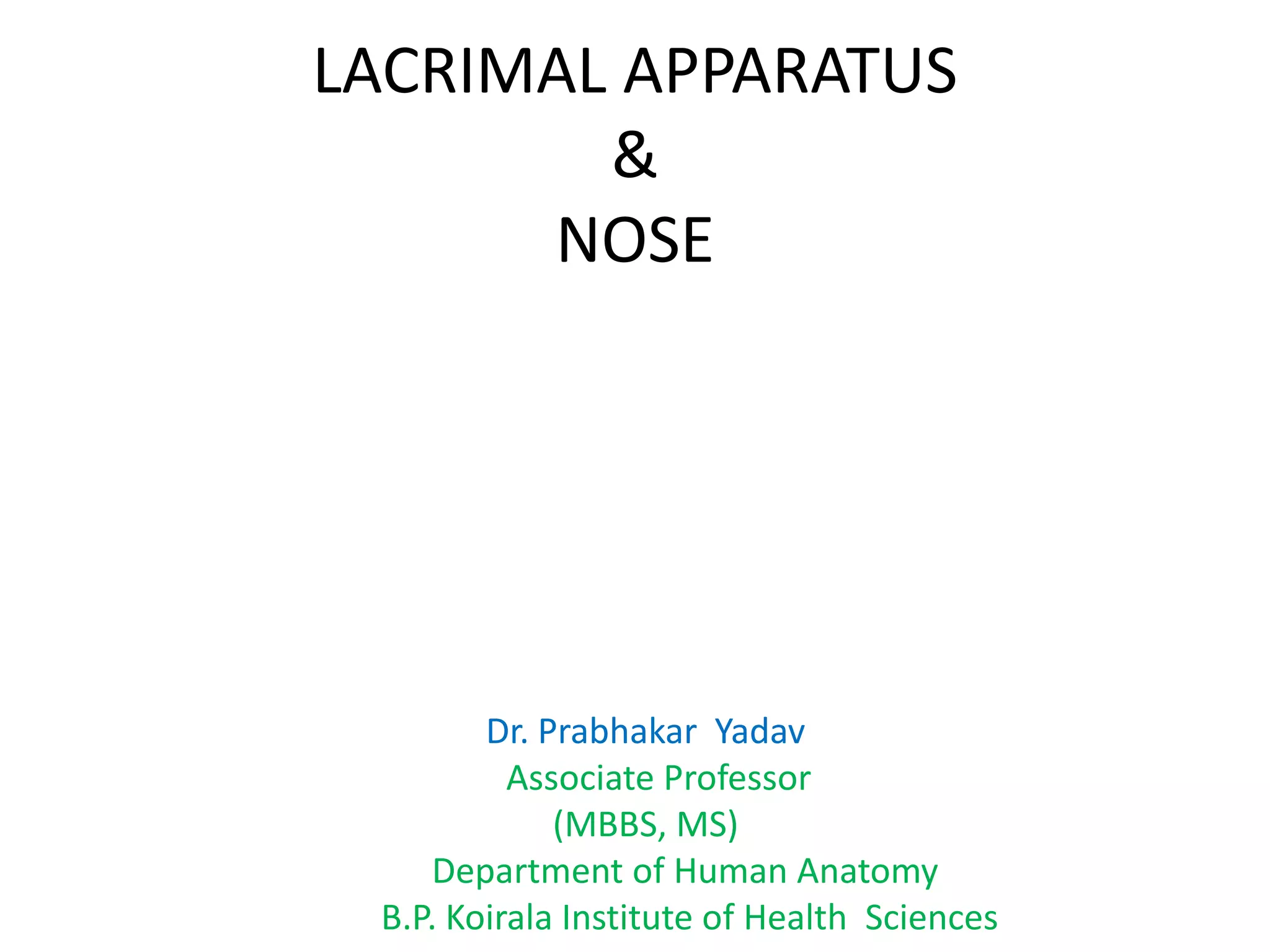
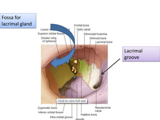
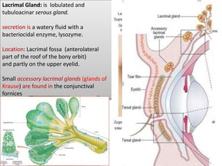
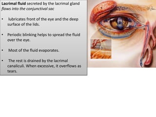
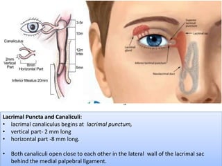
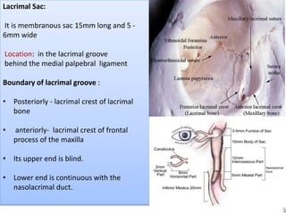
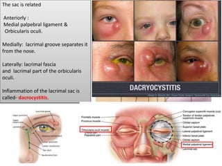
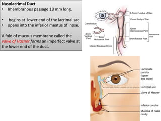
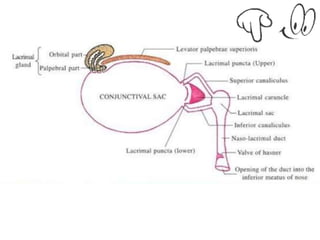
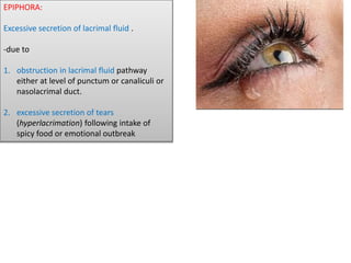
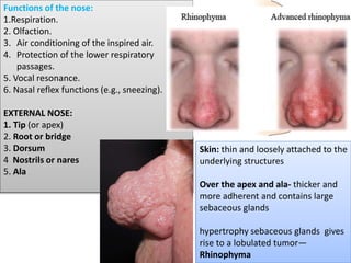
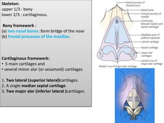
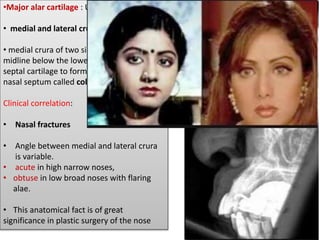
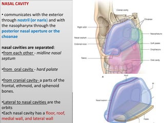
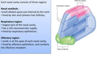
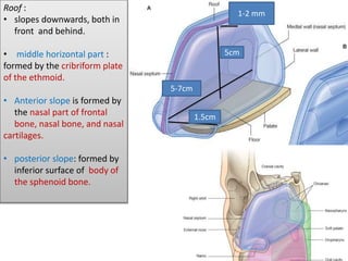
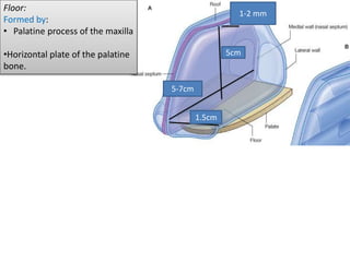
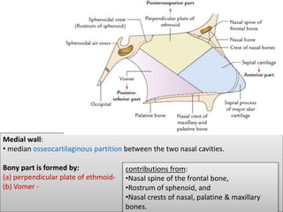
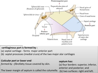
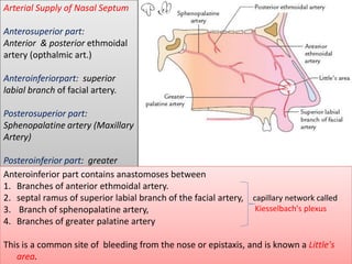
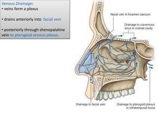
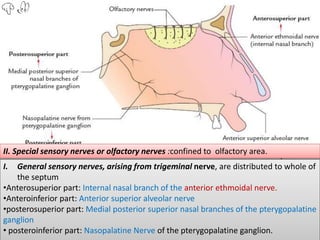
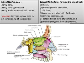
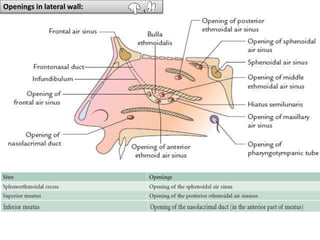
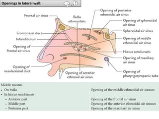
LACRIMAL APPARATUS
&
NOSE
Dr. Prabhakar Yadav
Associate Professor
(MBBS, MS)
Department of Human Anatomy
B.P. Koirala Institute of Health Sciences
Lacrimal
groove
Fossa for
lacrimal gland
Lacrimal apparatus:
structures concerned with secretion and drainage
of the lacrimal or tear fluid constitute the lacrimal
apparatus.
I. Lacrimal gland and its ducts
II. Conjunctival sac
III. Lacrimal puncta and lacrimal canaliculi
IV. Lacrimal sac
V. Naso lacrimal duct
Lacrimal Gland: is lobulated and
tubuloacinar serous gland.
secretion is a watery fluid with a
bacteriocidal enzyme, lysozyme.
Location: Lacrimal fossa (anterolateral
part of the roof of the bony orbit)
and partly on the upper eyelid.
Small accessory lacrimal glands (glands of
Krause) are found in the conjunctival
fornices
'J' shaped
tendon of levator palpebrae superioris muscle.
(a) an orbital part which is larger and deeper
(b) a palpebral part smaller and superficial, lying
within the eyelid
dozen of lacrimal gland ducts pierce the
conjunctiva of the upper lid and open
into the conjunctival sac near superior
fornix.
Ducts of the orbital part pass through
the palpebral part.
Removal of the palpebral part is
functionally equivalent to removal of
the entire gland.
After removal, the conjunctiva and cornea are
moistened by accessory lacrimal glands (glands of
Krause) .
Blood supply: lacrimal branch of ophthalmic artery
Nerve supply: lacrimal nerve.
The nerve has both sensory and secretomotor fibres
Lacrimal fluid secreted by the lacrimal gland
flows into the conjunctival sac
• lubricates front of the eye and the deep
surface of the lids.
• Periodic blinking helps to spread the fluid
over the eye.
• Most of the fluid evaporates.
• The rest is drained by the lacrimal
canaliculi. When excessive, it overflows as
tears.
Lacrimal Puncta and Canaliculi:
• lacrimal canaliculus begins at lacrimal punctum,
• vertical part- 2 mm long
• horizontal part -8 mm long.
• Both canaliculi open close to each other in the lateral wall of the lacrimal sac
behind the medial palpebral ligament.
Lacrimal Sac:
It is membranous sac 15mm long and 5 -
6mm wide
Location: in the lacrimal groove
behind the medial palpebral ligament
Boundary of lacrimal groove :
• Posteriorly - lacrimal crest of lacrimal
bone
• anteriorly- lacrimal crest of frontal
process of the maxilla
• Its upper end is blind.
• Lower end is continuous with the
nasolacrimal duct.
The sac is related
Anteriorly :
Medial palpebral ligament &
Orbicularis oculi.
Medially: lacrimal groove separates it
from the nose.
Laterally: lacrimal fascia
and lacrimal part of the orbicularis
oculi.
Inflammation of the lacrimal sac is
called- dacrocystitis.
Nasolacrimal Duct
• Imembranous passage 18 mm long.
• begins at lower end of the lacrimal sac
• opens into the inferior meatus of nose.
A fold of mucous membrane called the
valve of Hasner forms an imperfect valve at
the lower end of the duct.
EPIPHORA:
Excessive secretion of lacrimal fluid .
-due to
1. obstruction in lacrimal fluid pathway
either at level of punctum or canaliculi or
nasolacrimal duct.
2. excessive secretion of tears
(hyperlacrimation) following intake of
spicy food or emotional outbreak
Functions of the nose:
1.Respiration.
2. Olfaction.
3. Air conditioning of the inspired air.
4. Protection of the lower respiratory
passages.
5. Vocal resonance.
6. Nasal reflex functions (e.g., sneezing).
EXTERNAL NOSE:
1. Tip (or apex)
2. Root or bridge
3. Dorsum
4 Nostrils or nares
5. Ala
Skin: thin and loosely attached to the
underlying structures
Over the apex and ala- thicker and
more adherent and contains large
sebaceous glands
hypertrophy sebaceous glands gives
rise to a lobulated tumor—
Rhinophyma
Skeleton:
upper 1/3 : bony
lower 2/3 : cartilaginous.
Bony framework :
(a) two nasal bones :form bridge of the nose
(b) frontal processes of the maxillae.
Cartilaginous framework:
• 5 main cartilages and
• several minor alar (or sesamoid) cartilages
1. Two lateral (superior lateral)cartilages.
2. A single median septal cartilage.
3. Two major alar (inferior lateral )cartilages.
•Major alar cartilage : U-shaped
• medial and lateral crus
• medial crura of two sides meet in the
midline below the lower margin of the
septal cartilage to form the lower part of the
nasal septum called columella.
Clinical correlation:
• Nasal fractures
• Angle between medial and lateral crura
is variable.
• acute in high narrow noses,
• obtuse in low broad noses with flaring
alae.
• This anatomical fact is of great
significance in plastic surgery of the nose
NASAL CAVITY
• communicates with the exterior
through nostril (or naris) and with
the nasopharynx through the
posterior nasal aperture or the
choanae
nasal cavities are separated:
•from each other - midline nasal
septum
•from oral cavity - hard palate
•from cranial cavity- a parts of the
frontal, ethmoid, and sphenoid
bones.
•Lateral to nasal cavities are the
orbits
•Each nasal cavity has a floor, roof,
medial wall, and lateral wall
Each nasal cavity consists of three regions
Nasal vestibule :
•small dilated space just internal to the naris
• lined by skin and contains hair follicles;
Respiratory region:
• largest part of the nasal cavity,
• has a rich neurovascular supply,
• lined by respiratory epithelium;
Olfactory region :
• small, is at the apex of each nasal cavity,
• lined by olfactory epithelium, and contains
the olfactory receptors
5cm
5-7cm
1.5cm
1-2 mm
Roof :
• slopes downwards, both in
front and behind.
• middle horizontal part :
formed by the cribriform plate
of the ethmoid.
• Anterior slope is formed by
the nasal part of frontal
bone, nasal bone, and nasal
cartilages.
• posterior slope: formed by
inferior surface of body of
the sphenoid bone.
5cm
5-7cm
1.5cm
1-2 mm
Floor:
Formed by:
• Palatine process of the maxilla
•Horizontal plate of the palatine
bone.
Medial wall:
• median osseocartilaginous partition between the two nasal cavities.
Bony part is formed by:
(a) perpendicular plate of ethmoid-
(b) Vomer -
contributions from:
•Nasal spine of the frontal bone,
•Rostrum of sphenoid, and
•Nasal crests of nasal, palatine & maxillary
bones.
cartilaginous part is formed by :
(a) septal cartilage - forms major anterior part
(b) septal processes (medial crura) of the two major alar cartilages
Cuticular part or lower end:
formed by :-fibrofatty tissue covered by skin.
The lower margin of septum is called the columella.
septum has:
(a) four borders: superior, inferior,
anterior and posterior; and
(b) two surfaces: right and left.
Arterial Supply of Nasal Septum
Anterosuperior part:
Anterior & posterior ethmoidal
artery (opthalmic art.)
Anteroinferiorpart: superior
labial branch of facial artery.
Posterosuperior part:
Sphenopalatine artery (Maxillary
Artery)
Posteroinferior part: greater
palatine artery (Maxillary
Artery)
Anteroinferior part contains anastomoses between
1. Branches of anterior ethmoidal artery.
2. septal ramus of superior labial branch of the facial artery,
3. Branch of sphenopalatine artery,
4. Branches of greater palatine artery
This is a common site of bleeding from the nose or epistaxis, and is known a Little's
area.
capillary network called
Kiesselbach's plexus
Venous Drainage:
• veins form a plexus
• drains anteriorly into facial vein
• posteriorly through shenopalatine
vein to pterygoid venous plexus.
I. General sensory nerves, arising from trigeminal nerve, are distributed to whole of
the septum
•Anterosuperior part: Internal nasal branch of the anterior ethmoidal nerve.
•Anteroinferior part: Anterior superior alveolar nerve
•posterosuperior part: Medial posterior superior nasal branches of the pterygopalatine
ganglion
• posteroinferior part: Nasopalatine Nerve of the pterygopalatine ganglion.
II. Special sensory nerves or olfactory nerves :confined to olfactory area.
Lateral Wall : Bones forming the lateral wall:
(a) nasal,
(b) frontal process of maxilla,
(c) lacrimal,
(d) conchae and labyrinth of ethmoid,
(e) inferior nasal concha,
(f) perpendicular plate of palatine, and
(g) medial pterygoid plate of sphenoid.
Lateral Wall of Nose :
-partly bony,
-partly cartilaginous and
-partly made up only of soft tissues
• conchae: increase surface area for
air-conditioning of inspired air.
Cartilages forming the lateral wall are:
(a) lateral cartilage (superior lateral nasal cartilage),
(b) major alar cartilage (inferior lateral nasal cartilage), and
(c) three to four tiny cartilages of the alae (minor alar cartilages)..
Cuticular lower part: formed by fibrofatty tissue covered with skin.
lateral wall - subdivided into three parts.
(a) Anterior part (vestibule): small depressed area, lined by modified skin
containing short, stiff, curved hairs called vibrissae.
(b) Middle part: is known as atrium of the middle meatus
(c) posterior part :contains conchae.
Spaces separating the conchae are called meatuses
curved mucocutaneous junction between the
atrium and vestibule is known as limen nasi
Chonchae and Meatuses:
• nasal conchae are curved bony projections directed downwards and medially.
1. Inferior concha (largest) is an independent bone.
2. Middle concha & superior concha(Smallest) : projection from the medial surface
of the ethmoidal labyrinth.
Sphenoethmoidal recess: triangular depression, above & behind the superior concha
Meatuses: passages beneath overhanging conchae.
1. Inferior meatus: largest and lies underneath the inferior nasal concha.
2. Middle meatus: lies underneath the middle concha.
Features:
a) Ethmoidal bulla (bulla ethmoidalis): round elevation produced by underlying
middle ethmoidal sinuses.
(b) Hiatus semilunaris: semicircular sulcus below the Ethmoidal bulla .
(c) Infundibulum: short passage at the anterior end of middle meatus.
3. Superior meatus : smallest and lies below the superior concha
Openings in lateral wall:
1. Anterosuperior quadrant: anterior ethmoidal artery, a branch of ophthalmic artery.
2. Anteroinferior quadrant: branches of facial and greater palatine arteries.
3. Posterosuperior quadrant: sphenopalatine artery, a branch of maxillary artery.
4. Posteroinferior quadrant: branches of greater palatine artery, which pierces the
perpendicular plate of palatine.
Venous Drainage:
Veins form plexus
Drain into
•facial vein,
• pterygoid venous plexus, and
• pharyngeal venous plexus.
Lymphatic Drainage:
(Of both medial and lateral walls)
• Anterior half : drained into submandibular lymph nodes
• Posterior half: into retropharyngeal lymph nodes
I. General sensory nerves derived from the branches of trigeminal nerve are
distributed to whole of the lateral wall:
1. Anterosuperior quadrant: Anterior ethmoidal nerve branch of ophthalmic nerve.
2. Anteroinferior quadrant:Anterior superior alveolar nerve, branch of maxillary nerve.
3. posteriorsuperior quadrant: Lateral posterior superior nasal branches of
pterygopalatine ganglion suspended by the maxillary nerve
4. posteroinferior quadrant: Nasal branch of greater palatine nerve branch from
pterygopalatine ganglion suspended by the maxillarynerve.
II. Special sensory nerves or olfactory Nerve:
distributed to the upper part of the lateral wall ,below the cribriform plate of the
ethmoid up to the superior concha
1. Rhinitis: Inflammation of mucus
membrane lining the nasal cavity.
2.allergic rhinitis. hypertrophy of
mucosa over inferior concha
Presentation: nasal blockage,
sneezing, and water discharge from
nose (rhinorrhea).
2.Sinusitis: paranasal air sinuses may
get infected from the nose.
Maxillary sinusitis is commonest
3.The relations of the nose to the
anterior cranial fossa through the
cribriform plate, and lacrimal
apparatus through the
nasolacrimal duct are important in
the spread of infection.
Examination of the nasal cavity (Rhinoscopy):
(a) Anterior rhinoscopy:
carried out by inserting a nasal speculum through a nostril.
features visualized:
– Middle and inferior conchae.
– Superior ,middle and inferior meatuses.
– Nasal septum.
– Floor of the nasal cavity.
(b) Posterior rhinoscopy:
carried out by inserting a mirror into the pharynx
Deviated nasal septum (DNS):
• important cause of nasal obstruction.
•If severe, leads to difficulty in breathing, sinusitis, headache,
excessive snoring, etc.
• corrected by submucous resection (SMR) or septoplasty.
THANK YOU

Lacrimal apparatus & nose

  • 1.
    LACRIMAL APPARATUS & NOSE Dr. PrabhakarYadav Associate Professor (MBBS, MS) Department of Human Anatomy B.P. Koirala Institute of Health Sciences
  • 2.
  • 3.
    Lacrimal apparatus: structures concernedwith secretion and drainage of the lacrimal or tear fluid constitute the lacrimal apparatus. I. Lacrimal gland and its ducts II. Conjunctival sac III. Lacrimal puncta and lacrimal canaliculi IV. Lacrimal sac V. Naso lacrimal duct
  • 4.
    Lacrimal Gland: islobulated and tubuloacinar serous gland. secretion is a watery fluid with a bacteriocidal enzyme, lysozyme. Location: Lacrimal fossa (anterolateral part of the roof of the bony orbit) and partly on the upper eyelid. Small accessory lacrimal glands (glands of Krause) are found in the conjunctival fornices
  • 5.
    'J' shaped tendon oflevator palpebrae superioris muscle. (a) an orbital part which is larger and deeper (b) a palpebral part smaller and superficial, lying within the eyelid
  • 6.
    dozen of lacrimalgland ducts pierce the conjunctiva of the upper lid and open into the conjunctival sac near superior fornix. Ducts of the orbital part pass through the palpebral part. Removal of the palpebral part is functionally equivalent to removal of the entire gland. After removal, the conjunctiva and cornea are moistened by accessory lacrimal glands (glands of Krause) . Blood supply: lacrimal branch of ophthalmic artery Nerve supply: lacrimal nerve. The nerve has both sensory and secretomotor fibres
  • 8.
    Lacrimal fluid secretedby the lacrimal gland flows into the conjunctival sac • lubricates front of the eye and the deep surface of the lids. • Periodic blinking helps to spread the fluid over the eye. • Most of the fluid evaporates. • The rest is drained by the lacrimal canaliculi. When excessive, it overflows as tears.
  • 9.
    Lacrimal Puncta andCanaliculi: • lacrimal canaliculus begins at lacrimal punctum, • vertical part- 2 mm long • horizontal part -8 mm long. • Both canaliculi open close to each other in the lateral wall of the lacrimal sac behind the medial palpebral ligament.
  • 10.
    Lacrimal Sac: It ismembranous sac 15mm long and 5 - 6mm wide Location: in the lacrimal groove behind the medial palpebral ligament Boundary of lacrimal groove : • Posteriorly - lacrimal crest of lacrimal bone • anteriorly- lacrimal crest of frontal process of the maxilla • Its upper end is blind. • Lower end is continuous with the nasolacrimal duct.
  • 11.
    The sac isrelated Anteriorly : Medial palpebral ligament & Orbicularis oculi. Medially: lacrimal groove separates it from the nose. Laterally: lacrimal fascia and lacrimal part of the orbicularis oculi. Inflammation of the lacrimal sac is called- dacrocystitis.
  • 12.
    Nasolacrimal Duct • Imembranouspassage 18 mm long. • begins at lower end of the lacrimal sac • opens into the inferior meatus of nose. A fold of mucous membrane called the valve of Hasner forms an imperfect valve at the lower end of the duct.
  • 14.
    EPIPHORA: Excessive secretion oflacrimal fluid . -due to 1. obstruction in lacrimal fluid pathway either at level of punctum or canaliculi or nasolacrimal duct. 2. excessive secretion of tears (hyperlacrimation) following intake of spicy food or emotional outbreak
  • 15.
    Functions of thenose: 1.Respiration. 2. Olfaction. 3. Air conditioning of the inspired air. 4. Protection of the lower respiratory passages. 5. Vocal resonance. 6. Nasal reflex functions (e.g., sneezing). EXTERNAL NOSE: 1. Tip (or apex) 2. Root or bridge 3. Dorsum 4 Nostrils or nares 5. Ala Skin: thin and loosely attached to the underlying structures Over the apex and ala- thicker and more adherent and contains large sebaceous glands hypertrophy sebaceous glands gives rise to a lobulated tumor— Rhinophyma
  • 16.
    Skeleton: upper 1/3 :bony lower 2/3 : cartilaginous. Bony framework : (a) two nasal bones :form bridge of the nose (b) frontal processes of the maxillae. Cartilaginous framework: • 5 main cartilages and • several minor alar (or sesamoid) cartilages 1. Two lateral (superior lateral)cartilages. 2. A single median septal cartilage. 3. Two major alar (inferior lateral )cartilages.
  • 17.
    •Major alar cartilage: U-shaped • medial and lateral crus • medial crura of two sides meet in the midline below the lower margin of the septal cartilage to form the lower part of the nasal septum called columella. Clinical correlation: • Nasal fractures • Angle between medial and lateral crura is variable. • acute in high narrow noses, • obtuse in low broad noses with flaring alae. • This anatomical fact is of great significance in plastic surgery of the nose
  • 18.
    NASAL CAVITY • communicateswith the exterior through nostril (or naris) and with the nasopharynx through the posterior nasal aperture or the choanae nasal cavities are separated: •from each other - midline nasal septum •from oral cavity - hard palate •from cranial cavity- a parts of the frontal, ethmoid, and sphenoid bones. •Lateral to nasal cavities are the orbits •Each nasal cavity has a floor, roof, medial wall, and lateral wall
  • 19.
    Each nasal cavityconsists of three regions Nasal vestibule : •small dilated space just internal to the naris • lined by skin and contains hair follicles; Respiratory region: • largest part of the nasal cavity, • has a rich neurovascular supply, • lined by respiratory epithelium; Olfactory region : • small, is at the apex of each nasal cavity, • lined by olfactory epithelium, and contains the olfactory receptors
  • 20.
    5cm 5-7cm 1.5cm 1-2 mm Roof : •slopes downwards, both in front and behind. • middle horizontal part : formed by the cribriform plate of the ethmoid. • Anterior slope is formed by the nasal part of frontal bone, nasal bone, and nasal cartilages. • posterior slope: formed by inferior surface of body of the sphenoid bone.
  • 21.
    5cm 5-7cm 1.5cm 1-2 mm Floor: Formed by: •Palatine process of the maxilla •Horizontal plate of the palatine bone.
  • 22.
    Medial wall: • medianosseocartilaginous partition between the two nasal cavities. Bony part is formed by: (a) perpendicular plate of ethmoid- (b) Vomer - contributions from: •Nasal spine of the frontal bone, •Rostrum of sphenoid, and •Nasal crests of nasal, palatine & maxillary bones.
  • 23.
    cartilaginous part isformed by : (a) septal cartilage - forms major anterior part (b) septal processes (medial crura) of the two major alar cartilages Cuticular part or lower end: formed by :-fibrofatty tissue covered by skin. The lower margin of septum is called the columella. septum has: (a) four borders: superior, inferior, anterior and posterior; and (b) two surfaces: right and left.
  • 24.
    Arterial Supply ofNasal Septum Anterosuperior part: Anterior & posterior ethmoidal artery (opthalmic art.) Anteroinferiorpart: superior labial branch of facial artery. Posterosuperior part: Sphenopalatine artery (Maxillary Artery) Posteroinferior part: greater palatine artery (Maxillary Artery) Anteroinferior part contains anastomoses between 1. Branches of anterior ethmoidal artery. 2. septal ramus of superior labial branch of the facial artery, 3. Branch of sphenopalatine artery, 4. Branches of greater palatine artery This is a common site of bleeding from the nose or epistaxis, and is known a Little's area. capillary network called Kiesselbach's plexus
  • 25.
    Venous Drainage: • veinsform a plexus • drains anteriorly into facial vein • posteriorly through shenopalatine vein to pterygoid venous plexus.
  • 26.
    I. General sensorynerves, arising from trigeminal nerve, are distributed to whole of the septum •Anterosuperior part: Internal nasal branch of the anterior ethmoidal nerve. •Anteroinferior part: Anterior superior alveolar nerve •posterosuperior part: Medial posterior superior nasal branches of the pterygopalatine ganglion • posteroinferior part: Nasopalatine Nerve of the pterygopalatine ganglion. II. Special sensory nerves or olfactory nerves :confined to olfactory area.
  • 27.
    Lateral Wall :Bones forming the lateral wall: (a) nasal, (b) frontal process of maxilla, (c) lacrimal, (d) conchae and labyrinth of ethmoid, (e) inferior nasal concha, (f) perpendicular plate of palatine, and (g) medial pterygoid plate of sphenoid. Lateral Wall of Nose : -partly bony, -partly cartilaginous and -partly made up only of soft tissues • conchae: increase surface area for air-conditioning of inspired air.
  • 28.
    Cartilages forming thelateral wall are: (a) lateral cartilage (superior lateral nasal cartilage), (b) major alar cartilage (inferior lateral nasal cartilage), and (c) three to four tiny cartilages of the alae (minor alar cartilages).. Cuticular lower part: formed by fibrofatty tissue covered with skin.
  • 29.
    lateral wall -subdivided into three parts. (a) Anterior part (vestibule): small depressed area, lined by modified skin containing short, stiff, curved hairs called vibrissae. (b) Middle part: is known as atrium of the middle meatus (c) posterior part :contains conchae. Spaces separating the conchae are called meatuses curved mucocutaneous junction between the atrium and vestibule is known as limen nasi
  • 30.
    Chonchae and Meatuses: •nasal conchae are curved bony projections directed downwards and medially. 1. Inferior concha (largest) is an independent bone. 2. Middle concha & superior concha(Smallest) : projection from the medial surface of the ethmoidal labyrinth. Sphenoethmoidal recess: triangular depression, above & behind the superior concha
  • 31.
    Meatuses: passages beneathoverhanging conchae. 1. Inferior meatus: largest and lies underneath the inferior nasal concha. 2. Middle meatus: lies underneath the middle concha. Features: a) Ethmoidal bulla (bulla ethmoidalis): round elevation produced by underlying middle ethmoidal sinuses. (b) Hiatus semilunaris: semicircular sulcus below the Ethmoidal bulla . (c) Infundibulum: short passage at the anterior end of middle meatus. 3. Superior meatus : smallest and lies below the superior concha
  • 32.
  • 33.
  • 34.
    1. Anterosuperior quadrant:anterior ethmoidal artery, a branch of ophthalmic artery. 2. Anteroinferior quadrant: branches of facial and greater palatine arteries. 3. Posterosuperior quadrant: sphenopalatine artery, a branch of maxillary artery. 4. Posteroinferior quadrant: branches of greater palatine artery, which pierces the perpendicular plate of palatine. Venous Drainage: Veins form plexus Drain into •facial vein, • pterygoid venous plexus, and • pharyngeal venous plexus.
  • 36.
    Lymphatic Drainage: (Of bothmedial and lateral walls) • Anterior half : drained into submandibular lymph nodes • Posterior half: into retropharyngeal lymph nodes
  • 37.
    I. General sensorynerves derived from the branches of trigeminal nerve are distributed to whole of the lateral wall: 1. Anterosuperior quadrant: Anterior ethmoidal nerve branch of ophthalmic nerve. 2. Anteroinferior quadrant:Anterior superior alveolar nerve, branch of maxillary nerve. 3. posteriorsuperior quadrant: Lateral posterior superior nasal branches of pterygopalatine ganglion suspended by the maxillary nerve 4. posteroinferior quadrant: Nasal branch of greater palatine nerve branch from pterygopalatine ganglion suspended by the maxillarynerve.
  • 38.
    II. Special sensorynerves or olfactory Nerve: distributed to the upper part of the lateral wall ,below the cribriform plate of the ethmoid up to the superior concha
  • 40.
    1. Rhinitis: Inflammationof mucus membrane lining the nasal cavity. 2.allergic rhinitis. hypertrophy of mucosa over inferior concha Presentation: nasal blockage, sneezing, and water discharge from nose (rhinorrhea). 2.Sinusitis: paranasal air sinuses may get infected from the nose. Maxillary sinusitis is commonest 3.The relations of the nose to the anterior cranial fossa through the cribriform plate, and lacrimal apparatus through the nasolacrimal duct are important in the spread of infection.
  • 42.
    Examination of thenasal cavity (Rhinoscopy): (a) Anterior rhinoscopy: carried out by inserting a nasal speculum through a nostril. features visualized: – Middle and inferior conchae. – Superior ,middle and inferior meatuses. – Nasal septum. – Floor of the nasal cavity. (b) Posterior rhinoscopy: carried out by inserting a mirror into the pharynx
  • 43.
    Deviated nasal septum(DNS): • important cause of nasal obstruction. •If severe, leads to difficulty in breathing, sinusitis, headache, excessive snoring, etc. • corrected by submucous resection (SMR) or septoplasty.
  • 44.