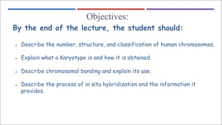
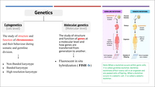
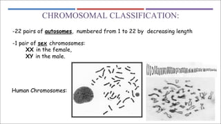
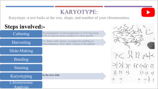
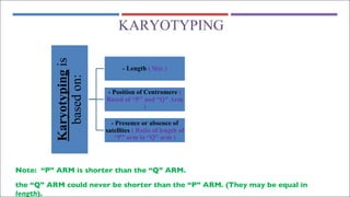
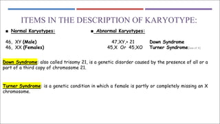
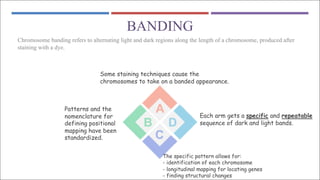
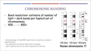
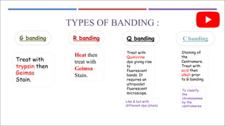
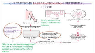
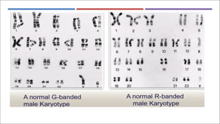
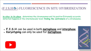
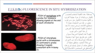
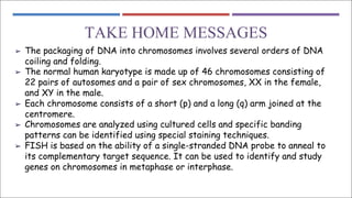
Index color:
• Important
• Slides
• Notes
• Extra information
Lecture 1
Human
chromosomes
OBJECTIVES:
Objectives:
By the end of the lecture, the student should:
● Describe the number, structure, and classification of human chromosomes.
● Explain what a Karyotype is and how it is obtained.
● Describe chromosomal banding and explain its use.
● Describe the process of in situ hybridization and the information it
provides.
TITLE
Eukaryotic cell
Genetics
Cytogenetics
(cell level)
Molecular genetics
(Molecular level)
The study of structure and
function of chromosomes
and their behaviour during
somatic and germline
division.
● Non-Banded karyotype
● Banded karyotype
● High resolution karyotype
The study of structure
and function of genes at
a molecular level and
how genes are
transferred from
generation to another.
● Fluorescent in situ
hybridization ( FISH 🐟 ) Note:When a mutation occurs within germ cells,
it is called germline mutation. Germline
mutations affect every cell in an organism and
are passed onto offspring. When a mutation
occurs in a somatic cell, it is called a somatic
mutation .
CYTOGENETICS
Human cytogenetics: The study of human chromosomes in health and disease.
Chromosome studies are an important laboratory diagnosis in:
Prenatal diagnosis
Certain patients with mental retardation and multiple birth defects
Patients with abnormal sexual development
Some cases of infertility or multiple miscarriages
In the study and treatment of patients with malignancies and hematologic disorders
CHROMOSOMES:
■ Carry genetic material
■ Heredity: each pair of homologues consists of one paternal and
one maternal chromosome.
■ The intact set is passed to each daughter cell at every mitosis.
Electron Microscope(EM)
OF HUMAN
CHROMOSOMES
Note:Genes are transmitted to
daughter cells while they are loose
(decondensed)
CHROMOSOMAL CLASSIFICATION:
-22 pairs of autosomes, numbered from 1 to 22 by decreasing length
-1 pair of sex chromosomes:
XX in the female,
XY in the male.
Human Chromosomes:
STRUCTURE OF CHROMOSOME
➔ Primary coiling : DNA double helix
➔ Secondary coiling : around histones
( basic protiens ) > nucleosomes
➔ Tertiary coiling : chromatin fiber
➔ Chromatin fibers form long loops
on non-histone protien > tighter
coils > chromosomes
MITOTIC CELL CYCLE :
METAPHASE CHROMOSOMES:
- The 2 sister chromatids are held together at the centromeric region
-Each chromosome has a centromere region (CEN) which contains the
kinetochore
-CEN divides the chromosome into two arms
1-short arm (P arm)
2-long arm (Q arm)
- Each arm terminates in a telomere.
A single complete set of
chromosomes. (N=23 for humans)
CENTROMERIC POSITION AND ARM LENGTH:
1 2 3
● The ratio of the lengths of the two arms is constant for each chromosome.
● This ratio is an important parameter for chromosome identification and allows
classification of chromosomes into several basic morphologic types:
● 1-metacentric
● 2-sub-metacentric
● 3-acrocentric
● In the human karyotype chromosome pairs 13, 14, 15, 21, 22 are acrocentric.
KARYOTYPE:
the propagation of microorganisms or of living tissue
cells in special media conducive to their growth.
Culturing
To obtain cells, tissues, or organs for grafting or
transplantation, from either a donor or the patient.
Harvesting
Slide-Making
Banding
Staining
In the next slide
Karyotyping
Chromosome
Analysis
Steps involved:-
Karyotype :a test looks at the size, shape, and number of your chromosomes.
KARYOTYPING
Note: “P” ARM is shorter than the “Q” ARM.
the “Q” ARM could never be shorter than the “P” ARM. (They may be equal in
length).
Karyotyping
is
based
on:
- Length ( Size )
- Position of Centromere (
Based of “P” and “Q” Arm
)
- Presence or absence of
satellites ( Ratio of length of
“P” arm to “Q” arm )
ITEMS IN THE DESCRIPTION OF KARYOTYPE:
■ Normal Karyotypes: ■ Abnormal Karyotypes:
46, XY (Male) 47,XY,+ 21 Down Syndrome
46, XX (Females) 45,X Or 45,XO Turner Syndrome(loss of X)
Down Syndrome: also called trisomy 21, is a genetic disorder caused by the presence of all or a
part of a third copy of chromosome 21.
Turner Syndrome: is a genetic condition in which a female is partly or completely missing an X
chromosome.
BANDING
Chromosome banding refers to alternating light and dark regions along the length of a chromosome, produced after
staining with a dye.
Some staining techniques cause the
chromosomes to take on a banded appearance.
Patterns and the
nomenclature for
defining positional
mapping have been
standardized.
The specific pattern allows for:
- identification of each chromosome
- longitudinal mapping for locating genes
- finding structural changes
Each arm gets a specific and repeatable
sequence of dark and light bands.
CHROMOSOME BANDING
• Band resolution= estimate of number of
light + dark bands per haploid set of
chromosomes.
• 400 850+
- Disadvantage of low(400 resolution) is inability to detect
abnormalities
- As resolution increases detection increases
- light regions are active, Dark regions are inactive
TYPES OF BANDING :
C banding
Q banding
R banding
G banding
Treat with
trypsin then
Geimsa
Stain.
Heat then
treat with
Geimsa
Stain.
Treat with
Quinicrine
dye giving rise
to
fluorescent
bands. It
requires an
ultraviolet
fluorescent
microscope.
Staining of
the
Centromere.
Treat with
acid then
alkali prior
to G banding.
To classify
the
chromosomes
by the
centromeres
Like G but with
different dye (stain)
CHROMOSOME PREPARATION FROM PERIPHERAL
BLOOD
Why do we use phytohemagglutinin?
We use it to increase their(cells)
number by increasing the rate of
division
Fixative substance fixes
them to a particular state
metaphase
(F.I.S.H🐟 ) FLUORESCENCE IN SITU HYBRIDIZATION
in-situ= in its place , determines the chromosomes and its position Extremely accurate
detection happens at the intermolecular level: finding the centromere of a chromosome.
● F.I.S.H can be used in both metaphase and interphase.
● Karyotyping can only be used for metaphase.
) FLUORESCENCE IN SITU HYBRIDIZATION
🐟
F.I.S.H
with
metaphase
FISH of
-
a probe for telomere
showing signals at the end
of each chromatid.
- FISH of interphase
nuclei with a chromosome
21 centromeric probe
showing 3 signals
consistent with trisomy
21.
‫ﺑ‬
‫ﺒ‬
‫ﺴ‬
‫ﺎ‬
‫ط‬
‫ﺔ‬
:
‫ھ‬
‫ﺬ‬
‫ه‬
‫ا‬
‫ﻟ‬
‫ﺘ‬
‫ﻘ‬
‫ﻨ‬
‫ﯿ‬
‫ﺔ‬
‫ﺗ‬
ُ
‫ﺴ‬
‫ﺘ‬
‫ﺨ‬
‫ﺪ‬
‫م‬
‫ﻟ‬
‫ﺘ‬
‫ﺤ‬
‫ﺪ‬
‫ﯾ‬
‫ﺪ‬
‫و‬
‫إ‬
‫ظ‬
‫ﮭ‬
‫ﺎ‬
‫ر‬
‫ﺑ‬
‫ﺮ‬
‫و‬
‫ﺗ‬
‫ﯿ‬
‫ﻨ‬
‫ﺎ‬
‫ت‬
‫أ‬
‫و‬
‫ﻣ‬
‫ﻮ‬
‫ا‬
‫د‬
‫ﺟ‬
‫ﯿ‬
‫ﻨ‬
‫ﯿ‬
‫ﺔ‬
‫أ‬
‫ﺧ‬
‫ﺮ‬
‫ى‬
،
‫و‬
‫ﻟ‬
‫ﻜ‬
‫ﻨ‬
ّ
‫ﮭ‬
‫ﺎ‬
‫ﻣ‬
‫ﺤ‬
‫ﺪ‬
‫و‬
‫د‬
‫ة‬
‫ﻹ‬
‫ظ‬
‫ﮭ‬
‫ﺎ‬
‫ر‬
‫ﺟ‬
‫ﺰ‬
‫ء‬
‫ﻣ‬
‫ﺮ‬
‫ﻏ‬
‫ﻮ‬
‫ب‬
‫ﻣ‬
‫ﻌ‬
‫ﯿ‬
ّ
‫ﻦ‬
،
‫و‬
‫ھ‬
‫ﺬ‬
‫ه‬
‫ﻣ‬
‫ﻦ‬
‫ا‬
‫ﻟ‬
‫ﺴ‬
‫ﻠ‬
‫ﺒ‬
‫ﯿ‬
‫ﺎ‬
‫ت‬
‫ﺣ‬
‫ﯿ‬
‫ﺚ‬
‫ﻻ‬
‫ﻧ‬
‫ﺴ‬
‫ﺘ‬
‫ﻄ‬
‫ﯿ‬
‫ﻊ‬
‫ا‬
‫ﻛ‬
‫ﺘ‬
‫ﺸ‬
‫ﺎ‬
‫ف‬
‫ﻣ‬
‫ﺮ‬
‫ض‬
‫ﻣ‬
‫ﺤ‬
‫ﺪ‬
‫د‬
‫إ‬
‫ﻻ‬
‫ﺑ‬
‫ﻌ‬
‫ﺪ‬
‫ا‬
‫ﺳ‬
‫ﺘ‬
‫ﺨ‬
‫ﺪ‬
‫ا‬
‫م‬
‫ا‬
‫ﻟ‬
‫ﺘ‬
‫ﻘ‬
‫ﻨ‬
‫ﯿ‬
‫ﺔ‬
‫ا‬
‫ﻟ‬
‫ﻤ‬
‫ﺤ‬
‫ﺪ‬
‫د‬
‫ة‬
‫ﻟ‬
‫ﮭ‬
‫ﺬ‬
‫ا‬
‫ا‬
‫ﻟ‬
‫ﻤ‬
‫ﺮ‬
‫ض‬
‫أ‬
‫و‬
‫ھ‬
‫ﺬ‬
‫ا‬
‫ا‬
‫ﻟ‬
‫ﺠ‬
‫ﺰ‬
‫ء‬
‫ا‬
‫ﻟ‬
‫ﻤ‬
‫ﺮ‬
‫ﻏ‬
‫ﻮ‬
‫ب‬ .
‫ﻛ‬
‫ﯿ‬
‫ﻒ‬
‫؟‬
‫ھ‬
‫ﺬ‬
‫ه‬
‫ا‬
‫ﻟ‬
‫ﻌ‬
‫ﻤ‬
‫ﻠ‬
‫ﯿ‬
‫ﺔ‬
‫ا‬
‫ﻟ‬
‫ﺴ‬
‫ﺮ‬
‫ﯾ‬
‫ﻌ‬
‫ﺔ‬
‫ﻧ‬
‫ﺴ‬
‫ﺒ‬
‫ﯿ‬
‫ﺎ‬
ً
‫و‬
‫ﺗ‬
‫ﻜ‬
‫ﻮ‬
‫ن‬
‫ﻋ‬
‫ﻦ‬
‫ط‬
‫ﺮ‬
‫ﯾ‬
‫ﻖ‬
‫د‬
‫ﻣ‬
‫ﺞ‬
‫أ‬
‫ﻋ‬
‫ﻮ‬
‫ا‬
‫د‬
‫ﻣ‬
‫ﻀ‬
‫ﯿ‬
‫ﺌ‬
‫ﺔ‬
‫ﻣ‬
‫ﺼ‬
‫ﻨ‬
ّ
‫ﻔ‬
‫ﺔ‬
،
‫ﻣ‬
‫ﻊ‬
‫ﺷ‬
‫ﺮ‬
‫ﯾ‬
‫ﻂ‬
‫و‬
‫ا‬
‫ﺣ‬
‫ﺪ‬
‫ﻣ‬
‫ﻦ‬
‫ا‬
‫ﻟ‬
‫ﺪ‬
‫ي‬
‫إ‬
‫ن‬
‫إ‬
‫ي‬
‫ﻣ‬
‫ﺜ‬
‫ﻼ‬
ً
‫؛‬
‫ﻟ‬
‫ﺘ‬
‫ﺤ‬
‫ﺪ‬
‫ﯾ‬
‫ﺪ‬
:
‫ﻣ‬
‫ﻨ‬
‫ﺎ‬
‫ط‬
‫ﻖ‬
‫ﻣ‬
‫ﻌ‬
‫ﯿ‬
ّ
‫ﻨ‬
‫ﺔ‬
‫أ‬
‫و‬
‫ﻣ‬
‫ﻨ‬
‫ﺎ‬
‫ط‬
‫ﻖ‬
‫ﺣ‬
‫ﺬ‬
‫ف‬
‫أ‬
‫و‬
‫ﺗ‬
‫ﺒ‬
‫ﺪ‬
‫ﯾ‬
‫ﻞ‬
(
‫ﺳ‬
‫ﯿ‬
‫ﺘ‬
‫ﻢ‬
‫ﺷ‬
‫ﺮ‬
‫ح‬
‫ھ‬
‫ﺬ‬
‫ه‬
‫ا‬
‫ﻟ‬
‫ﻤ‬
‫ﻨ‬
‫ﺎ‬
‫ط‬
‫ﻖ‬
‫ﻓ‬
‫ﻲ‬
‫ا‬
‫ﻟ‬
‫ﻤ‬
‫ﺤ‬
‫ﺎ‬
‫ﺿ‬
‫ﺮ‬
‫ة‬
‫ا‬
‫ﻟ‬
‫ﻘ‬
‫ﺎ‬
‫د‬
‫ﻣ‬
‫ﺔ‬
)
437 *
TAKE HOME MESSAGES
➢ The packaging of DNA into chromosomes involves several orders of DNA
coiling and folding.
➢ The normal human karyotype is made up of 46 chromosomes consisting of
22 pairs of autosomes and a pair of sex chromosomes, XX in the female,
and XY in the male.
➢ Each chromosome consists of a short (p) and a long (q) arm joined at the
centromere.
➢ Chromosomes are analyzed using cultured cells and specific banding
patterns can be identified using special staining techniques.
➢ FISH is based on the ability of a single-stranded DNA probe to anneal to
its complementary target sequence. It can be used to identify and study
genes on chromosomes in metaphase or interphase.
MCQS
Q1:It’s used to to stimulate T lymphocytes to divide
D) Phytohemaggluti nin
(PHA)
C) Geimsa Stain B) trypsin A) Colchicine
Q2:………………… is based on the ability of a single-stranded DNA probe to anneal to its complementary target sequence.
It can be used to identify and study genes on chromosomes in metaphase or interphase :
D) Quinidine C) FISH B) Banding A) Karotype
Q3:The 2 sister-chromatids held together at:
D) Isoelectric region C) Centromeric region B) Stop region A) Promoter region
Q4: …………… is used to arrest cell division during metaphase for karyotyping
D) Phytohemagglutinin (PHA) C) Trypsin B) C Banding A) Colchicine
MCQs
MCQs
answers
1)
D
2)
C
3)
C
4)
A
MCQs
Q5: The end of each chromosome is
D) FICH C) telomere B)colchicine A) PROPHASE
Q6:The genotype 47,XY,+ 21 is for …………….. Syndrome
D) Apert syndrome C) Normal B) Turner Syndrome A) Down Syndrome
5)
C
6)
A
MCQs
answers
Boys team:
• Nawaf Alghamdi
• Ahmed Alkhawashki
• Bassam Alasmari
• Rayan Alzahrani
• Khalid AlOsaimii
• Abdulrahman Alswat
• Faisal AlFadel
• Hadi AlHemsi
• Hesham Alsqabi
• Yazeed Alomar
• Mohammed Benhjji
• Badr Alshahrani
• Homoud Algadheb
Girls team:
• Ghaida Alasiri
• Arwa Alqahtani
• Albandri Ahmad
• Aljohara Albnyan
• Aljohara alshathry
• Alanoud Alshahrani
• Raghad Alasiri
• Renad Alhmidi
• Sara Alharbi
• Taif Almutari
• Abeer Awad
• Ghada Alabdi
• Noura Almassad
• Hind Almut
Team Leaders:
• Sumo Abdulrahman
• Abdulmalik Mokhtar
: HumanGenetics439@gmail.com Editing File

CHROMOSOME CORRE.pdf cell division DNA & RNA

  • 1.
    Index color: • Important •Slides • Notes • Extra information Lecture 1 Human chromosomes
  • 2.
    OBJECTIVES: Objectives: By the endof the lecture, the student should: ● Describe the number, structure, and classification of human chromosomes. ● Explain what a Karyotype is and how it is obtained. ● Describe chromosomal banding and explain its use. ● Describe the process of in situ hybridization and the information it provides.
  • 3.
  • 4.
    Genetics Cytogenetics (cell level) Molecular genetics (Molecularlevel) The study of structure and function of chromosomes and their behaviour during somatic and germline division. ● Non-Banded karyotype ● Banded karyotype ● High resolution karyotype The study of structure and function of genes at a molecular level and how genes are transferred from generation to another. ● Fluorescent in situ hybridization ( FISH 🐟 ) Note:When a mutation occurs within germ cells, it is called germline mutation. Germline mutations affect every cell in an organism and are passed onto offspring. When a mutation occurs in a somatic cell, it is called a somatic mutation .
  • 5.
    CYTOGENETICS Human cytogenetics: Thestudy of human chromosomes in health and disease. Chromosome studies are an important laboratory diagnosis in: Prenatal diagnosis Certain patients with mental retardation and multiple birth defects Patients with abnormal sexual development Some cases of infertility or multiple miscarriages In the study and treatment of patients with malignancies and hematologic disorders
  • 6.
    CHROMOSOMES: ■ Carry geneticmaterial ■ Heredity: each pair of homologues consists of one paternal and one maternal chromosome. ■ The intact set is passed to each daughter cell at every mitosis. Electron Microscope(EM) OF HUMAN CHROMOSOMES Note:Genes are transmitted to daughter cells while they are loose (decondensed)
  • 7.
    CHROMOSOMAL CLASSIFICATION: -22 pairsof autosomes, numbered from 1 to 22 by decreasing length -1 pair of sex chromosomes: XX in the female, XY in the male. Human Chromosomes:
  • 8.
    STRUCTURE OF CHROMOSOME ➔Primary coiling : DNA double helix ➔ Secondary coiling : around histones ( basic protiens ) > nucleosomes ➔ Tertiary coiling : chromatin fiber ➔ Chromatin fibers form long loops on non-histone protien > tighter coils > chromosomes
  • 9.
  • 10.
    METAPHASE CHROMOSOMES: - The2 sister chromatids are held together at the centromeric region -Each chromosome has a centromere region (CEN) which contains the kinetochore -CEN divides the chromosome into two arms 1-short arm (P arm) 2-long arm (Q arm) - Each arm terminates in a telomere. A single complete set of chromosomes. (N=23 for humans)
  • 11.
    CENTROMERIC POSITION ANDARM LENGTH: 1 2 3 ● The ratio of the lengths of the two arms is constant for each chromosome. ● This ratio is an important parameter for chromosome identification and allows classification of chromosomes into several basic morphologic types: ● 1-metacentric ● 2-sub-metacentric ● 3-acrocentric ● In the human karyotype chromosome pairs 13, 14, 15, 21, 22 are acrocentric.
  • 12.
    KARYOTYPE: the propagation ofmicroorganisms or of living tissue cells in special media conducive to their growth. Culturing To obtain cells, tissues, or organs for grafting or transplantation, from either a donor or the patient. Harvesting Slide-Making Banding Staining In the next slide Karyotyping Chromosome Analysis Steps involved:- Karyotype :a test looks at the size, shape, and number of your chromosomes.
  • 13.
    KARYOTYPING Note: “P” ARMis shorter than the “Q” ARM. the “Q” ARM could never be shorter than the “P” ARM. (They may be equal in length). Karyotyping is based on: - Length ( Size ) - Position of Centromere ( Based of “P” and “Q” Arm ) - Presence or absence of satellites ( Ratio of length of “P” arm to “Q” arm )
  • 14.
    ITEMS IN THEDESCRIPTION OF KARYOTYPE: ■ Normal Karyotypes: ■ Abnormal Karyotypes: 46, XY (Male) 47,XY,+ 21 Down Syndrome 46, XX (Females) 45,X Or 45,XO Turner Syndrome(loss of X) Down Syndrome: also called trisomy 21, is a genetic disorder caused by the presence of all or a part of a third copy of chromosome 21. Turner Syndrome: is a genetic condition in which a female is partly or completely missing an X chromosome.
  • 15.
    BANDING Chromosome banding refersto alternating light and dark regions along the length of a chromosome, produced after staining with a dye. Some staining techniques cause the chromosomes to take on a banded appearance. Patterns and the nomenclature for defining positional mapping have been standardized. The specific pattern allows for: - identification of each chromosome - longitudinal mapping for locating genes - finding structural changes Each arm gets a specific and repeatable sequence of dark and light bands.
  • 16.
    CHROMOSOME BANDING • Bandresolution= estimate of number of light + dark bands per haploid set of chromosomes. • 400 850+ - Disadvantage of low(400 resolution) is inability to detect abnormalities - As resolution increases detection increases - light regions are active, Dark regions are inactive
  • 17.
    TYPES OF BANDING: C banding Q banding R banding G banding Treat with trypsin then Geimsa Stain. Heat then treat with Geimsa Stain. Treat with Quinicrine dye giving rise to fluorescent bands. It requires an ultraviolet fluorescent microscope. Staining of the Centromere. Treat with acid then alkali prior to G banding. To classify the chromosomes by the centromeres Like G but with different dye (stain)
  • 18.
    CHROMOSOME PREPARATION FROMPERIPHERAL BLOOD Why do we use phytohemagglutinin? We use it to increase their(cells) number by increasing the rate of division Fixative substance fixes them to a particular state metaphase
  • 20.
    (F.I.S.H🐟 ) FLUORESCENCEIN SITU HYBRIDIZATION in-situ= in its place , determines the chromosomes and its position Extremely accurate detection happens at the intermolecular level: finding the centromere of a chromosome. ● F.I.S.H can be used in both metaphase and interphase. ● Karyotyping can only be used for metaphase.
  • 21.
    ) FLUORESCENCE INSITU HYBRIDIZATION 🐟 F.I.S.H with metaphase FISH of - a probe for telomere showing signals at the end of each chromatid. - FISH of interphase nuclei with a chromosome 21 centromeric probe showing 3 signals consistent with trisomy 21. ‫ﺑ‬ ‫ﺒ‬ ‫ﺴ‬ ‫ﺎ‬ ‫ط‬ ‫ﺔ‬ : ‫ھ‬ ‫ﺬ‬ ‫ه‬ ‫ا‬ ‫ﻟ‬ ‫ﺘ‬ ‫ﻘ‬ ‫ﻨ‬ ‫ﯿ‬ ‫ﺔ‬ ‫ﺗ‬ ُ ‫ﺴ‬ ‫ﺘ‬ ‫ﺨ‬ ‫ﺪ‬ ‫م‬ ‫ﻟ‬ ‫ﺘ‬ ‫ﺤ‬ ‫ﺪ‬ ‫ﯾ‬ ‫ﺪ‬ ‫و‬ ‫إ‬ ‫ظ‬ ‫ﮭ‬ ‫ﺎ‬ ‫ر‬ ‫ﺑ‬ ‫ﺮ‬ ‫و‬ ‫ﺗ‬ ‫ﯿ‬ ‫ﻨ‬ ‫ﺎ‬ ‫ت‬ ‫أ‬ ‫و‬ ‫ﻣ‬ ‫ﻮ‬ ‫ا‬ ‫د‬ ‫ﺟ‬ ‫ﯿ‬ ‫ﻨ‬ ‫ﯿ‬ ‫ﺔ‬ ‫أ‬ ‫ﺧ‬ ‫ﺮ‬ ‫ى‬ ، ‫و‬ ‫ﻟ‬ ‫ﻜ‬ ‫ﻨ‬ ّ ‫ﮭ‬ ‫ﺎ‬ ‫ﻣ‬ ‫ﺤ‬ ‫ﺪ‬ ‫و‬ ‫د‬ ‫ة‬ ‫ﻹ‬ ‫ظ‬ ‫ﮭ‬ ‫ﺎ‬ ‫ر‬ ‫ﺟ‬ ‫ﺰ‬ ‫ء‬ ‫ﻣ‬ ‫ﺮ‬ ‫ﻏ‬ ‫ﻮ‬ ‫ب‬ ‫ﻣ‬ ‫ﻌ‬ ‫ﯿ‬ ّ ‫ﻦ‬ ، ‫و‬ ‫ھ‬ ‫ﺬ‬ ‫ه‬ ‫ﻣ‬ ‫ﻦ‬ ‫ا‬ ‫ﻟ‬ ‫ﺴ‬ ‫ﻠ‬ ‫ﺒ‬ ‫ﯿ‬ ‫ﺎ‬ ‫ت‬ ‫ﺣ‬ ‫ﯿ‬ ‫ﺚ‬ ‫ﻻ‬ ‫ﻧ‬ ‫ﺴ‬ ‫ﺘ‬ ‫ﻄ‬ ‫ﯿ‬ ‫ﻊ‬ ‫ا‬ ‫ﻛ‬ ‫ﺘ‬ ‫ﺸ‬ ‫ﺎ‬ ‫ف‬ ‫ﻣ‬ ‫ﺮ‬ ‫ض‬ ‫ﻣ‬ ‫ﺤ‬ ‫ﺪ‬ ‫د‬ ‫إ‬ ‫ﻻ‬ ‫ﺑ‬ ‫ﻌ‬ ‫ﺪ‬ ‫ا‬ ‫ﺳ‬ ‫ﺘ‬ ‫ﺨ‬ ‫ﺪ‬ ‫ا‬ ‫م‬ ‫ا‬ ‫ﻟ‬ ‫ﺘ‬ ‫ﻘ‬ ‫ﻨ‬ ‫ﯿ‬ ‫ﺔ‬ ‫ا‬ ‫ﻟ‬ ‫ﻤ‬ ‫ﺤ‬ ‫ﺪ‬ ‫د‬ ‫ة‬ ‫ﻟ‬ ‫ﮭ‬ ‫ﺬ‬ ‫ا‬ ‫ا‬ ‫ﻟ‬ ‫ﻤ‬ ‫ﺮ‬ ‫ض‬ ‫أ‬ ‫و‬ ‫ھ‬ ‫ﺬ‬ ‫ا‬ ‫ا‬ ‫ﻟ‬ ‫ﺠ‬ ‫ﺰ‬ ‫ء‬ ‫ا‬ ‫ﻟ‬ ‫ﻤ‬ ‫ﺮ‬ ‫ﻏ‬ ‫ﻮ‬ ‫ب‬ . ‫ﻛ‬ ‫ﯿ‬ ‫ﻒ‬ ‫؟‬ ‫ھ‬ ‫ﺬ‬ ‫ه‬ ‫ا‬ ‫ﻟ‬ ‫ﻌ‬ ‫ﻤ‬ ‫ﻠ‬ ‫ﯿ‬ ‫ﺔ‬ ‫ا‬ ‫ﻟ‬ ‫ﺴ‬ ‫ﺮ‬ ‫ﯾ‬ ‫ﻌ‬ ‫ﺔ‬ ‫ﻧ‬ ‫ﺴ‬ ‫ﺒ‬ ‫ﯿ‬ ‫ﺎ‬ ً ‫و‬ ‫ﺗ‬ ‫ﻜ‬ ‫ﻮ‬ ‫ن‬ ‫ﻋ‬ ‫ﻦ‬ ‫ط‬ ‫ﺮ‬ ‫ﯾ‬ ‫ﻖ‬ ‫د‬ ‫ﻣ‬ ‫ﺞ‬ ‫أ‬ ‫ﻋ‬ ‫ﻮ‬ ‫ا‬ ‫د‬ ‫ﻣ‬ ‫ﻀ‬ ‫ﯿ‬ ‫ﺌ‬ ‫ﺔ‬ ‫ﻣ‬ ‫ﺼ‬ ‫ﻨ‬ ّ ‫ﻔ‬ ‫ﺔ‬ ، ‫ﻣ‬ ‫ﻊ‬ ‫ﺷ‬ ‫ﺮ‬ ‫ﯾ‬ ‫ﻂ‬ ‫و‬ ‫ا‬ ‫ﺣ‬ ‫ﺪ‬ ‫ﻣ‬ ‫ﻦ‬ ‫ا‬ ‫ﻟ‬ ‫ﺪ‬ ‫ي‬ ‫إ‬ ‫ن‬ ‫إ‬ ‫ي‬ ‫ﻣ‬ ‫ﺜ‬ ‫ﻼ‬ ً ‫؛‬ ‫ﻟ‬ ‫ﺘ‬ ‫ﺤ‬ ‫ﺪ‬ ‫ﯾ‬ ‫ﺪ‬ : ‫ﻣ‬ ‫ﻨ‬ ‫ﺎ‬ ‫ط‬ ‫ﻖ‬ ‫ﻣ‬ ‫ﻌ‬ ‫ﯿ‬ ّ ‫ﻨ‬ ‫ﺔ‬ ‫أ‬ ‫و‬ ‫ﻣ‬ ‫ﻨ‬ ‫ﺎ‬ ‫ط‬ ‫ﻖ‬ ‫ﺣ‬ ‫ﺬ‬ ‫ف‬ ‫أ‬ ‫و‬ ‫ﺗ‬ ‫ﺒ‬ ‫ﺪ‬ ‫ﯾ‬ ‫ﻞ‬ ( ‫ﺳ‬ ‫ﯿ‬ ‫ﺘ‬ ‫ﻢ‬ ‫ﺷ‬ ‫ﺮ‬ ‫ح‬ ‫ھ‬ ‫ﺬ‬ ‫ه‬ ‫ا‬ ‫ﻟ‬ ‫ﻤ‬ ‫ﻨ‬ ‫ﺎ‬ ‫ط‬ ‫ﻖ‬ ‫ﻓ‬ ‫ﻲ‬ ‫ا‬ ‫ﻟ‬ ‫ﻤ‬ ‫ﺤ‬ ‫ﺎ‬ ‫ﺿ‬ ‫ﺮ‬ ‫ة‬ ‫ا‬ ‫ﻟ‬ ‫ﻘ‬ ‫ﺎ‬ ‫د‬ ‫ﻣ‬ ‫ﺔ‬ ) 437 *
  • 22.
    TAKE HOME MESSAGES ➢The packaging of DNA into chromosomes involves several orders of DNA coiling and folding. ➢ The normal human karyotype is made up of 46 chromosomes consisting of 22 pairs of autosomes and a pair of sex chromosomes, XX in the female, and XY in the male. ➢ Each chromosome consists of a short (p) and a long (q) arm joined at the centromere. ➢ Chromosomes are analyzed using cultured cells and specific banding patterns can be identified using special staining techniques. ➢ FISH is based on the ability of a single-stranded DNA probe to anneal to its complementary target sequence. It can be used to identify and study genes on chromosomes in metaphase or interphase.
  • 23.
    MCQS Q1:It’s used toto stimulate T lymphocytes to divide D) Phytohemaggluti nin (PHA) C) Geimsa Stain B) trypsin A) Colchicine Q2:………………… is based on the ability of a single-stranded DNA probe to anneal to its complementary target sequence. It can be used to identify and study genes on chromosomes in metaphase or interphase : D) Quinidine C) FISH B) Banding A) Karotype Q3:The 2 sister-chromatids held together at: D) Isoelectric region C) Centromeric region B) Stop region A) Promoter region Q4: …………… is used to arrest cell division during metaphase for karyotyping D) Phytohemagglutinin (PHA) C) Trypsin B) C Banding A) Colchicine MCQs MCQs answers 1) D 2) C 3) C 4) A
  • 24.
    MCQs Q5: The endof each chromosome is D) FICH C) telomere B)colchicine A) PROPHASE Q6:The genotype 47,XY,+ 21 is for …………….. Syndrome D) Apert syndrome C) Normal B) Turner Syndrome A) Down Syndrome 5) C 6) A MCQs answers
  • 25.
    Boys team: • NawafAlghamdi • Ahmed Alkhawashki • Bassam Alasmari • Rayan Alzahrani • Khalid AlOsaimii • Abdulrahman Alswat • Faisal AlFadel • Hadi AlHemsi • Hesham Alsqabi • Yazeed Alomar • Mohammed Benhjji • Badr Alshahrani • Homoud Algadheb Girls team: • Ghaida Alasiri • Arwa Alqahtani • Albandri Ahmad • Aljohara Albnyan • Aljohara alshathry • Alanoud Alshahrani • Raghad Alasiri • Renad Alhmidi • Sara Alharbi • Taif Almutari • Abeer Awad • Ghada Alabdi • Noura Almassad • Hind Almut Team Leaders: • Sumo Abdulrahman • Abdulmalik Mokhtar : HumanGenetics439@gmail.com Editing File