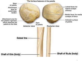
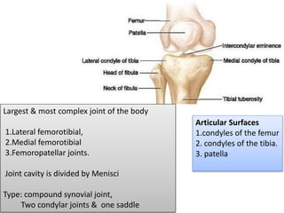
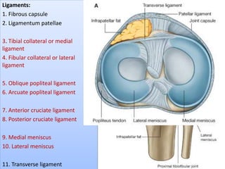
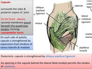
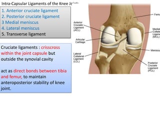
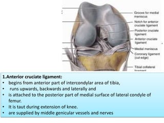
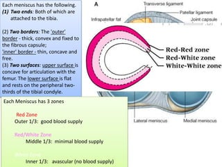
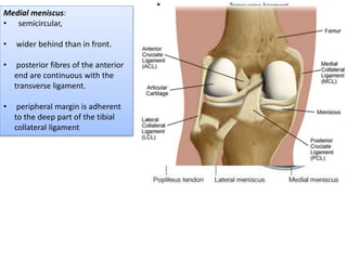
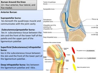
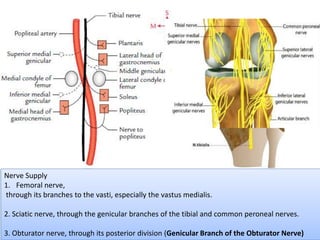
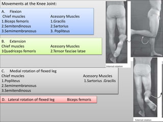
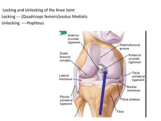
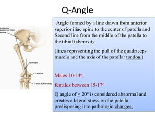
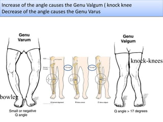
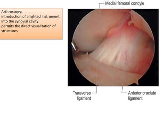
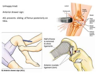
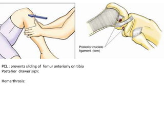
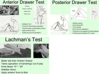
Knee Joint
Dr. Prabhakar Yadav
Associate Professor
Department of Human Anatomy
B.P. Koirala Institute of Health Sciences
Largest & most complex joint of the body
1.Lateral femorotibial,
2.Medial femorotibial
3.Femoropatellar joints.
Joint cavity is divided by Menisci
Type: compound synovial joint,
Two condylar joints & one saddle
Articular Surfaces
1.condyles of the femur
2. condyles of the tibia.
3. patella
Ligaments:
1. Fibrous capsule
2. Ligamentum patellae
3. Tibial collateral or medial
ligament
4. Fibular collateral or lateral
ligament
5. Oblique popliteal ligament
6. Arcuate popliteal ligament
7. Anterior cruciate ligament
8. Posterior cruciate ligament
9. Medial meniscus
10. Lateral meniscus
11. Transverse ligament
Capsule:
Attached ½ to 1 cm to the margins of the articular surfaces
Capsule:
surrounds the sides &
posterior aspect of joint.
On the front - absent,
synovial membrane pouch
beneath the quadriceps
tendon, forming
suprapatellar bursa
On each side of patella,
capsule is strengthened by
expansions from tendons of
vastus lateralis & medialis
Posteriorly- capsule is strengthened by oblique popliteal ligament
An opening in the capsule behind the lateral tibial condyle permits the tendon
of popliteus
Synovial Membrane:
• Attached to margins of
articular surfaces
• Lines the capsule
On the front & above
membrane pouchs, -
suprapatellar bursa--Held
in position by attachment
of a small portion of
vastus intermedius muscle,
-- Articularis genus muscle.
Posteriorly:
synovial membrane is
prolonged downward on
the deep surface of
tendon of popliteus,
forming popliteal bursa.
semimembranosus bursa – bursa interposed between medial head of
gastrocnemius & medial femoral condyle & semimembranosus tendon-
it communicates with the synovial cavity of the joint.
Synovial membrane is
reflected forward from
the posterior part of
capsule around the front
of cruciate ligaments.
Cruciate ligaments lie :-
behind the synovial cavity.
Anteriorly synovial membrane is reflected backward from the posterior surface
of ligamentum patellae to form infrapatellar fold;
free borders of the fold are termed - alar folds
Weak capsular ligament is
strengthened by
Anteriorly by:
Medial & Lateral patellar
retinacula.
Laterally by : Iliotibial tract
Posteriorly by:
Oblique polpliteal ligament
Medially by:
Tendons of sartorius and
Semimembranosus
Extracapsular Ligaments
joint capsule is strengthened by five
extracapsular ligaments:
1. Ligamentum patellae
2. Tibial collateral or medial ligament
3. Fibular collateral or lateral ligament
4. Oblique popliteal ligament
5. Arcuate popliteal ligament
1.Ligamentum patellae:
Attachment
Above –margins and apex of patella
Below- Tibial tuberosity.
Related to: infrapatella pad of fat ;
superficial & deep infrapatella bursa
2.oblique popliteal ligament:
Tendinous expansion of semimembranosus
muscle, runs upwards & laterally
Strengthens - posterior aspect of capsule
Attached to intracondylar line and lateral
condyle of femur
Have a close relation with popliteal artery
.3.Tibial collateral ligament (TCL):
(degenerated tendon of the
adductor magnus)
Extension:
Medial epicondyle of femur to
medial condyle and the superior
part of medial surface of tibia .
Deep fibers - firmly attached to
medial meniscus.
TCL, weaker than FCL, is more
often damaged. As a result, the
TCL and medial meniscus are
commonly torn.
4. Fibular collateral ligament (FCL):
(degenerated tendon of peroneus
longus.)
Extension: lateral epicondyle of
femur to lateral surface of fibular
head.
Tendon of popliteus passes deep to
FCL, separating it from lateral
meniscus.
Tendon of the biceps femoris is split
into two parts by this ligament
Collateral ligaments of knee are taut
when knee is fully extended,
contributing to stability while standing .
As flexion proceeds, they become
increasingly slack, permitting and limiting
(serving as check ligaments for) rotation
at the knee
5. Arcuate popliteal ligament:
• strengthens the joint capsule posterolaterally.
• arises from the posterior aspect of the fibular head,
• passes superomedially over the tendon of popliteus,
• and spreads over the posterior surface of the knee joint
Intra-Capsular Ligaments of the Knee Joint:
1. Anterior cruciate ligament
2. Posterior cruciate ligament
3 Medial meniscus
4. Lateral meniscus
5. Transverse ligament
Cruciate ligaments : crisscross
within the joint capsule but
outside the synovial cavity
act as direct bonds between tibia
and femur, to maintain
anteroposterior stability of knee
joint.
1.Anterior cruciate ligament:
• begins from anterior part of intercondylar area of tibia,
• runs upwards, backwards and laterally and
• is attached to the posterior part of medial surface of lateral condyle of
femur.
• It is taut during extension of knee.
• are supplied by middle genicular vessels and nerves
2.Posterior cruciate ligament :
• begins from the posterior part of intercondylar area of tibia,
• runs upwards, forwards and medially
• is attached to the anterior part of the lateral surface of medial condyle of
femur.
• It is taut during flexion of the knee.
• are supplied by middle genicular vessels and nerves
Factors maintaining the stability of knee joint:
1.Cruciate ligament – anteroposterior stability
2.Collateral ligaments- side to side stability
3.Factors strengthening the capsule
• Anteriorly by:Medial & Lateral patellar retinacula.
• Laterally by : Iliotibial tract
• Posteriorly by: Oblique polpliteal ligament
• Medially by: Tendons of sartorius and Semimembranosus
4.iliotibial tract
Menisci or Semilunar Cartilages :
• are two fibrocartilaginous discs.
• crescent shaped.
• deepen articular surfaces of the condyles of the tibia,
• partially divide the joint cavity into upper and lower compartments.
• Flexion and extension of the knee take place in the upper compartment,
• rotation takes place, in the lower compartment
Each meniscus has the following.
(1) Two ends: Both of which are
attached to the tibia.
(2) Two borders: The 'outer'
border - thick, convex and fixed to
the fibrous capsule;
'inner' border - thin, concave and
free.
(3) Two surfaces: upper surface is
concave for articulation with the
femur. The lower surface is flat
and rests on the peripheral two-
thirds of the tibial condyle.
Each Meniscus has 3 zones
Red Zone
Outer 1/3: good blood supply
Red/White Zone
Middle 1/3: minimal blood supply
White Zone
Inner 1/3: avascular (no blood supply)
Medial meniscus:
• semicircular,
• wider behind than in front.
• posterior fibres of the anterior
end are continuous with the
transverse ligament.
• peripheral margin is adherent
to the deep part of the tibial
collateral ligament
lateral meniscus
• circular.
• posterior end of the meniscus is
attached to the femur through
meniscofemoral ligaments.
• Tendon of popliteus & capsule
separate the meniscus from fibular
collateral ligament.
• Medial part of the tendon of the
popliteus is attached to the lateral
meniscus.
• Mobility of the posterior end of lateral
meniscus is controlled by popliteus
and by meniscofemoral ligaments.
Transverse Ligament :
• connects the anterior ends of medial &
lateral menisci
Functions of Menisci
(1) make the articular surfaces more congruent..
(2) acts as shock absorbers.
(3) help in lubricating the joint cavity.
(4) Because of their nerve supply, they give rise to
proprioceptive impulses
Bursae Around the Knee
13—four anterior, four lateral, and
five medial.
Anterior Bursae:
Suprapatellar bursa:
lies beneath the quadriceps muscle and
communicates with the joint cavity
(Subcutaneous)prepatellar bursa:
lies in subcutaneous tissue between the
skin and the front of the lower half of the
patella and the upper part of the
ligamentum patellae
Superficial (Subcutaneous) infrapatellar
bursa:
lies in the subcutaneous tissue between
the skin and the front of the lower part of
the ligamentum patellae .
Deep infrapatellar bursa: lies between
the ligamentum patellae and tibia .
Lateral
1. Gastrocnemius bursa: deep to the
lateral head of the gastrocnemius.
2. A bursa between FCL & biceps
femoris.
3. A bursa between FCL& tendon of
popliteus.
4. A bursa between tendon of
popliteus & lateral condyle of tibia.
Medial
1. Gastrocnemius bursa :
deep to the medial head of gastro-
cnemius.
2. Anserine bursa:
separates the tendons of the
sartorius, gracilis & semitendinosus
from one another, from the tibia,
and from tibial collateral ligament.
3. A bursa deep to the tibial
collateral ligament.
4. A bursa deep to
semimembranosus.
Relations of Knee Joint
• Anteriorly
(1) Anterior bursae, (2) ligamentum patellae, and (3) patellar plexus of nerves
• Posteriorly
(A) At the middle: (1) Popliteal vessels, (2) tibial nerve, and (3) middle genicular vessels
and nerve.
(B) Posterolaterally: (1) Lateral head of gastro-cnemius, (2) plantaris, and (3) common
peroneal nerve.
(C) Posteromedially: (1) Medial head of gastro-cnemius, (2) semitendinosus,
(3) semimem-branosus, (4) gracilis, and (5) popliteus at its insertion
Medially
• (1) Sartorius, gracilis and semitendinosus.
• (2) Great saphenous vein with saphenous nerve and vessels.
• (3) Semimembranosus, and (4) inferior medial genicular vessels and nerve
Laterally
• (1) Biceps femoris, (2) tendon of origin of popliteus, and (3) inferior lateral
genicular vessels and nerve
Blood Supply
by the anastomoses around knee.
The chief sources of blood supply are:
(1) Five genicular branches of the
popliteal artery,
(2) Descending genicular branch of
femoral artery,
(3) Descending branch of the lateral
circumflex femoral artery,
(4) Two recurrent branches of the
anterior tibial artery, and
(5) Circumflex fibular branch of
posterior tibial artery
descending
branch of t lateral
circumflex
femoral artery
Nerve Supply
1. Femoral nerve,
through its branches to the vasti, especially the vastus medialis.
2. Sciatic nerve, through the genicular branches of the tibial and common peroneal nerves.
3. Obturator nerve, through its posterior division (Genicular Branch of the Obturator Nerve)
Femoral nerve
genicular branches of the tibial Nr.
genicular branches of common peroneal nerves
Movements at the Knee Joint:
A. Flexion
Chief muscles Acessory Muscles
1.Biceps femoris 1.Gracilis
2.Semitendinosus 2.Sartorius
3.Semimembranosus 3. Popliteus
B. Extension
Chief muscles Acessory Muscles
1Quadriceps femoris 2.Tensor fasciae latae
C. Medial rotation of flexed leg
Chief muscles Acessory Muscles
1.Popliteus 1.Sartorius .Gracilis
2.Semimembranosus
3.Semitendinosus
D. Lateral rotation of flexed leg Biceps femoris
Locking and Unlocking of the Knee Joint
Locking --- (Quadriceps femoris)vastus Medialis
Unlocking ----Popliteus
osteoarthritis:
cartilage and bony tissues are
involved, with limited change
within the synovial membrane.
Findings:
• reduction in the joint space,
• eburnation (joint sclerosis),
• osteophytosis, and bony
cyst formation.
.
Q-Angle
Angle formed by a line drawn from anterior
superior iliac spine to the center of patella and
Second line from the middle of the patella to
the tibial tuberosity.
(lines representing the pull of the quadriceps
muscle and the axis of the patellar tendon.)
Males 10-14o,
females between 15-17o
Q angle of ≥ 20º is considered abnormal and
creates a lateral stress on the patella,
predisposing it to pathologic changes;
Increase of the angle causes the Genu Valgum ( knock knee
Decrease of the angle causes the Genu Varus
bowleg
knock-knees
Baker’s cyst:
synovial membrane protudes through a hole in posterior part of capsule of
knee joint.
Arthroscopy:
introduction of a lighted instrument
into the synovial cavity
permits the direct visualization of
structures
Unhappy triad:
Anterior drawer sign:
ACL prevents sliding of femur posteriorly on
tibia.
PCL : prevents sliding of femur anteriorly on tibia
Posterior drawer sign:
Hemarthrosis:
All of the following are false except:
a. Medial condyle is smaller than lateral condyle of femur.
b. Prepatellar bursa continues into the joint capsule of knee t.
c. Medial collateral ligament is taut in flexion.
d. Popliteus muscle intervenes between LCL and lateral meniscus
Posterior dislocation of femur on tibia is prevented by:
a. ACL.
b. PCL.
c. MCL.
d. LCL
Which of the following muscle has intra-articular tendon : (AIIMS-2006)
a. Sartorius.
b. Semitendinosus
c. Anconeus.
d. popliteus
All of the following are extracapsular ligament except:
a. TCL
b. FCL
c. Arcuate ligament.
d. Transverse ligament
Knee joint

Knee joint

  • 1.
    Knee Joint Dr. PrabhakarYadav Associate Professor Department of Human Anatomy B.P. Koirala Institute of Health Sciences
  • 3.
    Largest & mostcomplex joint of the body 1.Lateral femorotibial, 2.Medial femorotibial 3.Femoropatellar joints. Joint cavity is divided by Menisci Type: compound synovial joint, Two condylar joints & one saddle Articular Surfaces 1.condyles of the femur 2. condyles of the tibia. 3. patella
  • 4.
    Ligaments: 1. Fibrous capsule 2.Ligamentum patellae 3. Tibial collateral or medial ligament 4. Fibular collateral or lateral ligament 5. Oblique popliteal ligament 6. Arcuate popliteal ligament 7. Anterior cruciate ligament 8. Posterior cruciate ligament 9. Medial meniscus 10. Lateral meniscus 11. Transverse ligament
  • 5.
    Capsule: Attached ½ to1 cm to the margins of the articular surfaces
  • 6.
    Capsule: surrounds the sides& posterior aspect of joint. On the front - absent, synovial membrane pouch beneath the quadriceps tendon, forming suprapatellar bursa On each side of patella, capsule is strengthened by expansions from tendons of vastus lateralis & medialis Posteriorly- capsule is strengthened by oblique popliteal ligament An opening in the capsule behind the lateral tibial condyle permits the tendon of popliteus
  • 7.
    Synovial Membrane: • Attachedto margins of articular surfaces • Lines the capsule On the front & above membrane pouchs, - suprapatellar bursa--Held in position by attachment of a small portion of vastus intermedius muscle, -- Articularis genus muscle. Posteriorly: synovial membrane is prolonged downward on the deep surface of tendon of popliteus, forming popliteal bursa.
  • 8.
    semimembranosus bursa –bursa interposed between medial head of gastrocnemius & medial femoral condyle & semimembranosus tendon- it communicates with the synovial cavity of the joint.
  • 9.
    Synovial membrane is reflectedforward from the posterior part of capsule around the front of cruciate ligaments. Cruciate ligaments lie :- behind the synovial cavity. Anteriorly synovial membrane is reflected backward from the posterior surface of ligamentum patellae to form infrapatellar fold; free borders of the fold are termed - alar folds
  • 10.
    Weak capsular ligamentis strengthened by Anteriorly by: Medial & Lateral patellar retinacula. Laterally by : Iliotibial tract Posteriorly by: Oblique polpliteal ligament Medially by: Tendons of sartorius and Semimembranosus
  • 11.
    Extracapsular Ligaments joint capsuleis strengthened by five extracapsular ligaments: 1. Ligamentum patellae 2. Tibial collateral or medial ligament 3. Fibular collateral or lateral ligament 4. Oblique popliteal ligament 5. Arcuate popliteal ligament 1.Ligamentum patellae: Attachment Above –margins and apex of patella Below- Tibial tuberosity. Related to: infrapatella pad of fat ; superficial & deep infrapatella bursa
  • 12.
    2.oblique popliteal ligament: Tendinousexpansion of semimembranosus muscle, runs upwards & laterally Strengthens - posterior aspect of capsule Attached to intracondylar line and lateral condyle of femur Have a close relation with popliteal artery
  • 13.
    .3.Tibial collateral ligament(TCL): (degenerated tendon of the adductor magnus) Extension: Medial epicondyle of femur to medial condyle and the superior part of medial surface of tibia . Deep fibers - firmly attached to medial meniscus. TCL, weaker than FCL, is more often damaged. As a result, the TCL and medial meniscus are commonly torn.
  • 14.
    4. Fibular collateralligament (FCL): (degenerated tendon of peroneus longus.) Extension: lateral epicondyle of femur to lateral surface of fibular head. Tendon of popliteus passes deep to FCL, separating it from lateral meniscus. Tendon of the biceps femoris is split into two parts by this ligament Collateral ligaments of knee are taut when knee is fully extended, contributing to stability while standing . As flexion proceeds, they become increasingly slack, permitting and limiting (serving as check ligaments for) rotation at the knee
  • 15.
    5. Arcuate poplitealligament: • strengthens the joint capsule posterolaterally. • arises from the posterior aspect of the fibular head, • passes superomedially over the tendon of popliteus, • and spreads over the posterior surface of the knee joint
  • 16.
    Intra-Capsular Ligaments ofthe Knee Joint: 1. Anterior cruciate ligament 2. Posterior cruciate ligament 3 Medial meniscus 4. Lateral meniscus 5. Transverse ligament Cruciate ligaments : crisscross within the joint capsule but outside the synovial cavity act as direct bonds between tibia and femur, to maintain anteroposterior stability of knee joint.
  • 17.
    1.Anterior cruciate ligament: •begins from anterior part of intercondylar area of tibia, • runs upwards, backwards and laterally and • is attached to the posterior part of medial surface of lateral condyle of femur. • It is taut during extension of knee. • are supplied by middle genicular vessels and nerves
  • 18.
    2.Posterior cruciate ligament: • begins from the posterior part of intercondylar area of tibia, • runs upwards, forwards and medially • is attached to the anterior part of the lateral surface of medial condyle of femur. • It is taut during flexion of the knee. • are supplied by middle genicular vessels and nerves
  • 19.
    Factors maintaining thestability of knee joint: 1.Cruciate ligament – anteroposterior stability 2.Collateral ligaments- side to side stability 3.Factors strengthening the capsule • Anteriorly by:Medial & Lateral patellar retinacula. • Laterally by : Iliotibial tract • Posteriorly by: Oblique polpliteal ligament • Medially by: Tendons of sartorius and Semimembranosus 4.iliotibial tract
  • 20.
    Menisci or SemilunarCartilages : • are two fibrocartilaginous discs. • crescent shaped. • deepen articular surfaces of the condyles of the tibia, • partially divide the joint cavity into upper and lower compartments. • Flexion and extension of the knee take place in the upper compartment, • rotation takes place, in the lower compartment
  • 21.
    Each meniscus hasthe following. (1) Two ends: Both of which are attached to the tibia. (2) Two borders: The 'outer' border - thick, convex and fixed to the fibrous capsule; 'inner' border - thin, concave and free. (3) Two surfaces: upper surface is concave for articulation with the femur. The lower surface is flat and rests on the peripheral two- thirds of the tibial condyle. Each Meniscus has 3 zones Red Zone Outer 1/3: good blood supply Red/White Zone Middle 1/3: minimal blood supply White Zone Inner 1/3: avascular (no blood supply)
  • 22.
    Medial meniscus: • semicircular, •wider behind than in front. • posterior fibres of the anterior end are continuous with the transverse ligament. • peripheral margin is adherent to the deep part of the tibial collateral ligament
  • 23.
    lateral meniscus • circular. •posterior end of the meniscus is attached to the femur through meniscofemoral ligaments. • Tendon of popliteus & capsule separate the meniscus from fibular collateral ligament. • Medial part of the tendon of the popliteus is attached to the lateral meniscus. • Mobility of the posterior end of lateral meniscus is controlled by popliteus and by meniscofemoral ligaments.
  • 24.
    Transverse Ligament : •connects the anterior ends of medial & lateral menisci Functions of Menisci (1) make the articular surfaces more congruent.. (2) acts as shock absorbers. (3) help in lubricating the joint cavity. (4) Because of their nerve supply, they give rise to proprioceptive impulses
  • 25.
    Bursae Around theKnee 13—four anterior, four lateral, and five medial. Anterior Bursae: Suprapatellar bursa: lies beneath the quadriceps muscle and communicates with the joint cavity (Subcutaneous)prepatellar bursa: lies in subcutaneous tissue between the skin and the front of the lower half of the patella and the upper part of the ligamentum patellae Superficial (Subcutaneous) infrapatellar bursa: lies in the subcutaneous tissue between the skin and the front of the lower part of the ligamentum patellae . Deep infrapatellar bursa: lies between the ligamentum patellae and tibia .
  • 27.
    Lateral 1. Gastrocnemius bursa:deep to the lateral head of the gastrocnemius. 2. A bursa between FCL & biceps femoris. 3. A bursa between FCL& tendon of popliteus. 4. A bursa between tendon of popliteus & lateral condyle of tibia.
  • 28.
    Medial 1. Gastrocnemius bursa: deep to the medial head of gastro- cnemius. 2. Anserine bursa: separates the tendons of the sartorius, gracilis & semitendinosus from one another, from the tibia, and from tibial collateral ligament. 3. A bursa deep to the tibial collateral ligament. 4. A bursa deep to semimembranosus.
  • 29.
    Relations of KneeJoint • Anteriorly (1) Anterior bursae, (2) ligamentum patellae, and (3) patellar plexus of nerves • Posteriorly (A) At the middle: (1) Popliteal vessels, (2) tibial nerve, and (3) middle genicular vessels and nerve. (B) Posterolaterally: (1) Lateral head of gastro-cnemius, (2) plantaris, and (3) common peroneal nerve. (C) Posteromedially: (1) Medial head of gastro-cnemius, (2) semitendinosus, (3) semimem-branosus, (4) gracilis, and (5) popliteus at its insertion
  • 30.
    Medially • (1) Sartorius,gracilis and semitendinosus. • (2) Great saphenous vein with saphenous nerve and vessels. • (3) Semimembranosus, and (4) inferior medial genicular vessels and nerve Laterally • (1) Biceps femoris, (2) tendon of origin of popliteus, and (3) inferior lateral genicular vessels and nerve
  • 31.
    Blood Supply by theanastomoses around knee. The chief sources of blood supply are: (1) Five genicular branches of the popliteal artery, (2) Descending genicular branch of femoral artery, (3) Descending branch of the lateral circumflex femoral artery, (4) Two recurrent branches of the anterior tibial artery, and (5) Circumflex fibular branch of posterior tibial artery descending branch of t lateral circumflex femoral artery
  • 32.
    Nerve Supply 1. Femoralnerve, through its branches to the vasti, especially the vastus medialis. 2. Sciatic nerve, through the genicular branches of the tibial and common peroneal nerves. 3. Obturator nerve, through its posterior division (Genicular Branch of the Obturator Nerve) Femoral nerve genicular branches of the tibial Nr. genicular branches of common peroneal nerves
  • 33.
    Movements at theKnee Joint: A. Flexion Chief muscles Acessory Muscles 1.Biceps femoris 1.Gracilis 2.Semitendinosus 2.Sartorius 3.Semimembranosus 3. Popliteus B. Extension Chief muscles Acessory Muscles 1Quadriceps femoris 2.Tensor fasciae latae C. Medial rotation of flexed leg Chief muscles Acessory Muscles 1.Popliteus 1.Sartorius .Gracilis 2.Semimembranosus 3.Semitendinosus D. Lateral rotation of flexed leg Biceps femoris
  • 34.
    Locking and Unlockingof the Knee Joint Locking --- (Quadriceps femoris)vastus Medialis Unlocking ----Popliteus
  • 35.
    osteoarthritis: cartilage and bonytissues are involved, with limited change within the synovial membrane. Findings: • reduction in the joint space, • eburnation (joint sclerosis), • osteophytosis, and bony cyst formation. .
  • 36.
    Q-Angle Angle formed bya line drawn from anterior superior iliac spine to the center of patella and Second line from the middle of the patella to the tibial tuberosity. (lines representing the pull of the quadriceps muscle and the axis of the patellar tendon.) Males 10-14o, females between 15-17o Q angle of ≥ 20º is considered abnormal and creates a lateral stress on the patella, predisposing it to pathologic changes;
  • 37.
    Increase of theangle causes the Genu Valgum ( knock knee Decrease of the angle causes the Genu Varus bowleg knock-knees
  • 38.
    Baker’s cyst: synovial membraneprotudes through a hole in posterior part of capsule of knee joint.
  • 39.
    Arthroscopy: introduction of alighted instrument into the synovial cavity permits the direct visualization of structures
  • 40.
    Unhappy triad: Anterior drawersign: ACL prevents sliding of femur posteriorly on tibia.
  • 41.
    PCL : preventssliding of femur anteriorly on tibia Posterior drawer sign: Hemarthrosis:
  • 43.
    All of thefollowing are false except: a. Medial condyle is smaller than lateral condyle of femur. b. Prepatellar bursa continues into the joint capsule of knee t. c. Medial collateral ligament is taut in flexion. d. Popliteus muscle intervenes between LCL and lateral meniscus Posterior dislocation of femur on tibia is prevented by: a. ACL. b. PCL. c. MCL. d. LCL Which of the following muscle has intra-articular tendon : (AIIMS-2006) a. Sartorius. b. Semitendinosus c. Anconeus. d. popliteus All of the following are extracapsular ligament except: a. TCL b. FCL c. Arcuate ligament. d. Transverse ligament