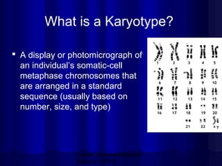
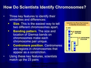
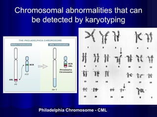
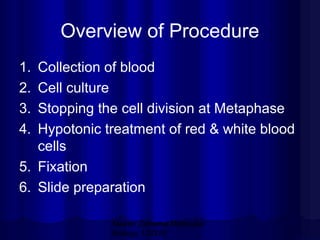
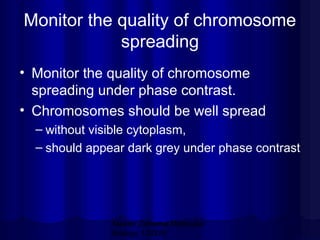
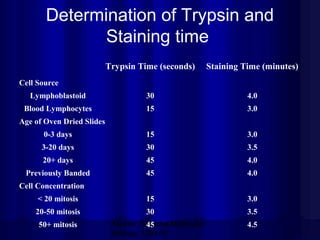
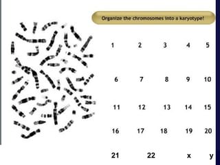
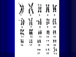
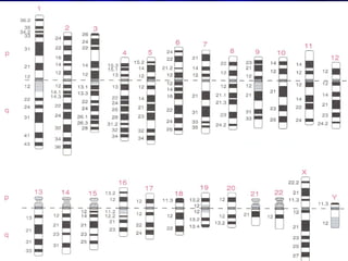
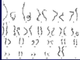
This document describes the process of karyotyping and identifying human chromosomes through G-banding. Key points include:
- Karyotyping involves preparing, staining, and observing human chromosomes to diagnose genetic disorders and identify chromosomal abnormalities.
- Humans normally have 23 pairs of chromosomes, including 22 autosomes and 1 sex chromosome. G-banding allows identification of chromosomes based on size, banding pattern, and centromere position.
- The karyotyping process involves culturing cells, arresting cell division, staining chromosomes, and analyzing spreads under a microscope to identify numerical or structural abnormalities. G-banding produces dark and light bands that make each chromosome pair unique.
- Common chrom
A cytological technique to detect the nature of adjacent chromosomal regions by using different staining technique assisted with some pre treatment of metaphase chromosomes prepared on the slides
Definition
Centromere Particular chromosome complement of an individual or a related group of individuals, as defined by the chromosome size, morphology, and number –Karyotype.
Karyotype
CLASSIFICATION OF CHROMOSOMES FORKARYOTYPING
Types of karyotype
Asymmetric Karyotype
• Show larger difference
between smaller and
larger chromosome in a
set.
• Have more acrocentric
chromosomes.
• Have relatively
advanced feature.
Symmetric Karyotype
Show lesser difference
between smaller and
larger chromosome in a
set.
• Have more metacentric
chromosomes.
• Have no relatively
advanced feature
Procedure of karyotyping
SPECIMENS USED
Types of banding
G-banding
R-banding
c-banding
Q-banding
T-banding
Karyotype Detects Various Chromosome Abnormalities
Aneuploidy
Deletions
Duplications
Translocations
Idiogram
Advantages of Karyotyping
Disadvantages:
Karyotyping is the process by which photographs of chromosomes are taken in order to determine the chromosome complement of an individual, including the number of chromosomes and any abnormalities.
The term is also used for the complete set of chromosomes in a species or in an individual organism and for a test that detects this complement or measures the number.
This presentation is about the chromose structure, it's banding & painting. It includes the physical structure of chromosome, then karyotype & idiogram. Different types of chromosome banding & painting in details. FISH & GISH.
Introduction to HUMAN CHROMOSOME ANALYSIS: Conventional Karyotyping Method (G...SABARI KRISHNAN B. B.
This text is a report made on behalf of the training session organised by Dr M. Jeevan Kumar, PhD., Research Assistant Professor, Department of Genetic Engineering, SRM Institute of Science and Technology, Kattankulathur. The report covers the laboratory practices involved in karyotyping (GTG Banding) human chromosome from whole blood, the explanation to each step of karyotyping, the details about the functions of each reagent, reagent preparation protocols, etc. Karyotyping was done using GenASIs BandView software. The text involves invaluable information about the landmarks of each chromosome in a much simplified and organised way and several symbols approved by the ISCN used in karyotyping routine. Wishing all the very best to the readers and young scientists, for whom this text will find worthful.
Chromosomes are bundles of tightly coiled DNA located within the nucleus of almost every cell in our body. A chromosome is a DNA molecule with part or all of the genetic material (genome) of an organism. Chromosomes are normally visible under a light microscope only when the cell is undergoing the metaphase of cell division. Before this happens, every chromosome is copied once (S phase), and the copy is joined to the original by a centromere, resulting in an X-shaped structure. The original chromosome and the copy are now called sister chromatids. During metaphase, when a chromosome is in its most condensed state, the X-shape structure is called a metaphase chromosome.
June 3, 2024 Anti-Semitism Letter Sent to MIT President Kornbluth and MIT Cor...Levi Shapiro
Letter from the Congress of the United States regarding Anti-Semitism sent June 3rd to MIT President Sally Kornbluth, MIT Corp Chair, Mark Gorenberg
Dear Dr. Kornbluth and Mr. Gorenberg,
The US House of Representatives is deeply concerned by ongoing and pervasive acts of antisemitic
harassment and intimidation at the Massachusetts Institute of Technology (MIT). Failing to act decisively to ensure a safe learning environment for all students would be a grave dereliction of your responsibilities as President of MIT and Chair of the MIT Corporation.
This Congress will not stand idly by and allow an environment hostile to Jewish students to persist. The House believes that your institution is in violation of Title VI of the Civil Rights Act, and the inability or
unwillingness to rectify this violation through action requires accountability.
Postsecondary education is a unique opportunity for students to learn and have their ideas and beliefs challenged. However, universities receiving hundreds of millions of federal funds annually have denied
students that opportunity and have been hijacked to become venues for the promotion of terrorism, antisemitic harassment and intimidation, unlawful encampments, and in some cases, assaults and riots.
The House of Representatives will not countenance the use of federal funds to indoctrinate students into hateful, antisemitic, anti-American supporters of terrorism. Investigations into campus antisemitism by the Committee on Education and the Workforce and the Committee on Ways and Means have been expanded into a Congress-wide probe across all relevant jurisdictions to address this national crisis. The undersigned Committees will conduct oversight into the use of federal funds at MIT and its learning environment under authorities granted to each Committee.
• The Committee on Education and the Workforce has been investigating your institution since December 7, 2023. The Committee has broad jurisdiction over postsecondary education, including its compliance with Title VI of the Civil Rights Act, campus safety concerns over disruptions to the learning environment, and the awarding of federal student aid under the Higher Education Act.
• The Committee on Oversight and Accountability is investigating the sources of funding and other support flowing to groups espousing pro-Hamas propaganda and engaged in antisemitic harassment and intimidation of students. The Committee on Oversight and Accountability is the principal oversight committee of the US House of Representatives and has broad authority to investigate “any matter” at “any time” under House Rule X.
• The Committee on Ways and Means has been investigating several universities since November 15, 2023, when the Committee held a hearing entitled From Ivory Towers to Dark Corners: Investigating the Nexus Between Antisemitism, Tax-Exempt Universities, and Terror Financing. The Committee followed the hearing with letters to those institutions on January 10, 202
Thinking of getting a dog? Be aware that breeds like Pit Bulls, Rottweilers, and German Shepherds can be loyal and dangerous. Proper training and socialization are crucial to preventing aggressive behaviors. Ensure safety by understanding their needs and always supervising interactions. Stay safe, and enjoy your furry friends!
Exploiting Artificial Intelligence for Empowering Researchers and Faculty, In...Dr. Vinod Kumar Kanvaria
Exploiting Artificial Intelligence for Empowering Researchers and Faculty,
International FDP on Fundamentals of Research in Social Sciences
at Integral University, Lucknow, 06.06.2024
By Dr. Vinod Kumar Kanvaria
Unit 8 - Information and Communication Technology (Paper I).pdfThiyagu K
This slides describes the basic concepts of ICT, basics of Email, Emerging Technology and Digital Initiatives in Education. This presentations aligns with the UGC Paper I syllabus.
Biological screening of herbal drugs: Introduction and Need for
Phyto-Pharmacological Screening, New Strategies for evaluating
Natural Products, In vitro evaluation techniques for Antioxidants, Antimicrobial and Anticancer drugs. In vivo evaluation techniques
for Anti-inflammatory, Antiulcer, Anticancer, Wound healing, Antidiabetic, Hepatoprotective, Cardio protective, Diuretics and
Antifertility, Toxicity studies as per OECD guidelines
2024.06.01 Introducing a competency framework for languag learning materials ...Sandy Millin
http://sandymillin.wordpress.com/iateflwebinar2024
Published classroom materials form the basis of syllabuses, drive teacher professional development, and have a potentially huge influence on learners, teachers and education systems. All teachers also create their own materials, whether a few sentences on a blackboard, a highly-structured fully-realised online course, or anything in between. Despite this, the knowledge and skills needed to create effective language learning materials are rarely part of teacher training, and are mostly learnt by trial and error.
Knowledge and skills frameworks, generally called competency frameworks, for ELT teachers, trainers and managers have existed for a few years now. However, until I created one for my MA dissertation, there wasn’t one drawing together what we need to know and do to be able to effectively produce language learning materials.
This webinar will introduce you to my framework, highlighting the key competencies I identified from my research. It will also show how anybody involved in language teaching (any language, not just English!), teacher training, managing schools or developing language learning materials can benefit from using the framework.
This slide is special for master students (MIBS & MIFB) in UUM. Also useful for readers who are interested in the topic of contemporary Islamic banking.
This presentation includes basic of PCOS their pathology and treatment and also Ayurveda correlation of PCOS and Ayurvedic line of treatment mentioned in classics.
How to Build a Module in Odoo 17 Using the Scaffold MethodCeline George
Odoo provides an option for creating a module by using a single line command. By using this command the user can make a whole structure of a module. It is very easy for a beginner to make a module. There is no need to make each file manually. This slide will show how to create a module using the scaffold method.
Read| The latest issue of The Challenger is here! We are thrilled to announce that our school paper has qualified for the NATIONAL SCHOOLS PRESS CONFERENCE (NSPC) 2024. Thank you for your unwavering support and trust. Dive into the stories that made us stand out!
2. Experiment Objectives
• Preparing, Staining and Observing Gbanding human chromosomes
• Develop an understanding of karyotyping
and the association of various
chromosomal abnormalities to diseases.
Mazen Zaharna Molecular
Biology 1/2009
3. Human Chromosomes
• A “normal” human carries 23 PAIRS of
chromosomes (1 set came from the
mother, 1 set came from the father)
– 22 of these sets are called autosomes (or
“self chromosomes”)
– 1 set are the sex chromosomes
• A female carries two X chromosomes (XX)
• A male carries an X chromosome and a Y
chromosome (XY)
Mazen Zaharna Molecular
Biology 1/2009
4. Why do scientists look at
chromosomes?
• Scientists can diagnose or predict genetic
disorders by looking at chromosomes.
• This kind of analysis is used in prenatal
testing and in diagnosing certain
disorders, such as
– Down syndrome,
– or in diagnosing a specific types of leukemia.
Mazen Zaharna Molecular
Biology 1/2009
5. Chromosome abnormalities
• Chromosome abnormalities can be
– numerical, as in the presence of
• extra
• or missing chromosomes,
– or structural as in translocations, inversions,
large scale deletions or duplications.
Mazen Zaharna Molecular
Biology 1/2009
6. Situations where analysis is
strongly recommended
Problems with early growth &
development
Fertility problems
Neoplasia
Pregnancy in older women
Mazen Zaharna Molecular
Biology 1/2009
7. What is a Karyotype?
A display or photomicrograph of
an individual’s somatic-cell
metaphase chromosomes that
are arranged in a standard
sequence (usually based on
number, size, and type)
Mazen Zaharna Molecular
Biology 1/2009
8. Performing a Karyotype
• The slides are scanned for metaphase spreads
and usually 10 to 30 cells are analyzed under
the microscope by a cytogeneticist.
• When a good spread (minimum number of
overlapping chromosomes) is found, a
photograph is taken or the analysis is done by a
computer.
• The chromosomes are arranged in a standard
presentation format of longest to shortest.
Mazen Zaharna Molecular
Biology 1/2009
9. How Do Scientists Identify Chromosomes?
•
•
Three key features to identify their
similarities and differences:
Size. This is the easiest way to tell
two different chromosomes apart.
Banding pattern. The size and
location of Giemsa bands on
chromosomes make each
chromosome pair unique.
Centromere position. Centromeres
are regions in chromosomes that
appear as a constriction.
Using these key features, scientists
match up the 23 pairs
Mazen Zaharna Molecular
Biology 1/2009
10. In metacentric chromosomes, the centromere lies near the
center of the chromosome.
Submetacentric & very Submetacentric chromosomes,
have a centromere that is off-center, so that one
chromosome arm is longer than the other.
In acrocentric chromosomes, the centromere resides very
near one end.
Mazen Zaharna Molecular
Biology 1/2009
11. Chromosome banding
• Chromosomes are stained with various
dyes enabling the chromosome segments
to be identified
• Most methods can distinguish 550 bands/
haploid set
• High resolution methods can distinguish
up to 850 bands/ haploid set that can
allow identification of small interstitial
deletions
Mazen Zaharna Molecular
Biology 1/2009
12. G-Banding
Dye gives chromosomes a striped appearance
because it stains the regions of DNA that are rich in
adenine (A) and thymine (T) base pairs.
Mazen Zaharna Molecular
Biology 1/2009
13. G-Banding
• Regions that stain as dark G bands
replicate late in S phase of the cell cycle
and contain more condensed chromatin,
• While light G bands generally replicate
early in S phase, and have less
condensed chromatin.
Mazen Zaharna Molecular
Biology 1/2009
14. Chromosome Groups
Group Chromosomes
Description
A
1–3
Largest; 1 and 3 are metacentric but 2 is submetacentric
B
4,5
Large; submetacentric with two arms very different in
size
C
6–12,X
Medium size; submetacentric
D
13–15
Medium size; acrocentric with satellites
E
16–18
Small; 16 is metacentric but 17 and 18 are
submetacentric
F
19,20
Small; metacentric
G
21,22,Y
Small; acrocentric, with satellites on 21 and 22 but not
on the Y
Autosomes are numbered from largest to smallest, except that chromosome 21 is
smaller than chromosome 22.
Mazen Zaharna Molecular
Biology 1/2009
15. Chromosomal Abnormalities
•
Alterations in chromosome number.
– Euploid - normal set (2n)
– Polyploidy – extra set of the entire genome.
•
(3n, 4n etc)
– Aneuploidy – the number of chromosomes is
not a multiple of the normal haploid number.
•
Monosomy
– one member of a chromosome pair is missing, (2n-1)
•
Trisomy
– one chromosome set consists of 3 copies of a
chromosome, (2n+1)
Mazen Zaharna Molecular
Biology 1/2009
17. Chromosomal abnormalities that can
be detected by karyotyping
Mazen Zaharna Molecular
Philadelphia Chromosome - CML
Biology 1/2009
18. Overview of Procedure
1.
2.
3.
4.
Collection of blood
Cell culture
Stopping the cell division at Metaphase
Hypotonic treatment of red & white blood
cells
5. Fixation
6. Slide preparation
Mazen Zaharna Molecular
Biology 1/2009
19. Overview of Procedure
7. Slide dehydration
8. Treatment with enzyme
9. Staining
Mazen Zaharna Molecular
Biology 1/2009
20. Monitor the quality of chromosome
spreading
• Monitor the quality of chromosome
spreading under phase contrast.
• Chromosomes should be well spread
– without visible cytoplasm,
– should appear dark grey under phase contrast
Mazen Zaharna Molecular
Biology 1/2009
21. 7- Slide dehydration
• Place fixed, dry slides on slide rack in 60 oC
oven
• Bake for 3 days
• Allow to cool before proceeding to the next
step
Mazen Zaharna Molecular
Biology 1/2009
22. 8- Treatment with enzyme
• Prepare 0.025% trypsin solution fresh, by
mixing 5 ml of 0.25% trypsin with 45 ml
Hank’s solution
• Immerse slide in 0.025 % trypsin for 10120 seconds
• Remove slide from trypsin and
immediately immerse in phosphate buffer
to stop trypsin action
Mazen Zaharna Molecular
Biology 1/2009
23. Determination of Trypsin and
Staining time
Trypsin Time (seconds)
Staining Time (minutes)
Lymphoblastoid
30
4.0
Blood Lymphocytes
15
3.0
0-3 days
15
3.0
3-20 days
30
3.5
20+ days
45
4.0
Previously Banded
45
4.0
< 20 mitosis
15
3.0
20-50 mitosis
30
3.5
Cell Source
Age of Oven Dried Slides
Cell Concentration
50+ mitosis
Mazen Zaharna Molecular
45
Biology 1/2009
4.5
24. 9- Staining
• Prepare a dilution of Giemsa stain by
mixing 1 part of Giemsa stain with 3 parts
of Phosphate buffer
• Flood slide with Giemsa stain for 2
minutes
• Rinse slides thoroughly with distilled water
• Allow slides to drain, then place on 60 oC
slide warming tray until completely dry
Mazen Zaharna Molecular
Biology 1/2009
Chorionic villus sampling (CVS) is a form of prenatal diagnosis to determine chromosomal or genetic disorders in the fetus. It entails getting a sample of the chorionic villus ( placental tissue) and testing it. The advantage of CVS is that it can be carried out 10-13 weeks after the last period, earlier than amniocentesis (which is carried out at 15-18 weeks).