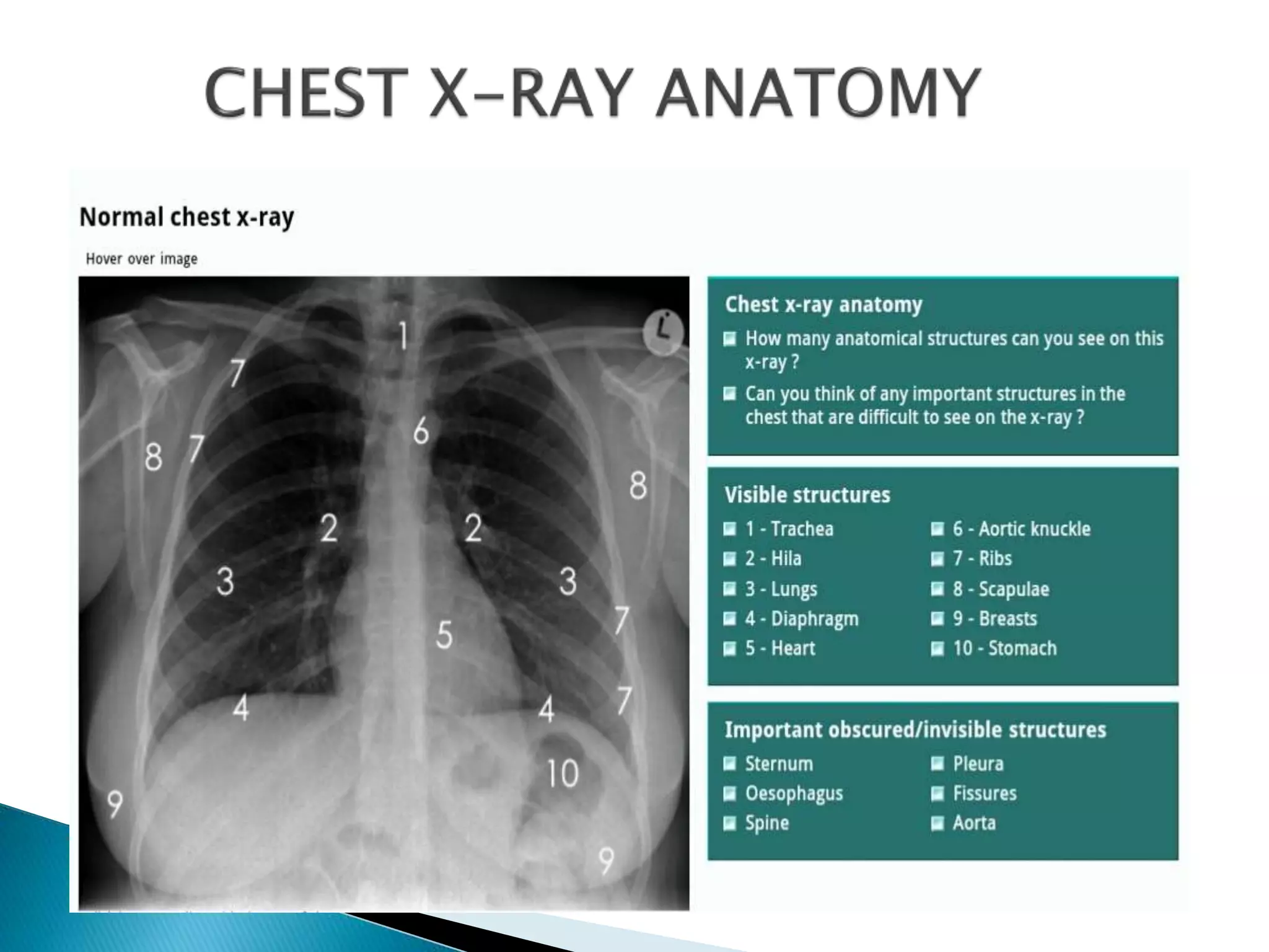
Chest X-ray is a commonly used and inexpensive imaging test. The document provides guidance on evaluating various aspects of a chest X-ray such as quality, positioning, inspiration, and penetration. It describes how to analyze key anatomical structures including the lungs, heart, trachea, diaphragm, bones and soft tissues. Any abnormalities should be described in detail and the rest of the image checked after identifying the most prominent abnormality.


![Radiological_diagnosis_of_TB_ECHO_MOH[1].pptx](https://cdn.slidesharecdn.com/ss_thumbnails/radiologicaldiagnosisoftbechomoh1-240905083452-eb26e5f9-thumbnail.jpg?width=640&height=640&fit=bounds)


