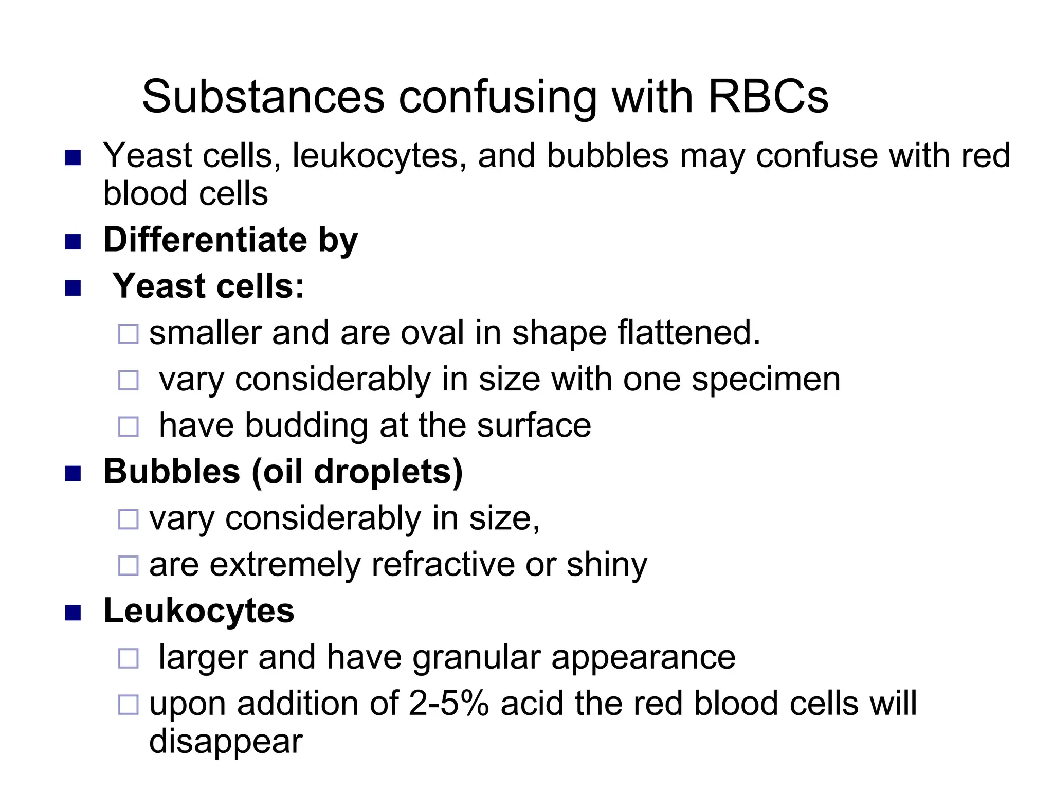
CHAPTER FIVE
Microscopic Examination Of
Urine
Chapter Objective
At the end of this chapter the students will be able to
describe
 Microscopic examination for urine sediment
 Normal and abnormal organized urine sediments with
their diagnostic features.
 Formation and significance of casts
 Normal and abnormal crystals encounter in urine
sediments
 Relationship between sediments, chemical, physical
findings in urine
 Reporting of urinary sediments
Chapter Outline
5. Microscopic Examination Of Urine
5.1. Procedure for microscopic examination
5.2. Source of errors in the microscopic examination of urine
5.3 Urinary Sediments
5.4 Organized Urinary Sediments
5.5 Parasite, fungus and bacteria in urine
5.6 Non-Organized Urinary Sediments Urine Crystals
5.7 Body cells, crystals, casts, yeasts, bacteria, sperm
5.8 Methods of reporting formed elements
Introduction
 In examining urinary sediment it is necessary to learn the
distinguishing characteristics of elements which have
significance .
 If the urine sample is properly collected, the test is
carefully performed, and the person performing the test
is knowledgeable, skillful valuable information can be
obtained from microscopic examination of urine
specimen.
Standardization
 Aspects of microscopic examination that
should be standardizes:
1. Volume of urine analyzed
2. Length and force of centrifugal
3. Re-suspending volume and concentration of
sediment
4. Volume and amount of sediment examined
5. Terminology and reporting format
Procedure for Microscopic Examination
Assemble all necessary materials used for the collection,
centrifugation and examination.
 Conical centrifuge tubes, or regular test tubes up to
15 ml.
 centrifuge.
 Pasture pipette with .
 Slides and cover slides 20 x 20 mm.
 microscope
Procedure for Microscopic Examination
cont’d…
1. Mix the urine specimen
2. Transfer about 10 ml of urine into a labelled centrifuge
tube.
3. Centrifuge the specimen at a medium speed (from 1500
– 2000 rpm) for 3-5 minutes
4. Discard the supernatant by quick inversion of the tube
5. Re suspend the sediment that is at the bottom of the
tube, by tapping the tube by your fingers
6. Take the sediment by Pasteur pipette from the tube and
transfer a drop into the clean and dry slide.
Procedure for Microscopic Examination
 Apply cover slide on the urine sediment that is on the
slide.
 Put on the microscope and look under 10x objective of
the microscope.
 Then after looking through the low power objective,
change the objective in to 40x objective .
 Then report what you get under low power and high
power objective on the laboratory request form of the
patient.
Source of Errors
 Drying of the specimen on the slide.
 If the supernatant fluid after centrifugation is not poured
off properly, it may decrease concentration of urine
sediments and false result may be reported
 If the whole sediment with supernatant is discarded during
inverting down the tube for long period, the whole
sediments will be discarded and so again false negative
result will be reported.
 Thus another sample should be collected and the test
repeated.
Classification of Urinary Sediments
 Urine sediments can grossly be categorized into
organized and non-organized sediments based
on the substances they are composed of.
Organized (Formed ) elements
 RBCs/HPF
 WBCs/HPF
 Epithelial cells / LPF
 Casts / LPF
 Parasites/LPF
 Bacteria / HPF
 Yeast Cells /`LPF
 Mucus trade/LPF
 Spermatozoa
 Miscellaneous substances
Non-organized (Non-living Material)
 acidic urine crystals
 Amorphous Urates,
 Uric acid crystals
 Calcium carbonate
 Calcium phosphate
 Acidic, Neutral, or slightly alkaline Urine crystal
 Calcium Oxalate crystals
 Alkaline, Neutral, or Slightly acidic urine
 Triple phosphates
Alkaline Urine Crystals
 Amorphous phosphate
 Calcium carbonate
 Calcium phosphate
Organized Urinary Sediments
 RED BLOOD CELLS
 Red blood cells are not usually present in normal urine.
 Appearance:
 Normally RBCs appear in the fresh sample as intact, small
and faint yellowish discs, darker at the edges
 Measure 7-8 m
 In concentrated urine may be crenated and became small
(5-6 m)
 In diluted urine, RBCs may be turgid and increase in size (9-
10 m)
 In alkaline urine, they may be small or entirely destroyed
forming massive of brownish granules
 In diluted and alkaline urine, the red cell will rupture and
release the hemoglobin, leaving faint colorless cell
membrane, and are known as “ ghost” cells (shadow of
original cells)
 This field contains mostly
RBC’s, (hp)
 Notice many of them
show biconcavity
 Some show variability in
shape
 Some times it is easy to
mistake fungi for RBC’s
 To get rid of RBC’s so
that WBC’s are more
visible – acetic acid is
very helpful, Why?
Microscopic Exam
 Red blood cells
 presence of a few is
normal
 higher numbers are
indicator of renal
disease
 result of bleeding at
any point in urinary
system
40x objective
Clinical significance
 When the number of RBCs is found more than their
normal range, usually greater than 5 RBCs/HPF it may
indicate:
 Presence of disease conditions in the urinary tract, such
as:
 Acute and chronic glomerulonephritis
 Tumor that erode any part of the urinary tract
 Renal stone
 Cystitis
 Prostates
 Trauma of the kidney
 traumatic catheterization
Substances confusing with RBCs
 Yeast cells, leukocytes, and bubbles may confuse with red
blood cells
 Differentiate by
 Yeast cells:
 smaller and are oval in shape flattened.
 vary considerably in size with one specimen
 have budding at the surface
 Bubbles (oil droplets)
 vary considerably in size,
 are extremely refractive or shiny
 Leukocytes
 larger and have granular appearance
 upon addition of 2-5% acid the red blood cells will
disappear
 These can easily be
mistaken for RBC’s –
 They are budding yeast,
notice the almost cactus
like appearance of those
in the box
 They will not rupture in
acetic acid, RBC’s will
 These may truly be from
the bladder or they may
be a contamination
 This structure, (hp)
marked by the arrow,
could be mistaken
for a RBC
 See the next slide
 One of the options in
identifying this
structure is to use
polarizing microscopy
 In this case, the
maltese shaped
cross indicates that
this structure is an oil
droplet
Interfering factors:
 Factors that may result falsely in high number of RBCs, i.e.
without the presence of actual renal or other normal
physiological disturbances included:
 Menstrual bleeding
 Vaginal bleeding
 Trauma to peranal area in female patients
 Following traumatic cateterization
 Some drugs:
 Aspirin ingestion or over dose
 Anticoagulant therapy over dose
LEUKOCYTES (WBCs)
 Normal range: 0-4 WBC/HPF.
 Appearance: normally, clear granular disc shaped,
 Measure 10-15 m, the nuclei may be visible.
 In alkaline urine, they may increase their size and become
irregular.
 Predominantly, polymorph nuclear neutrophils are seen.
 Sometimes because of predominance of neutrophils and
the occurrence of bacterial cell together with
polymorphonuclear cells, WBCs are called pus cells.
 WBCs (pus cells) may be seen in clumps.
Microscopic Exam
 White blood cells
 a few are normal
 high numbers indicate
inflammation or infection
somewhere along the
urinary or genital tract
40x objective
How to report the result of WBCs
 After observing the distribution of leukocytes under 40x
objective, at least 10 fields of microscope
 When 0-5 leukocytes / HPF are seen-- normal
 5-10 leukocytes / HPF are seen-- few leukocytes /
HPF
 10-20 leukocytes/HPF are seen--->moderate
leukocytes/ HPF
 20-30 leukocytes /HPF are seen ----> many leukocytes /
HPF
 Above 30 leukocytes / HPF / are seen - full/field
Clinical significance
 Increased number of leukocyte urine are seen in case of:
 Urinary tract infection such as renal tuberculosis
 All renal disease
 Bladder tumor
 Cystitis
 Prostates
 Temporarily increased number of leukocytes are also seen
during:
 Fever
 After strenuous exercise
EPITHELIAL CELLS
 Those coming from renal cells:
 Size is small as compared to other epithelial cells
 It measures 10 to 18 m in length, i.e., slightly larger
than leukocytes
 Very granular
 Have refractive and clearly visible nucleus
 Usually seen in association with proteins or casts .
EPITHELIAL CELLS (cont…)
 Cells from pelvis and urethra of the kidney
 Size is larger than renal epithelia’s
 Those from pelvis area are granular with sort of tail,
while those from urethra are oval in shape
 Most of the time urethral epithelia is seen with together
of leukocytes and filaments (mucus trades and large in
number)
 Pelvic epithelia’s seen usually with no leukocyte and
mucus trade, and are few in number
EPITHELIAL CELLS (cont..)
 Bladder cells
 Are squamous epithelial cells
 Very large in size.
 Shape seems rectangular and often with irregular
border.
 Have single nucleus.
 These are 2 Squamous
Epithelial cells shown on
high power (hp)
 They are usually large, flat,
colorless cells
 However, there can be
some granularity to the
cytoplasm & the edges
may be rolled
 The nucleus is usually
distinct & centered
Microscopic Exam
 Epithelial cells
 cells are large and
flat
 normal cells that line
the urinary and
genital tract or renal
tubules
 These Epithelial cells, hp,
are shown with phase
microscopy
 Notice how much sharper
the details are on an
unstained cell with a low
refractive index
 The edge in the rectangle
appears rolled which
suggests a vaginal origin
 The details of these
Transitional Epithelial cells
(3) are somewhat obscured by
the large number of bacteria
present
 Originate in proximal 2/3’s of
urethra, the bladder, ureters,
calices & pelvis of the kidney
 They are usually round with a
large round nucleus
 Notice they are smaller than
the Squamous Epithelial cell
 While these could be
Renal Tubular cells, hp,
they more likely are
Transitional cells
 The key here is to notice
that they do not have the
appearance of Squamous
Epithelial & are too large
to be WBC’s
 In a wet prep you could
compare them to other
cells to help identify
 The cell in the box has
the tail associated with
Transitional cells
 Notice this cell is much
larger than a WBC – it is
almost ½ as big as the
rolled Squamous
Epithelial
 Transitional cells line the
tract from the pelvis to the
upper portions of the
urethra
Clinical significance
 Presence of epithelial cells in large number, mostly renal
types may indicate:
 Acute tubular damage
 Acute glomerulonephritis
 Silicate over dose
Note: The presence of large number of epithelial cells with
large number of Leukocytes and mucus trades
(filaments) may indicate Urinary Tract Infections (UTI).
Reporting of epithelial cells
 Epithelial cells distribution reported after looking under 10x
objective of the microscope.
 Usually they are reported semi quantitatively by saying
 1-3 epithelial cells /LPF
 2-4 epithelial / LPF
 6-14 epithelial / LPF
 15-25 epithelial/ LPF
 Full of epithelial cells / LPF when the whole field of 10 x
objective covered by epithelial cells.
Casts
 Introduction:
 Casts are long cylindrical structures that result from the
solidification of material within the lumen of the kidney
tubules.
 Formed by precipitation of proteins, and aggregation of cells
within the renal tubules.
 Most of them dissociate in alkaline urine, and diluted urine
(specific gravity  1.010) even in the presence of
Proteinuria.
 Most of them are transparent.
 Pathological Conditions that favors for the creation of casts
include
 The presence of protein constituents in the tubular urine
 Increase acidification
 increase osmolar concentration
 Most urinary casts are formed either in the distal
convoluted tubules or in the collecting ducts, because
urine more concentrated and maximally acidified here.
 But rare conditions such as ,in myeloma, casts may be
found in the proximal convoluted tubules.
 Casts formed in the collecting tubules tends to be very
broad, and usually indicates the significant reduction in
the functional capacity of the nephron and indicate
severe renal damage.
Casts cont’d…
CASTS (cont…)
 Major casts types:
 hyaline
 epithelial
 white blood cell, and. red blood cell casts
 granular (coarse and fine). waxes, Fatty
Casts in Urinary Sediment
 Casts in urinary sediment is an important aid in the
differential diagnosis of renal disease
 Pure Hyaline casts may be seen in Proteinuria from a
variety of causes.
 Small Hyaline cast seen transiently may occur with
marked exercise or febrile conditions
 Casts with inclusions, such as RBC’s or WBC’s may be
formed without a protein matrix
Casts in Urinary
Sediment
Hyaline Casts
 All hyaline cast have a precipitated protein matrix, so
there has to be renal Proteinuria for these to be
formed
 The Proteinuria is predominately Tamm-Horsfall
mucoprotein, the later being secreted by cells lining
the distal parts of the nephron
 Low pH & increased electrolyte concentration
readily precipitate Tamm-Horsfall protein
 Hyaline cast,
high power
 These tend to be
colorless &
almost invisible
unless the
microscope is
optimally
focused
 Hyaline casts,
high power
using phase
microscopy
 Same set of
casts as in
previous
frame, made
more visible by
use of phase
 Hyaline cast at
same
magnification as
before
 Using an
interference filter
 Can you find the
pieces of the
cast?
Clinical Implication
 Presence of large number of hyaline casts may show
possible damage of glomerular capillary membrane.
This damage permits leakage of protein through
glomerulus and result in precipitate and gel formation
(i.e. hyaline casts) in the tubule. Thus this may indicate:
 Nephritis
 Meningitis
 Chronic renal disease
 Congenital heart failure
 Diabetic nephropathy
Granular cast
 More similar in appearance with hyaline casts and in
which homogenous, course granules are seen.
 More dense (opaque) than hyaline cast.
 Shorter and broader than hyaline casts.
 May represent the first stage of epithelial cell cast
degeneration.
 Based on the amount and type of granules, divided into:
 fine (which may appear grey or pale yellow in color)
 course granular casts ( which may appear as darker).
Granular Casts
 If cellular casts stay within the tubules of the
nephron due to prolonged stasis, the cells will
begin to degenerate
 These casts are referred to as Finely Granular
or Coarsely Granular Casts
 Granular cast
under brightlight
microscopy
 If this were on you
scope you would
want to reduce the
amount of light by
closing the
substage
condenser, this
really shows the
importance of your
lighting!
 Same Granular
cast as in
previous frame,
with phase
microscopy
 Notice the
increased detail
missed due to the
poor lighting in the
previous frame!
 Should this cast
be classified as
broad,
convoluted, or
narrow?
 This is using
interference filter
& is a frame of
the same
convoluted fine
granular cast
[granules from
degenerated
cells as in frames
6 & 7
 Notice the
beautiful RBC
 Mixed Cellular
Granular Cast,
high power
 Notice that the
cells are
degenerating
 This would
tend to be a
Course
Granular
 This is the same
cast as in frame
# 18
 It is with phase
microscopy
 This is the
same cast as
seen in frame
# 22 & 23
 Notice the
coarse
granularity is
very noticeable
 This is the
same cast as in
frame # 22,
using phase
 Notice the
coarse
granularity can
be seen
 Coarse
Granular
Cast next to
2 Epithelial
Cells, high
power
 The same cast
as in frame #
25, using
phase
Clinical significance
 Granular casts may be seen in:
 Acute tubular necrosis
 Advanced granulonephritis
 Pyelonephrites
 Malignant nephrosis
 Chronic lead poisoning
 In healthy individuals these casts may be seen
 after strenuous exercise
Cellular & Other Cast
 As the protein concentrates in the distal tubule &
becomes stickier, cells can become entrapped
 These become Hyaline Casts with Inclusions
& while the formal name would be for example
Hyaline-WBC Cast, they are frequently simply
referred to as WBC Cast
 Mixed Hyaline-
Cellular Cast,
high power
 Protein matrix is
clearly visible
 The cells are
probably WBC’s
& Renal Tubular
cells but is
difficult to be sure
 This is the same
frame as in #10
using phase
microscopy
 The material in
the black box is a
mucous thread,
notice it is dense
& has no light
center or cells in
its center
 This is the
same mixed
hyaline-
cellular cast
found in frame
# 10 & 11
 Interference
filter was used
White blood cell casts
 formed by aggregates of white blood cells that
trapped in protein matrix in the renal tubular
lumen.
 An excess of white blood cells, singly or in
clamps, in the urine may indicate inflammation.
 white blood cell casts definitely are renal origin.
 They characteristically seen in acute pyelonephrities and
occasionally in glomerulonephirites.
 WBC cast,
high power
 Some of the
nuclear lobes
can be seen
 The same
WBC cast as
in #13, using
phase
 Notice you
can clearly
see mucous
threads
around the
cast
 Same cast as
seen in # 13 &
14, using
interference filter
 Notice the
details of the
mucous threads
are more difficult
to see
Red blood cell casts
- Usually, they found in hematuria. Red blood cell casts
may appear broen to almost colorless and are usually
diagnostic of glomerular diseases.
- Normal range: normally not seen in normal individual
- Appearance
- Formed usually after accumulation of cellular element in
the renal tubules
 RBC cast, high
power
 Notice the cell
membranes are
clearly visible, but
there does not
appear to be a
nucleus
 Notice how
difficult the
mucous threads
are to see, this
might be improved
by reducing the
light a bit
 With the phase
microscopy it is
easier to see that
these are RBC’s in
the cast
 One even appears
to be biconcave
 The mucous
threads are also
easier to see
 Notice the large
nuclei can be
seen using an
interference filter
as it was with
the regular light
& with phase
Waxy Casts (Renal Failure Casts)
 Not seen in normal individual
 Shorter and broader than hyaline casts.
 Composed of homogeneous, yellowish materials.
 Broad waxy casts are from two to six times the width of
ordinary
 appear waxy and granular.
 Have high retractive index.
 May occur from cells (WBC, RBC, or Epithelial) casts,
hyaline casts.
 This is a Waxy
Cast, on high
power
 Notice the
crack in the
side of the
cast, which is
frequently seen
in Waxy Casts
 This is the
same Waxy
Cast under
phase
 The
‘thickened’
waxy exterior
is more easily
seen
 This is the
same cast as
seen in # 27 &
28, using an
interference
filter
 Notice again
the waxyness
of the
appearance
Waxy Casts
 Clinical significance
 Waxy casts are found in
 Chronic renal disease.
 Tubular inflammation and degeneration.
 Localized nephron obstruction.
 malignant hypertension
 in diabetic diseases
 * The presence of waxy casts indicates severity of renal
disease.
Fatty Casts
 -normally not seen in health individuals.
 These are casts, which contain fat droplets inside them.
 Fat droplets are formed after accumulation of fat in the
tubular vessels, especially tubular epithelial and finally
disintegrated.
 Clinical Implication:
 The occurrence of fat droplets, oval, fat bodies, or fat
casts is
 very important sign of nephritic syndrome.
 Chronic renal disease.
 Inflammation and degeneration of renal tubules.
 lupus and toxic renal poisoning
 This is a cast
containing ‘fat’
bodies, high
power
 On wet mount
the droplets are
highly refractile
[they bounce the
light back]
 This is the cast
containing ‘fat’
bodies under
polarizing light
 The ‘fat’ bodies
have a maltese
cross appearance
in polarized light,
which is one way
of confirming
 Confirmation can
also be by fat red
stain
 This is the
cast
containing
‘fat’ bodies
under phase
 This is the fatty
cast under
interference filter
Epithelial Casts
 Epithelial Casts are composed largely of tubular
epithelial cell desquamated within the tubule.
 They often appear as two rows if cells in protein cast
matrix.
 Inflammation of the kidney may cause greater sloughing
of renal epithelial cells, so large number of epithelial
casts is indicative of renal parenchymal disease with
tubular damage.
 Renal Tubular Cast,
high power
 Can be difficult to
differentiate from
other cellular casts
& at times the
decision must be
made on other cells
in the sample
 Notice in this cast
the cells have large
nuclei
Reporting of casts
 Casts are examined under 10x objective of the
microscope.
 Casts are reported quantitatively by saying:
 Few casts / LPF
 Moderate casts / LPF and
 Many casts / LPF
 During the report the, type of cast that is seen should
also be mentioned
 Example: few hyaline casts / LPF are seen
PARASITES
 Parasites that can be seen in urine microscopy are:
 Trichomonas vaginalis
 Schistosoma haematobium
 Wuchereria bancroftie
 * Other parasites such as Entrobious vermicularies
also may occur due to contamination of the urine with
stool.
 Here is another frame
of the Trichomonas
(hp), both of these are
shown with phase
microscopy which
enhances the details
of cells with low
refractive indices
 Trichomonas (hp) is a
small parasite that is very
active in a fresh
specimen
 They have multiple
flagella (white pointers)
as well as an undulating
membrane which
contribute to movement
 In the absence of
movement they can be
mistaken for other cells
 There are 2 sperm in
this frame
 In a fresh specimen,
they are visible due to
their movement
 In an older urine
specimen, they may
be difficult to visualize
 This frame contains 2
sperm also, with
phase microscopy (lp)
 The phase makes
both the head & tail
more visible
 Notice also the WBC
and the mucous
thread
 This structure, (hp)
marked by the arrow,
could be mistaken
for a RBC
 See the next slide
 One of the options in
identifying this
structure is to use
polarizing microscopy
 In this case, the
maltese shaped
cross indicates that
this structure is an oil
droplet
Schistosoma haematobium Egg
Enterobius vermicularis Eggs
YEAST CELL
 Yeast cells are fungi that are not normally seen in
health individuals.
 Appearance
 Variable in size
 Colorless.
 Oval in shape, and usually form budding.
 Have high refractive index.
 Usually confused with Red Blood Cells.
 These can easily be
mistaken for RBC’s –
 They are budding yeast,
notice the almost cactus
like appearance of those
in the box
 They will not rupture in
acetic acid, RBC’s will
 These may truly be from
the bladder or they may
be a contamination
 These are branching
pseudohyphae of a
fungus
 Their main
significance is that
they obscure more
important features of
the specimen & may
indicate that the
specimen is not a
clean catch
Clinical Significance
 They are usually of candida species (candida albicans)
and are common in patients with
 Urinary tract infection
 Vaginites
 Diabetic mellitus
 Intensive antibiotic or immunosuppressive therapy
BACTERIA
 Bacteria are commonly found in urine specimen because
of abundant normal microbial flora of the vagina or
external urinary meatus.
 Most common cause of UTI dipstick test can give indirect
clue.
 Further the observed bacterial cell can be identified by
bacteriological culture
Clinical Significance
 Presence of bacteria may indicate the presence of UTI
or contamination by genital or intestinal microflora.
 Report of the Result
 Few bacteria / HPF
 Moderate bacteria / HPF
 Many bacteria / HPF
 Full of bacteria / HPF
Crystals in Sediment
 Crystals
 precipitation of solutes
 are not normally present in freshly voided urine
 can precipitate on storage
 most are not clinically significant
 pH critical to differentiating some important
crystals
Contributing factors to Crystal
Formation
 Concentration of solute in specimen
 Decreased flow of urine through tubules
 This enhances precipitation of solutes
 Ultrafiltrate can become supersaturated in
tubules
Crystals Correlate With:
 pH of urine
 solutes differ in solubility
 inorganic salts: oxalate, phosphate, calcium,
ammonium & magnesium less soluble in neutral or
alkaline urine
 organic solutes: uric acid, bilirubin, & cystine less
soluble in acidic urine
Acidic Urine
Clinically significant crystal are found in acid urine
 Include: cystine, tyrosine, leucine & iatrogenic
crytsals: sulfonamide & ampicillin
Acidic Urine
 Amorphous Urates
 Non crystalline urate salts of sodium, potassium,
magnesium & calcium
 small & yellow-brown granules - - similar to sand
 enhanced by refrigeration
 can be in acidic or neutral urine
Amorphous Urates
 Will dissolve in alkaline or heated to 600C
 If add acetic acid, uric acid crystals will
precipitate out
 Uroerythrin deposits on urate crystals giving
pink-organish color -- referred to as “brick dust”
Uric Acid Crystals
 Urine pH usually around 5.0 to 5.5
 Most common form is diamond shape but may
be cube shaped or cluster in rosettes
Uric Acid Crystals
 Diamond shape may cluster in rosettes
 Sometimes 6 sided & must be differentiated from
clinically significant cystine
Uric Acid Crystals and
Pathology
 Usually yellow to orange-
brown
 Are birefringent under
polarizing light
 Can appear normally BUT
 See large #s in gout &
increased purine
metabolism such as
cytotoxic drugs
Acid Urine: Calcium Oxalate
Crystals
 Calcium oxalate
 Usually octahedral or
look like envelope
 Dihydrate form is 2
pyramids joined at the
basewhen
 Squares with lines
intersecting the center
can be seen on the end
Calcium Oxalate Crystals
 Monohydrate form - small ovoid or dumb bell
 rare & can mistake for RBC’s
 are birefringent under polarizing light
 are colorless & vary in size - usually small and may
be in either neutral or acidic urines
 Monohydrate form - small ovoid or dumb bell
 often see in normal urine, 2nd to ascorbic acid,
ingesting tomatoes, asparagus, spinach & oranges
Bilirubin
 Appear as fine needles, granules, or plates
 urine is acidic
 always yellow-brown
 the bile stains the other components of the sediment
 presence of the crystals indicate high concentrations
of bilirubin in the urine
Bilirubin Crystals: Abnormal
State
 If you suspect bilirubin crystals are present, the
strip reaction must confirm the presence of
bilirubin
 Otherwise the identification is incorrect
 The presence of the positive bilirubin strip &/or
the crystals indicate a pathologic process - are
always considered an abnormal crystal
 May see in liver disease
Cystine: Always Abnormal
 Colorless hexagonal
plates
 sides may be uneven
 Crystals appear layered
 tend to clump
 primarily seen in acidic urine
 Must be counted
 Can be confused with uric
acid crystals, must confirm
identification with sodium
cyanide
Cystine: Always Abnormal
 Clincally significant, seen in congenital
cystinosis or cystinuria
 Deposit out in tubules as calculi/stone causing
damage
Amino Acid Crystals
 Tyrosine
 fine, delicate needles,
colorless or yellow
 frequently in clusters or
sheaves [as in stacks of
wheat]
 see singly or in small groups
 in acidic urine
 less soluble than leucine, so
found more often
Leucine
 Highly refractile yellow to brown
spheres in acid urine.
 Have concentric/radial
striations on their surface
 Can be mistaken for fat
globules [or vice versa]
 But will not stain with fat stains
or appear as maltese cross
under polarization
 Can be seen in urine containing
tyrosine crystals if use alcohol
to ‘precipitate’
Bactrim has similar appearance
check patient history
Amino Acid Crystals and
Pathology
 Amino acid crystals are abnormal & seen in
overflow aminoaciduria
 can be seen in rare cases of liver disease, more likely
to reflect inherited metabolic disorder
 before reporting should be confirmed by confirmatory
tests such as chromatography
Cholesterol
 Clear flat rectangular plates with
notched corners
 in acidic urine
 are soluble in chloroform & in
ether
 Rarely seen
 Presence indicates both ideal
conditions for precipitation &
supersaturation:
 Always see with positive protein
+ fat droplets, fatty casts or oval
fat bodies
 Seen in nephrotic syndrome &
other renal damage
Confounding Conditions
 Diatrizoate meglumine [radiopaque contrast medium]
can be mistaken for cholesterol
 contrast medium will give abnormally high S.G. >1.040
 not associated with proteinuria or lipiduria
 cholesterol crystals found with normal S.G.
 Medications
 can be excreted in high concentrations, resulting in precipitation
 these crystals are termed ‘iatrogenic’
 proper identification of drug crystals important in alerting to
potential renal tubular damage
Ampicillin
 Appear as long thin
colorless prisms or
needles
 May aggregate in small
clusters or if refrigerated
may form large clusters
 Appear in acidic urine
 Require large dosage for
formation, so rarely seen
Sulfonamides
 Highly refractile & birefringent
 In acidic urine,
 Should be confirmed before
reporting
 Closely resemble ammonium
biurate but differentiated on
 pH & solubility
 chemical confirmatory test
 Type varies with form of drug
prescribed
 Sulfa drugs have been modified
to be more soluble & so crystals
rarely seen
Alkaline Urine Crystals
 Ammonium Phosphate
 alkaline or neutral urine
 microscopically not distinguishable from amorphous
urates
 distinguishable on urine pH & solubility
 precipitate white rather than pink-orange of amorphous
urates
 are soluble in acid & will not dissolve when heated to 60C
 fine colorless grains with tendency to obscure other
more significant sediment
 presence enhanced by refrigeration
Triple Phosphate
 Colorless & in different
forms
 most common are 3 & 6
sided ‘coffin lids’
 vary greatly in size
 may also see a ‘fern leaf’
form, feathery
 See in normal healthy
individuals but are often
present in formation of
calculi &
 are associated with UTI
Calcium Phosphate
 In 2 forms dicalcium & calcium
 Dicalcium colorless thin prisms in rosettes or
star-shaped ‘stellar phosphates’
 tend to have 1 tapered or pointed end & the other
squared off
 calcium phosphates are irregular granular sheets or
plates - - often resemble degenerating squamous
epithelial cells
Ammonium Biurate
 Yellow brown spheres
with striations
 Can have irregular
spicules ‘thorny apple’
 In alkaline or neutral urine
 Not significant unless
seen in fresh urine
 Usually seen in old
specimens
 Dissolve in acetic acid or
heating to 600C
Just forming
Calcium Carbonate
 Very small granular
crystals
 Can be misidentified
as bacteria
 Birefringent with
polarizing light
 Usually found in pairs
‘dumbbell shape’
Cystine Crystals
 Rarely found.
 Flat, hexagonal plates with well defined edges.
 Colorless, and highly refractile.
 Size is 30-60 m.
 Found only in fresh urine, because if there is delay,
they are soluble and not seen.
 Appeared during cystinosis, which is a hereditary
disease (Wilson disease), or during transient acute
phase of pyelonephritis. Its appearance in the urine is
called cystinuria.
Calcium Sulfate Crystals
 Have large prism or flat bladder shaped.
 Seen separately or in bundles.
 Size 50-100 m.
 Can be distinguished from calcium phosphate
crystals by measuring pH of urine.
MISCELLANEOUS
Spermatozoa
 Are small structures consisting of a head and
tail, connected by a short middle piece (neck).
 Easily recognized especially if they are motile.
 Frequently seen in the urine of males.
 They may see in the urine of females, when the
urine collected after coitus usually not reported,
unless the physician has special interest in it.
 There are 2 sperm in
this frame
 In a fresh specimen,
they are visible due to
their movement
 In an older urine
specimen, they may
be difficult to visualize
 This frame contains 2
sperm also, with
phase microscopy (lp)
 The phase makes
both the head & tail
more visible
 Notice also the WBC
and the mucous
thread
Mucus Trades
 Formed by the precipitation of mucoprotein in cooled
urine.
 Have fine, fiber like appearance.
 Wavy in shape and tapered at ends.
 If not examined carefully may confuse with hyaline casts.
Contaminates and Artifact Structure
 Muscle fibers
 Vegetable cells
 Structure from slide or cover slide
 Fat droplets (other bubbles)
 Oil droplets
 Pollen greens
 Starch granules
 This structure, (hp)
marked by the arrow,
could be mistaken
for a RBC
 See the next slide
 One of the options in
identifying this
structure is to use
polarizing microscopy
 In this case, the
maltese shaped
cross indicates that
this structure is an oil
droplet
Methods for Examining Urine Sediments
(1)Unstained Urine Sediment
 Bright field microscopy of the unstained urine
sediment
 Phase Contrasts (PC)
Stained Preparation
(a) A crystal violet safranin stain (sternheimer and malbin)
is useful in the identification of cellular elements.
Staining reaction to crystal – violet safranin stain:
 RBC – Purple to dark purple.
 WBC – Cytoplasm -violet to blue.
 Nucleus – reddish purple.
 Glitter cells – blue .
Stained Preparation (cont…)
(b) Methyl blue (Loeffler's stain)
(c) CytoDiachrome stains
Automations in Urinalysis
 automations are utilized in urinalysis laboratories.
 These machines can be applied for physical, chemical,
and microscopical analysis of urine
Automations in Urinalysis (cont….)
 advantages of automations:
 the readings are more reproducible and unbiased
 help to analyze a great number of specimen in less
time
 help to develop standards about the sediments and
give better interpretation about the sediments in close
agreement between laboratories
The next chapter will
deal concerning quality
control of urine
What is the importance of keeping
quality control for urinalysis
Quality control in urinalysis.
 Quality assurance is a set of activates starting
from specimen collection to issuing test results
that ensure test results are accurate and precise
as possible.
 It is the sum of all the activates of the laboratory
that ensures test results are of good quality.
Quality control in urinalysis cont’d…
 Quality assurance includes
 inside and outside the laboratory performance
standards
 good laboratory practice and management skills that
are required by achieving and maintaining a quality
service and that provide for continuing improvement
Quality control in urinalysis (cont…)
 part of quality assurance, which primarily concern the
control of errors in the performance of tests and
verification of test results.
 must be practical, achievable, affordable, and above all
continuous.
 The purpose of quality control procedure is to monitor
analytical processes, analytical error and to correct result
of analysis.
two types of quality control programs
A) internal quality control
 Is carried out in the laboratory, an intra-lab program.
 encompasses all measurements made, technical skills
performed within an individual laboratory.
 use control samples, like pooled serum
 The purpose of quality control program is to insure
tests are performed reliably and reported correctly.
 An effective quality control system detect errors at an
early stage, before they lead to incorrect test results.
 B) external quality control.
 External quality control is observation of variance in
results when the same material is analyzed in different
laboratories
Cont…..
B) external quality control.
 External quality control is observation of variance in
results when the same material is analyzed in
different laboratories
 Quality control steps:
 Pre analytical steps
 Analytical steps
 Post analytical steps
Pre analytical Quality control in
urinalysis
 read and understand requested paper
 guide the patient to bring an appropriate urine sample
 labeling the urine container after collecting the sample
 cheek the material we are going to use whether they are
properly cleaned or not
 ask the patient whether the urine sample has been long
time ,more than two hours, after it is voided.
 do not accept contaminated requested paper
 cheek the slide, the microscope, and all needed material
before taking the next procedure.
 if the urine comes from far place ask or read the
preservative applied
Cot…
 concentrate and find out an abnormalities that is also
related from chemical and physical apperance.
 proper sample preparation is also most important.
 reduce possible source of errors
 do not open the centrifuge while it is not stopped
 proper balance of urine in the centrifuge
analytical Quality control in urinalysis
 Small urine sample how to be rejected
 follow exactly standard operation procedure (SOP)
 Check and read reagent strip chemical test according to
the instruction of the manual of the manufacturer, at the
right time
 write the physical appearances properly
 use the needed amount of urine for centrifugation
 when discarding the supernatant, it has to be quick and
vertical up side down in order not to loss the sediment
 examine as quickly as possible
Post analytical Quality control in
urinalysis
 improper written result
 incorrect calculation
 missing of requesting paper

Chapter 5. Microscopic examination of urine ppt

  • 1.
  • 2.
    Chapter Objective At theend of this chapter the students will be able to describe  Microscopic examination for urine sediment  Normal and abnormal organized urine sediments with their diagnostic features.  Formation and significance of casts  Normal and abnormal crystals encounter in urine sediments  Relationship between sediments, chemical, physical findings in urine  Reporting of urinary sediments
  • 3.
    Chapter Outline 5. MicroscopicExamination Of Urine 5.1. Procedure for microscopic examination 5.2. Source of errors in the microscopic examination of urine 5.3 Urinary Sediments 5.4 Organized Urinary Sediments 5.5 Parasite, fungus and bacteria in urine 5.6 Non-Organized Urinary Sediments Urine Crystals 5.7 Body cells, crystals, casts, yeasts, bacteria, sperm 5.8 Methods of reporting formed elements
  • 4.
    Introduction  In examiningurinary sediment it is necessary to learn the distinguishing characteristics of elements which have significance .  If the urine sample is properly collected, the test is carefully performed, and the person performing the test is knowledgeable, skillful valuable information can be obtained from microscopic examination of urine specimen.
  • 5.
    Standardization  Aspects ofmicroscopic examination that should be standardizes: 1. Volume of urine analyzed 2. Length and force of centrifugal 3. Re-suspending volume and concentration of sediment 4. Volume and amount of sediment examined 5. Terminology and reporting format
  • 6.
    Procedure for MicroscopicExamination Assemble all necessary materials used for the collection, centrifugation and examination.  Conical centrifuge tubes, or regular test tubes up to 15 ml.  centrifuge.  Pasture pipette with .  Slides and cover slides 20 x 20 mm.  microscope
  • 7.
    Procedure for MicroscopicExamination cont’d… 1. Mix the urine specimen 2. Transfer about 10 ml of urine into a labelled centrifuge tube. 3. Centrifuge the specimen at a medium speed (from 1500 – 2000 rpm) for 3-5 minutes 4. Discard the supernatant by quick inversion of the tube 5. Re suspend the sediment that is at the bottom of the tube, by tapping the tube by your fingers 6. Take the sediment by Pasteur pipette from the tube and transfer a drop into the clean and dry slide.
  • 8.
    Procedure for MicroscopicExamination  Apply cover slide on the urine sediment that is on the slide.  Put on the microscope and look under 10x objective of the microscope.  Then after looking through the low power objective, change the objective in to 40x objective .  Then report what you get under low power and high power objective on the laboratory request form of the patient.
  • 9.
    Source of Errors Drying of the specimen on the slide.  If the supernatant fluid after centrifugation is not poured off properly, it may decrease concentration of urine sediments and false result may be reported  If the whole sediment with supernatant is discarded during inverting down the tube for long period, the whole sediments will be discarded and so again false negative result will be reported.  Thus another sample should be collected and the test repeated.
  • 10.
    Classification of UrinarySediments  Urine sediments can grossly be categorized into organized and non-organized sediments based on the substances they are composed of.
  • 11.
    Organized (Formed )elements  RBCs/HPF  WBCs/HPF  Epithelial cells / LPF  Casts / LPF  Parasites/LPF  Bacteria / HPF  Yeast Cells /`LPF  Mucus trade/LPF  Spermatozoa  Miscellaneous substances
  • 12.
    Non-organized (Non-living Material) acidic urine crystals  Amorphous Urates,  Uric acid crystals  Calcium carbonate  Calcium phosphate  Acidic, Neutral, or slightly alkaline Urine crystal  Calcium Oxalate crystals  Alkaline, Neutral, or Slightly acidic urine  Triple phosphates Alkaline Urine Crystals  Amorphous phosphate  Calcium carbonate  Calcium phosphate
  • 13.
    Organized Urinary Sediments RED BLOOD CELLS  Red blood cells are not usually present in normal urine.  Appearance:  Normally RBCs appear in the fresh sample as intact, small and faint yellowish discs, darker at the edges  Measure 7-8 m  In concentrated urine may be crenated and became small (5-6 m)  In diluted urine, RBCs may be turgid and increase in size (9- 10 m)  In alkaline urine, they may be small or entirely destroyed forming massive of brownish granules  In diluted and alkaline urine, the red cell will rupture and release the hemoglobin, leaving faint colorless cell membrane, and are known as “ ghost” cells (shadow of original cells)
  • 14.
     This fieldcontains mostly RBC’s, (hp)  Notice many of them show biconcavity  Some show variability in shape  Some times it is easy to mistake fungi for RBC’s  To get rid of RBC’s so that WBC’s are more visible – acetic acid is very helpful, Why?
  • 15.
    Microscopic Exam  Redblood cells  presence of a few is normal  higher numbers are indicator of renal disease  result of bleeding at any point in urinary system 40x objective
  • 16.
    Clinical significance  Whenthe number of RBCs is found more than their normal range, usually greater than 5 RBCs/HPF it may indicate:  Presence of disease conditions in the urinary tract, such as:  Acute and chronic glomerulonephritis  Tumor that erode any part of the urinary tract  Renal stone  Cystitis  Prostates  Trauma of the kidney  traumatic catheterization
  • 17.
    Substances confusing withRBCs  Yeast cells, leukocytes, and bubbles may confuse with red blood cells  Differentiate by  Yeast cells:  smaller and are oval in shape flattened.  vary considerably in size with one specimen  have budding at the surface  Bubbles (oil droplets)  vary considerably in size,  are extremely refractive or shiny  Leukocytes  larger and have granular appearance  upon addition of 2-5% acid the red blood cells will disappear
  • 18.
     These caneasily be mistaken for RBC’s –  They are budding yeast, notice the almost cactus like appearance of those in the box  They will not rupture in acetic acid, RBC’s will  These may truly be from the bladder or they may be a contamination
  • 19.
     This structure,(hp) marked by the arrow, could be mistaken for a RBC  See the next slide
  • 20.
     One ofthe options in identifying this structure is to use polarizing microscopy  In this case, the maltese shaped cross indicates that this structure is an oil droplet
  • 21.
    Interfering factors:  Factorsthat may result falsely in high number of RBCs, i.e. without the presence of actual renal or other normal physiological disturbances included:  Menstrual bleeding  Vaginal bleeding  Trauma to peranal area in female patients  Following traumatic cateterization  Some drugs:  Aspirin ingestion or over dose  Anticoagulant therapy over dose
  • 22.
    LEUKOCYTES (WBCs)  Normalrange: 0-4 WBC/HPF.  Appearance: normally, clear granular disc shaped,  Measure 10-15 m, the nuclei may be visible.  In alkaline urine, they may increase their size and become irregular.  Predominantly, polymorph nuclear neutrophils are seen.  Sometimes because of predominance of neutrophils and the occurrence of bacterial cell together with polymorphonuclear cells, WBCs are called pus cells.  WBCs (pus cells) may be seen in clumps.
  • 23.
    Microscopic Exam  Whiteblood cells  a few are normal  high numbers indicate inflammation or infection somewhere along the urinary or genital tract 40x objective
  • 24.
    How to reportthe result of WBCs  After observing the distribution of leukocytes under 40x objective, at least 10 fields of microscope  When 0-5 leukocytes / HPF are seen-- normal  5-10 leukocytes / HPF are seen-- few leukocytes / HPF  10-20 leukocytes/HPF are seen--->moderate leukocytes/ HPF  20-30 leukocytes /HPF are seen ----> many leukocytes / HPF  Above 30 leukocytes / HPF / are seen - full/field
  • 25.
    Clinical significance  Increasednumber of leukocyte urine are seen in case of:  Urinary tract infection such as renal tuberculosis  All renal disease  Bladder tumor  Cystitis  Prostates  Temporarily increased number of leukocytes are also seen during:  Fever  After strenuous exercise
  • 26.
    EPITHELIAL CELLS  Thosecoming from renal cells:  Size is small as compared to other epithelial cells  It measures 10 to 18 m in length, i.e., slightly larger than leukocytes  Very granular  Have refractive and clearly visible nucleus  Usually seen in association with proteins or casts .
  • 27.
    EPITHELIAL CELLS (cont…) Cells from pelvis and urethra of the kidney  Size is larger than renal epithelia’s  Those from pelvis area are granular with sort of tail, while those from urethra are oval in shape  Most of the time urethral epithelia is seen with together of leukocytes and filaments (mucus trades and large in number)  Pelvic epithelia’s seen usually with no leukocyte and mucus trade, and are few in number
  • 28.
    EPITHELIAL CELLS (cont..) Bladder cells  Are squamous epithelial cells  Very large in size.  Shape seems rectangular and often with irregular border.  Have single nucleus.
  • 29.
     These are2 Squamous Epithelial cells shown on high power (hp)  They are usually large, flat, colorless cells  However, there can be some granularity to the cytoplasm & the edges may be rolled  The nucleus is usually distinct & centered
  • 30.
    Microscopic Exam  Epithelialcells  cells are large and flat  normal cells that line the urinary and genital tract or renal tubules
  • 31.
     These Epithelialcells, hp, are shown with phase microscopy  Notice how much sharper the details are on an unstained cell with a low refractive index  The edge in the rectangle appears rolled which suggests a vaginal origin
  • 32.
     The detailsof these Transitional Epithelial cells (3) are somewhat obscured by the large number of bacteria present  Originate in proximal 2/3’s of urethra, the bladder, ureters, calices & pelvis of the kidney  They are usually round with a large round nucleus  Notice they are smaller than the Squamous Epithelial cell
  • 33.
     While thesecould be Renal Tubular cells, hp, they more likely are Transitional cells  The key here is to notice that they do not have the appearance of Squamous Epithelial & are too large to be WBC’s  In a wet prep you could compare them to other cells to help identify
  • 34.
     The cellin the box has the tail associated with Transitional cells  Notice this cell is much larger than a WBC – it is almost ½ as big as the rolled Squamous Epithelial  Transitional cells line the tract from the pelvis to the upper portions of the urethra
  • 35.
    Clinical significance  Presenceof epithelial cells in large number, mostly renal types may indicate:  Acute tubular damage  Acute glomerulonephritis  Silicate over dose Note: The presence of large number of epithelial cells with large number of Leukocytes and mucus trades (filaments) may indicate Urinary Tract Infections (UTI).
  • 36.
    Reporting of epithelialcells  Epithelial cells distribution reported after looking under 10x objective of the microscope.  Usually they are reported semi quantitatively by saying  1-3 epithelial cells /LPF  2-4 epithelial / LPF  6-14 epithelial / LPF  15-25 epithelial/ LPF  Full of epithelial cells / LPF when the whole field of 10 x objective covered by epithelial cells.
  • 37.
    Casts  Introduction:  Castsare long cylindrical structures that result from the solidification of material within the lumen of the kidney tubules.  Formed by precipitation of proteins, and aggregation of cells within the renal tubules.  Most of them dissociate in alkaline urine, and diluted urine (specific gravity  1.010) even in the presence of Proteinuria.  Most of them are transparent.  Pathological Conditions that favors for the creation of casts include  The presence of protein constituents in the tubular urine  Increase acidification  increase osmolar concentration
  • 38.
     Most urinarycasts are formed either in the distal convoluted tubules or in the collecting ducts, because urine more concentrated and maximally acidified here.  But rare conditions such as ,in myeloma, casts may be found in the proximal convoluted tubules.  Casts formed in the collecting tubules tends to be very broad, and usually indicates the significant reduction in the functional capacity of the nephron and indicate severe renal damage. Casts cont’d…
  • 39.
    CASTS (cont…)  Majorcasts types:  hyaline  epithelial  white blood cell, and. red blood cell casts  granular (coarse and fine). waxes, Fatty
  • 40.
    Casts in UrinarySediment  Casts in urinary sediment is an important aid in the differential diagnosis of renal disease  Pure Hyaline casts may be seen in Proteinuria from a variety of causes.  Small Hyaline cast seen transiently may occur with marked exercise or febrile conditions  Casts with inclusions, such as RBC’s or WBC’s may be formed without a protein matrix
  • 41.
  • 42.
    Hyaline Casts  Allhyaline cast have a precipitated protein matrix, so there has to be renal Proteinuria for these to be formed  The Proteinuria is predominately Tamm-Horsfall mucoprotein, the later being secreted by cells lining the distal parts of the nephron  Low pH & increased electrolyte concentration readily precipitate Tamm-Horsfall protein
  • 43.
     Hyaline cast, highpower  These tend to be colorless & almost invisible unless the microscope is optimally focused
  • 44.
     Hyaline casts, highpower using phase microscopy  Same set of casts as in previous frame, made more visible by use of phase
  • 45.
     Hyaline castat same magnification as before  Using an interference filter  Can you find the pieces of the cast?
  • 46.
    Clinical Implication  Presenceof large number of hyaline casts may show possible damage of glomerular capillary membrane. This damage permits leakage of protein through glomerulus and result in precipitate and gel formation (i.e. hyaline casts) in the tubule. Thus this may indicate:  Nephritis  Meningitis  Chronic renal disease  Congenital heart failure  Diabetic nephropathy
  • 47.
    Granular cast  Moresimilar in appearance with hyaline casts and in which homogenous, course granules are seen.  More dense (opaque) than hyaline cast.  Shorter and broader than hyaline casts.  May represent the first stage of epithelial cell cast degeneration.  Based on the amount and type of granules, divided into:  fine (which may appear grey or pale yellow in color)  course granular casts ( which may appear as darker).
  • 48.
    Granular Casts  Ifcellular casts stay within the tubules of the nephron due to prolonged stasis, the cells will begin to degenerate  These casts are referred to as Finely Granular or Coarsely Granular Casts
  • 49.
     Granular cast underbrightlight microscopy  If this were on you scope you would want to reduce the amount of light by closing the substage condenser, this really shows the importance of your lighting!
  • 50.
     Same Granular castas in previous frame, with phase microscopy  Notice the increased detail missed due to the poor lighting in the previous frame!  Should this cast be classified as broad, convoluted, or narrow?
  • 51.
     This isusing interference filter & is a frame of the same convoluted fine granular cast [granules from degenerated cells as in frames 6 & 7  Notice the beautiful RBC
  • 52.
     Mixed Cellular GranularCast, high power  Notice that the cells are degenerating  This would tend to be a Course Granular
  • 53.
     This isthe same cast as in frame # 18  It is with phase microscopy
  • 54.
     This isthe same cast as seen in frame # 22 & 23  Notice the coarse granularity is very noticeable
  • 55.
     This isthe same cast as in frame # 22, using phase  Notice the coarse granularity can be seen
  • 56.
     Coarse Granular Cast nextto 2 Epithelial Cells, high power
  • 57.
     The samecast as in frame # 25, using phase
  • 58.
    Clinical significance  Granularcasts may be seen in:  Acute tubular necrosis  Advanced granulonephritis  Pyelonephrites  Malignant nephrosis  Chronic lead poisoning  In healthy individuals these casts may be seen  after strenuous exercise
  • 59.
    Cellular & OtherCast  As the protein concentrates in the distal tubule & becomes stickier, cells can become entrapped  These become Hyaline Casts with Inclusions & while the formal name would be for example Hyaline-WBC Cast, they are frequently simply referred to as WBC Cast
  • 60.
     Mixed Hyaline- CellularCast, high power  Protein matrix is clearly visible  The cells are probably WBC’s & Renal Tubular cells but is difficult to be sure
  • 61.
     This isthe same frame as in #10 using phase microscopy  The material in the black box is a mucous thread, notice it is dense & has no light center or cells in its center
  • 62.
     This isthe same mixed hyaline- cellular cast found in frame # 10 & 11  Interference filter was used
  • 63.
    White blood cellcasts  formed by aggregates of white blood cells that trapped in protein matrix in the renal tubular lumen.  An excess of white blood cells, singly or in clamps, in the urine may indicate inflammation.  white blood cell casts definitely are renal origin.  They characteristically seen in acute pyelonephrities and occasionally in glomerulonephirites.
  • 64.
     WBC cast, highpower  Some of the nuclear lobes can be seen
  • 65.
     The same WBCcast as in #13, using phase  Notice you can clearly see mucous threads around the cast
  • 66.
     Same castas seen in # 13 & 14, using interference filter  Notice the details of the mucous threads are more difficult to see
  • 67.
    Red blood cellcasts - Usually, they found in hematuria. Red blood cell casts may appear broen to almost colorless and are usually diagnostic of glomerular diseases. - Normal range: normally not seen in normal individual - Appearance - Formed usually after accumulation of cellular element in the renal tubules
  • 68.
     RBC cast,high power  Notice the cell membranes are clearly visible, but there does not appear to be a nucleus  Notice how difficult the mucous threads are to see, this might be improved by reducing the light a bit
  • 69.
     With thephase microscopy it is easier to see that these are RBC’s in the cast  One even appears to be biconcave  The mucous threads are also easier to see
  • 70.
     Notice thelarge nuclei can be seen using an interference filter as it was with the regular light & with phase
  • 71.
    Waxy Casts (RenalFailure Casts)  Not seen in normal individual  Shorter and broader than hyaline casts.  Composed of homogeneous, yellowish materials.  Broad waxy casts are from two to six times the width of ordinary  appear waxy and granular.  Have high retractive index.  May occur from cells (WBC, RBC, or Epithelial) casts, hyaline casts.
  • 72.
     This isa Waxy Cast, on high power  Notice the crack in the side of the cast, which is frequently seen in Waxy Casts
  • 73.
     This isthe same Waxy Cast under phase  The ‘thickened’ waxy exterior is more easily seen
  • 74.
     This isthe same cast as seen in # 27 & 28, using an interference filter  Notice again the waxyness of the appearance
  • 75.
    Waxy Casts  Clinicalsignificance  Waxy casts are found in  Chronic renal disease.  Tubular inflammation and degeneration.  Localized nephron obstruction.  malignant hypertension  in diabetic diseases  * The presence of waxy casts indicates severity of renal disease.
  • 76.
    Fatty Casts  -normallynot seen in health individuals.  These are casts, which contain fat droplets inside them.  Fat droplets are formed after accumulation of fat in the tubular vessels, especially tubular epithelial and finally disintegrated.  Clinical Implication:  The occurrence of fat droplets, oval, fat bodies, or fat casts is  very important sign of nephritic syndrome.  Chronic renal disease.  Inflammation and degeneration of renal tubules.  lupus and toxic renal poisoning
  • 77.
     This isa cast containing ‘fat’ bodies, high power  On wet mount the droplets are highly refractile [they bounce the light back]
  • 78.
     This isthe cast containing ‘fat’ bodies under polarizing light  The ‘fat’ bodies have a maltese cross appearance in polarized light, which is one way of confirming  Confirmation can also be by fat red stain
  • 79.
     This isthe cast containing ‘fat’ bodies under phase
  • 80.
     This isthe fatty cast under interference filter
  • 81.
    Epithelial Casts  EpithelialCasts are composed largely of tubular epithelial cell desquamated within the tubule.  They often appear as two rows if cells in protein cast matrix.  Inflammation of the kidney may cause greater sloughing of renal epithelial cells, so large number of epithelial casts is indicative of renal parenchymal disease with tubular damage.
  • 82.
     Renal TubularCast, high power  Can be difficult to differentiate from other cellular casts & at times the decision must be made on other cells in the sample  Notice in this cast the cells have large nuclei
  • 83.
    Reporting of casts Casts are examined under 10x objective of the microscope.  Casts are reported quantitatively by saying:  Few casts / LPF  Moderate casts / LPF and  Many casts / LPF  During the report the, type of cast that is seen should also be mentioned  Example: few hyaline casts / LPF are seen
  • 84.
    PARASITES  Parasites thatcan be seen in urine microscopy are:  Trichomonas vaginalis  Schistosoma haematobium  Wuchereria bancroftie  * Other parasites such as Entrobious vermicularies also may occur due to contamination of the urine with stool.
  • 85.
     Here isanother frame of the Trichomonas (hp), both of these are shown with phase microscopy which enhances the details of cells with low refractive indices
  • 86.
     Trichomonas (hp)is a small parasite that is very active in a fresh specimen  They have multiple flagella (white pointers) as well as an undulating membrane which contribute to movement  In the absence of movement they can be mistaken for other cells
  • 87.
     There are2 sperm in this frame  In a fresh specimen, they are visible due to their movement  In an older urine specimen, they may be difficult to visualize
  • 88.
     This framecontains 2 sperm also, with phase microscopy (lp)  The phase makes both the head & tail more visible  Notice also the WBC and the mucous thread
  • 89.
     This structure,(hp) marked by the arrow, could be mistaken for a RBC  See the next slide
  • 90.
     One ofthe options in identifying this structure is to use polarizing microscopy  In this case, the maltese shaped cross indicates that this structure is an oil droplet
  • 91.
  • 92.
  • 93.
    YEAST CELL  Yeastcells are fungi that are not normally seen in health individuals.  Appearance  Variable in size  Colorless.  Oval in shape, and usually form budding.  Have high refractive index.  Usually confused with Red Blood Cells.
  • 94.
     These caneasily be mistaken for RBC’s –  They are budding yeast, notice the almost cactus like appearance of those in the box  They will not rupture in acetic acid, RBC’s will  These may truly be from the bladder or they may be a contamination
  • 95.
     These arebranching pseudohyphae of a fungus  Their main significance is that they obscure more important features of the specimen & may indicate that the specimen is not a clean catch
  • 96.
    Clinical Significance  Theyare usually of candida species (candida albicans) and are common in patients with  Urinary tract infection  Vaginites  Diabetic mellitus  Intensive antibiotic or immunosuppressive therapy
  • 97.
    BACTERIA  Bacteria arecommonly found in urine specimen because of abundant normal microbial flora of the vagina or external urinary meatus.  Most common cause of UTI dipstick test can give indirect clue.  Further the observed bacterial cell can be identified by bacteriological culture
  • 98.
    Clinical Significance  Presenceof bacteria may indicate the presence of UTI or contamination by genital or intestinal microflora.  Report of the Result  Few bacteria / HPF  Moderate bacteria / HPF  Many bacteria / HPF  Full of bacteria / HPF
  • 99.
    Crystals in Sediment Crystals  precipitation of solutes  are not normally present in freshly voided urine  can precipitate on storage  most are not clinically significant  pH critical to differentiating some important crystals
  • 100.
    Contributing factors toCrystal Formation  Concentration of solute in specimen  Decreased flow of urine through tubules  This enhances precipitation of solutes  Ultrafiltrate can become supersaturated in tubules
  • 101.
    Crystals Correlate With: pH of urine  solutes differ in solubility  inorganic salts: oxalate, phosphate, calcium, ammonium & magnesium less soluble in neutral or alkaline urine  organic solutes: uric acid, bilirubin, & cystine less soluble in acidic urine
  • 102.
    Acidic Urine Clinically significantcrystal are found in acid urine  Include: cystine, tyrosine, leucine & iatrogenic crytsals: sulfonamide & ampicillin
  • 103.
    Acidic Urine  AmorphousUrates  Non crystalline urate salts of sodium, potassium, magnesium & calcium  small & yellow-brown granules - - similar to sand  enhanced by refrigeration  can be in acidic or neutral urine
  • 104.
    Amorphous Urates  Willdissolve in alkaline or heated to 600C  If add acetic acid, uric acid crystals will precipitate out  Uroerythrin deposits on urate crystals giving pink-organish color -- referred to as “brick dust”
  • 105.
    Uric Acid Crystals Urine pH usually around 5.0 to 5.5  Most common form is diamond shape but may be cube shaped or cluster in rosettes
  • 106.
    Uric Acid Crystals Diamond shape may cluster in rosettes  Sometimes 6 sided & must be differentiated from clinically significant cystine
  • 107.
    Uric Acid Crystalsand Pathology  Usually yellow to orange- brown  Are birefringent under polarizing light  Can appear normally BUT  See large #s in gout & increased purine metabolism such as cytotoxic drugs
  • 108.
    Acid Urine: CalciumOxalate Crystals  Calcium oxalate  Usually octahedral or look like envelope  Dihydrate form is 2 pyramids joined at the basewhen  Squares with lines intersecting the center can be seen on the end
  • 109.
    Calcium Oxalate Crystals Monohydrate form - small ovoid or dumb bell  rare & can mistake for RBC’s  are birefringent under polarizing light  are colorless & vary in size - usually small and may be in either neutral or acidic urines  Monohydrate form - small ovoid or dumb bell  often see in normal urine, 2nd to ascorbic acid, ingesting tomatoes, asparagus, spinach & oranges
  • 110.
    Bilirubin  Appear asfine needles, granules, or plates  urine is acidic  always yellow-brown  the bile stains the other components of the sediment  presence of the crystals indicate high concentrations of bilirubin in the urine
  • 111.
    Bilirubin Crystals: Abnormal State If you suspect bilirubin crystals are present, the strip reaction must confirm the presence of bilirubin  Otherwise the identification is incorrect  The presence of the positive bilirubin strip &/or the crystals indicate a pathologic process - are always considered an abnormal crystal  May see in liver disease
  • 112.
    Cystine: Always Abnormal Colorless hexagonal plates  sides may be uneven  Crystals appear layered  tend to clump  primarily seen in acidic urine  Must be counted  Can be confused with uric acid crystals, must confirm identification with sodium cyanide
  • 113.
    Cystine: Always Abnormal Clincally significant, seen in congenital cystinosis or cystinuria  Deposit out in tubules as calculi/stone causing damage
  • 114.
    Amino Acid Crystals Tyrosine  fine, delicate needles, colorless or yellow  frequently in clusters or sheaves [as in stacks of wheat]  see singly or in small groups  in acidic urine  less soluble than leucine, so found more often
  • 115.
    Leucine  Highly refractileyellow to brown spheres in acid urine.  Have concentric/radial striations on their surface  Can be mistaken for fat globules [or vice versa]  But will not stain with fat stains or appear as maltese cross under polarization  Can be seen in urine containing tyrosine crystals if use alcohol to ‘precipitate’ Bactrim has similar appearance check patient history
  • 116.
    Amino Acid Crystalsand Pathology  Amino acid crystals are abnormal & seen in overflow aminoaciduria  can be seen in rare cases of liver disease, more likely to reflect inherited metabolic disorder  before reporting should be confirmed by confirmatory tests such as chromatography
  • 117.
    Cholesterol  Clear flatrectangular plates with notched corners  in acidic urine  are soluble in chloroform & in ether  Rarely seen  Presence indicates both ideal conditions for precipitation & supersaturation:  Always see with positive protein + fat droplets, fatty casts or oval fat bodies  Seen in nephrotic syndrome & other renal damage
  • 118.
    Confounding Conditions  Diatrizoatemeglumine [radiopaque contrast medium] can be mistaken for cholesterol  contrast medium will give abnormally high S.G. >1.040  not associated with proteinuria or lipiduria  cholesterol crystals found with normal S.G.  Medications  can be excreted in high concentrations, resulting in precipitation  these crystals are termed ‘iatrogenic’  proper identification of drug crystals important in alerting to potential renal tubular damage
  • 119.
    Ampicillin  Appear aslong thin colorless prisms or needles  May aggregate in small clusters or if refrigerated may form large clusters  Appear in acidic urine  Require large dosage for formation, so rarely seen
  • 120.
    Sulfonamides  Highly refractile& birefringent  In acidic urine,  Should be confirmed before reporting  Closely resemble ammonium biurate but differentiated on  pH & solubility  chemical confirmatory test  Type varies with form of drug prescribed  Sulfa drugs have been modified to be more soluble & so crystals rarely seen
  • 121.
    Alkaline Urine Crystals Ammonium Phosphate  alkaline or neutral urine  microscopically not distinguishable from amorphous urates  distinguishable on urine pH & solubility  precipitate white rather than pink-orange of amorphous urates  are soluble in acid & will not dissolve when heated to 60C  fine colorless grains with tendency to obscure other more significant sediment  presence enhanced by refrigeration
  • 122.
    Triple Phosphate  Colorless& in different forms  most common are 3 & 6 sided ‘coffin lids’  vary greatly in size  may also see a ‘fern leaf’ form, feathery  See in normal healthy individuals but are often present in formation of calculi &  are associated with UTI
  • 123.
    Calcium Phosphate  In2 forms dicalcium & calcium  Dicalcium colorless thin prisms in rosettes or star-shaped ‘stellar phosphates’  tend to have 1 tapered or pointed end & the other squared off  calcium phosphates are irregular granular sheets or plates - - often resemble degenerating squamous epithelial cells
  • 124.
    Ammonium Biurate  Yellowbrown spheres with striations  Can have irregular spicules ‘thorny apple’  In alkaline or neutral urine  Not significant unless seen in fresh urine  Usually seen in old specimens  Dissolve in acetic acid or heating to 600C Just forming
  • 125.
    Calcium Carbonate  Verysmall granular crystals  Can be misidentified as bacteria  Birefringent with polarizing light  Usually found in pairs ‘dumbbell shape’
  • 126.
    Cystine Crystals  Rarelyfound.  Flat, hexagonal plates with well defined edges.  Colorless, and highly refractile.  Size is 30-60 m.  Found only in fresh urine, because if there is delay, they are soluble and not seen.  Appeared during cystinosis, which is a hereditary disease (Wilson disease), or during transient acute phase of pyelonephritis. Its appearance in the urine is called cystinuria.
  • 127.
    Calcium Sulfate Crystals Have large prism or flat bladder shaped.  Seen separately or in bundles.  Size 50-100 m.  Can be distinguished from calcium phosphate crystals by measuring pH of urine.
  • 128.
    MISCELLANEOUS Spermatozoa  Are smallstructures consisting of a head and tail, connected by a short middle piece (neck).  Easily recognized especially if they are motile.  Frequently seen in the urine of males.  They may see in the urine of females, when the urine collected after coitus usually not reported, unless the physician has special interest in it.
  • 129.
     There are2 sperm in this frame  In a fresh specimen, they are visible due to their movement  In an older urine specimen, they may be difficult to visualize
  • 130.
     This framecontains 2 sperm also, with phase microscopy (lp)  The phase makes both the head & tail more visible  Notice also the WBC and the mucous thread
  • 131.
    Mucus Trades  Formedby the precipitation of mucoprotein in cooled urine.  Have fine, fiber like appearance.  Wavy in shape and tapered at ends.  If not examined carefully may confuse with hyaline casts.
  • 132.
    Contaminates and ArtifactStructure  Muscle fibers  Vegetable cells  Structure from slide or cover slide  Fat droplets (other bubbles)  Oil droplets  Pollen greens  Starch granules
  • 133.
     This structure,(hp) marked by the arrow, could be mistaken for a RBC  See the next slide
  • 134.
     One ofthe options in identifying this structure is to use polarizing microscopy  In this case, the maltese shaped cross indicates that this structure is an oil droplet
  • 135.
    Methods for ExaminingUrine Sediments (1)Unstained Urine Sediment  Bright field microscopy of the unstained urine sediment  Phase Contrasts (PC)
  • 136.
    Stained Preparation (a) Acrystal violet safranin stain (sternheimer and malbin) is useful in the identification of cellular elements. Staining reaction to crystal – violet safranin stain:  RBC – Purple to dark purple.  WBC – Cytoplasm -violet to blue.  Nucleus – reddish purple.  Glitter cells – blue .
  • 137.
    Stained Preparation (cont…) (b)Methyl blue (Loeffler's stain) (c) CytoDiachrome stains
  • 138.
    Automations in Urinalysis automations are utilized in urinalysis laboratories.  These machines can be applied for physical, chemical, and microscopical analysis of urine
  • 139.
    Automations in Urinalysis(cont….)  advantages of automations:  the readings are more reproducible and unbiased  help to analyze a great number of specimen in less time  help to develop standards about the sediments and give better interpretation about the sediments in close agreement between laboratories
  • 140.
    The next chapterwill deal concerning quality control of urine What is the importance of keeping quality control for urinalysis
  • 141.
    Quality control inurinalysis.  Quality assurance is a set of activates starting from specimen collection to issuing test results that ensure test results are accurate and precise as possible.  It is the sum of all the activates of the laboratory that ensures test results are of good quality.
  • 142.
    Quality control inurinalysis cont’d…  Quality assurance includes  inside and outside the laboratory performance standards  good laboratory practice and management skills that are required by achieving and maintaining a quality service and that provide for continuing improvement
  • 143.
    Quality control inurinalysis (cont…)  part of quality assurance, which primarily concern the control of errors in the performance of tests and verification of test results.  must be practical, achievable, affordable, and above all continuous.  The purpose of quality control procedure is to monitor analytical processes, analytical error and to correct result of analysis.
  • 144.
    two types ofquality control programs A) internal quality control  Is carried out in the laboratory, an intra-lab program.  encompasses all measurements made, technical skills performed within an individual laboratory.  use control samples, like pooled serum  The purpose of quality control program is to insure tests are performed reliably and reported correctly.  An effective quality control system detect errors at an early stage, before they lead to incorrect test results.  B) external quality control.  External quality control is observation of variance in results when the same material is analyzed in different laboratories
  • 145.
    Cont….. B) external qualitycontrol.  External quality control is observation of variance in results when the same material is analyzed in different laboratories  Quality control steps:  Pre analytical steps  Analytical steps  Post analytical steps
  • 146.
    Pre analytical Qualitycontrol in urinalysis  read and understand requested paper  guide the patient to bring an appropriate urine sample  labeling the urine container after collecting the sample  cheek the material we are going to use whether they are properly cleaned or not  ask the patient whether the urine sample has been long time ,more than two hours, after it is voided.  do not accept contaminated requested paper  cheek the slide, the microscope, and all needed material before taking the next procedure.  if the urine comes from far place ask or read the preservative applied
  • 147.
    Cot…  concentrate andfind out an abnormalities that is also related from chemical and physical apperance.  proper sample preparation is also most important.  reduce possible source of errors  do not open the centrifuge while it is not stopped  proper balance of urine in the centrifuge
  • 148.
    analytical Quality controlin urinalysis  Small urine sample how to be rejected  follow exactly standard operation procedure (SOP)  Check and read reagent strip chemical test according to the instruction of the manual of the manufacturer, at the right time  write the physical appearances properly  use the needed amount of urine for centrifugation  when discarding the supernatant, it has to be quick and vertical up side down in order not to loss the sediment  examine as quickly as possible
  • 149.
    Post analytical Qualitycontrol in urinalysis  improper written result  incorrect calculation  missing of requesting paper