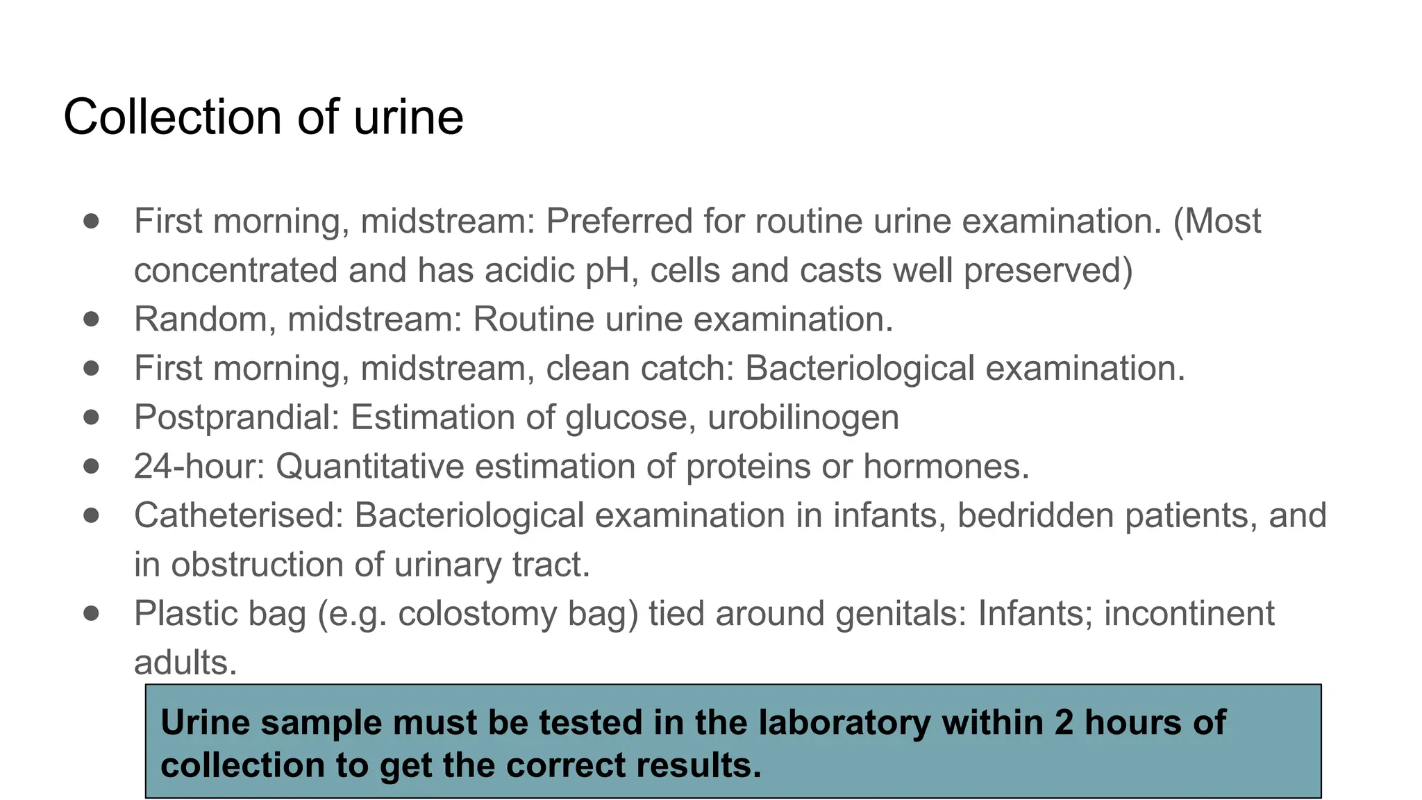
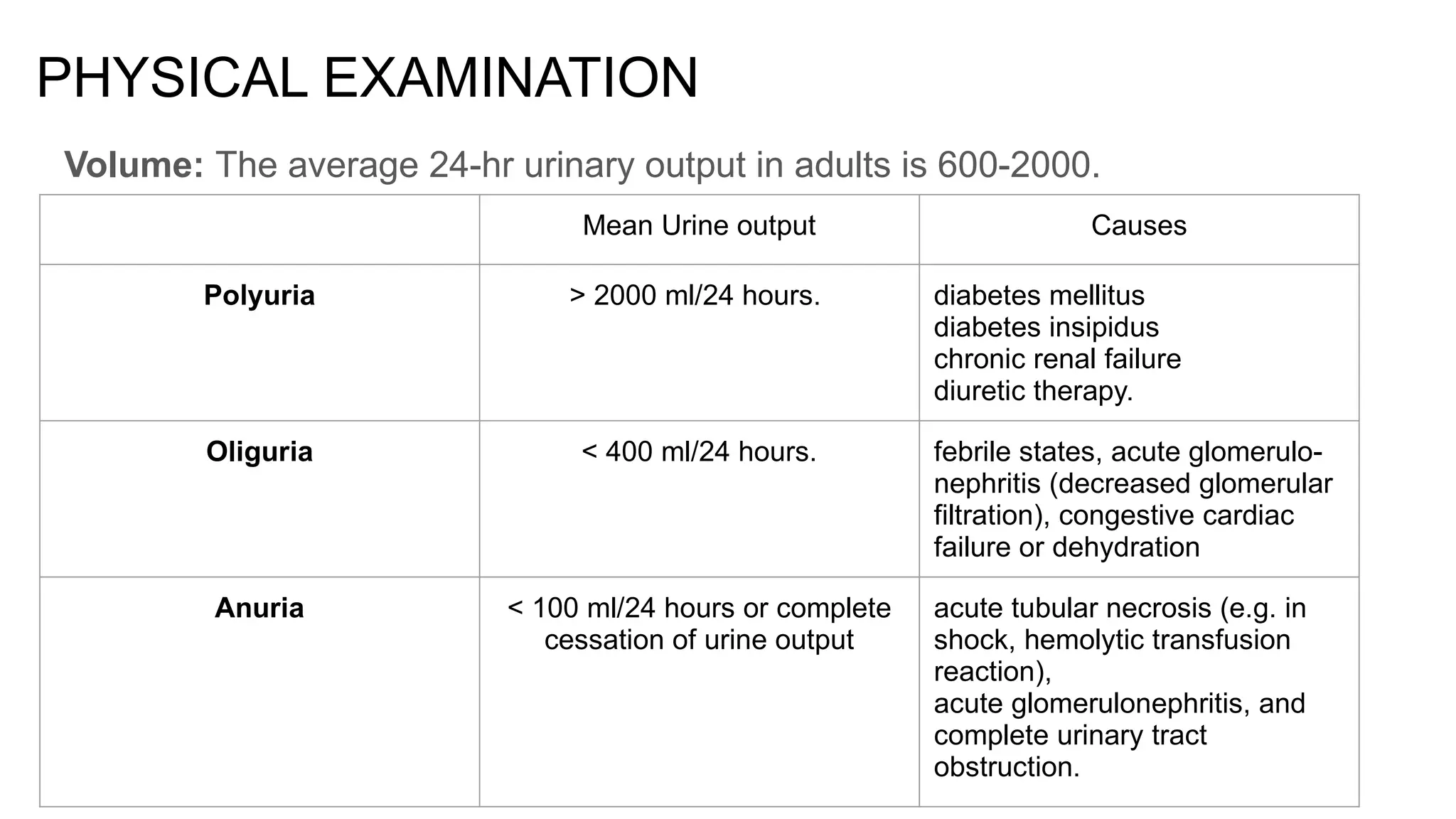
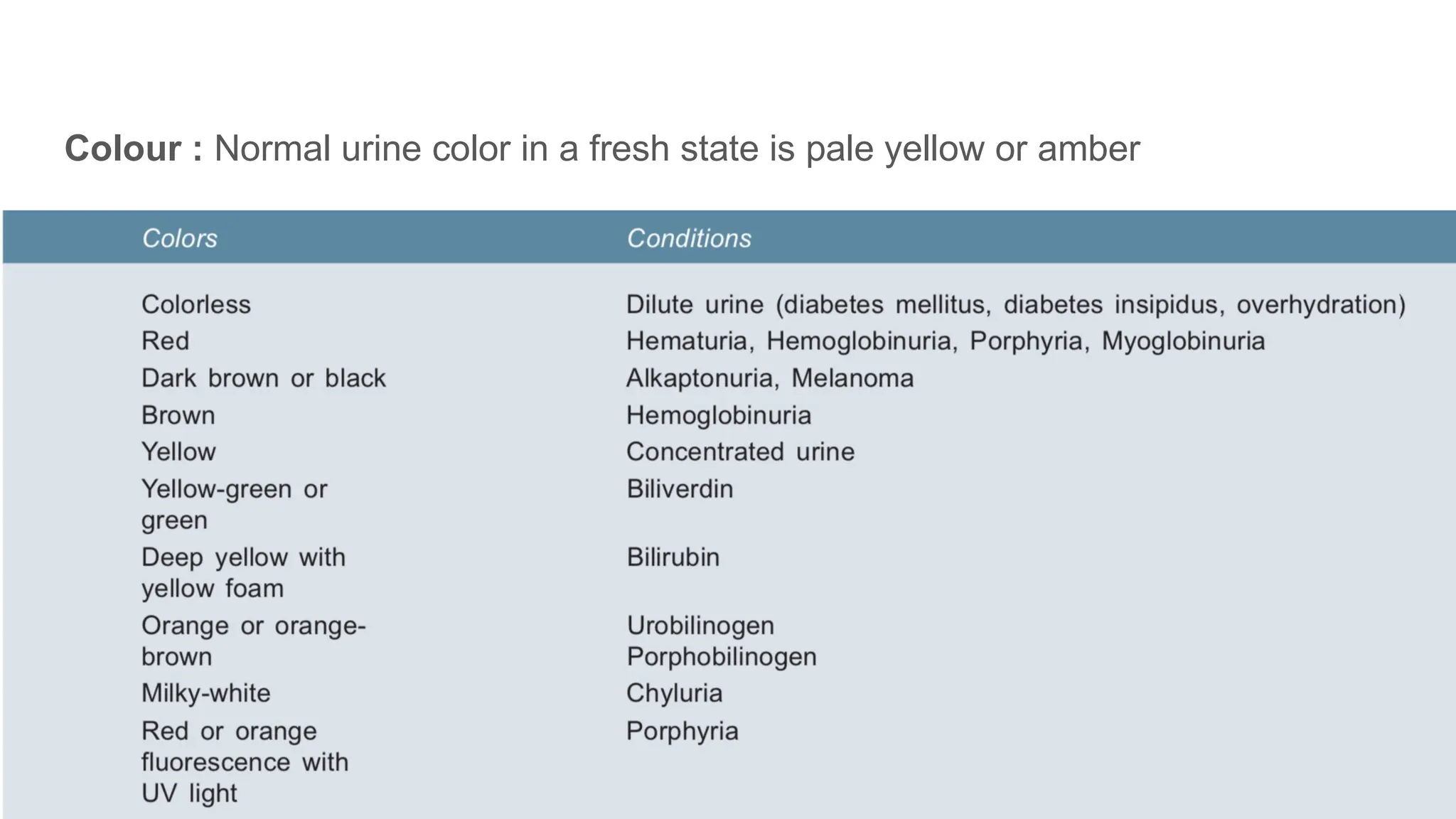
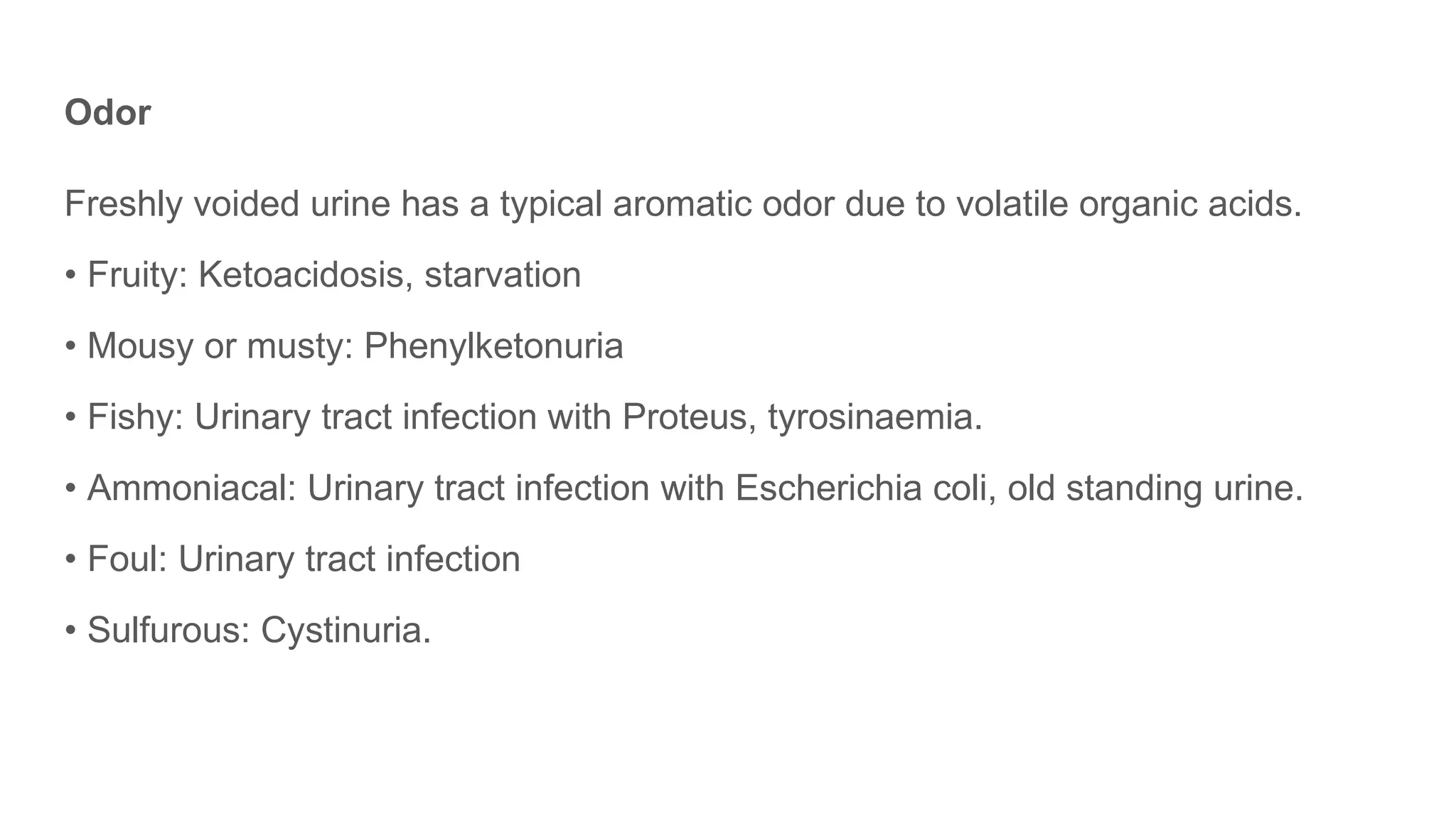
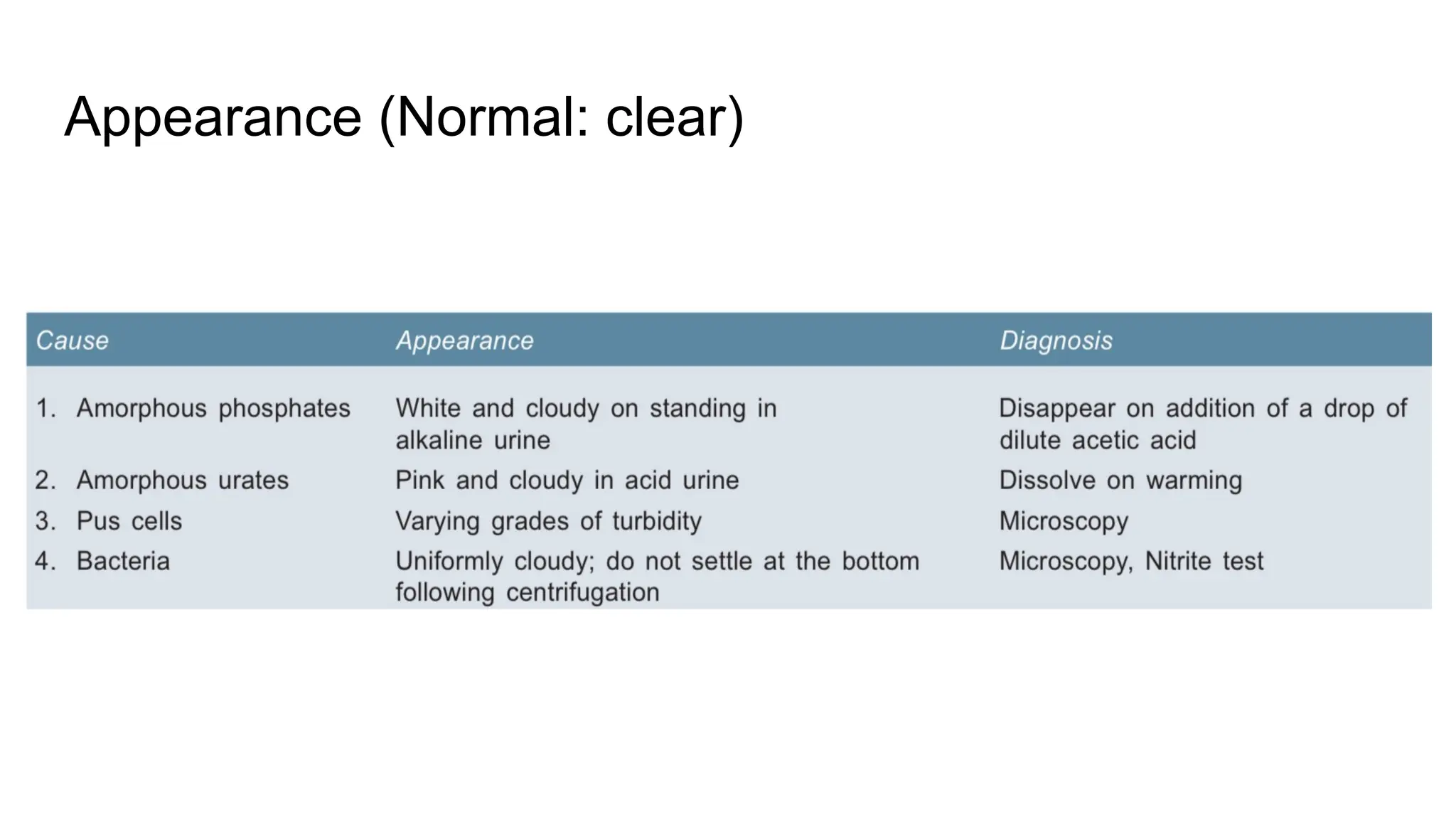
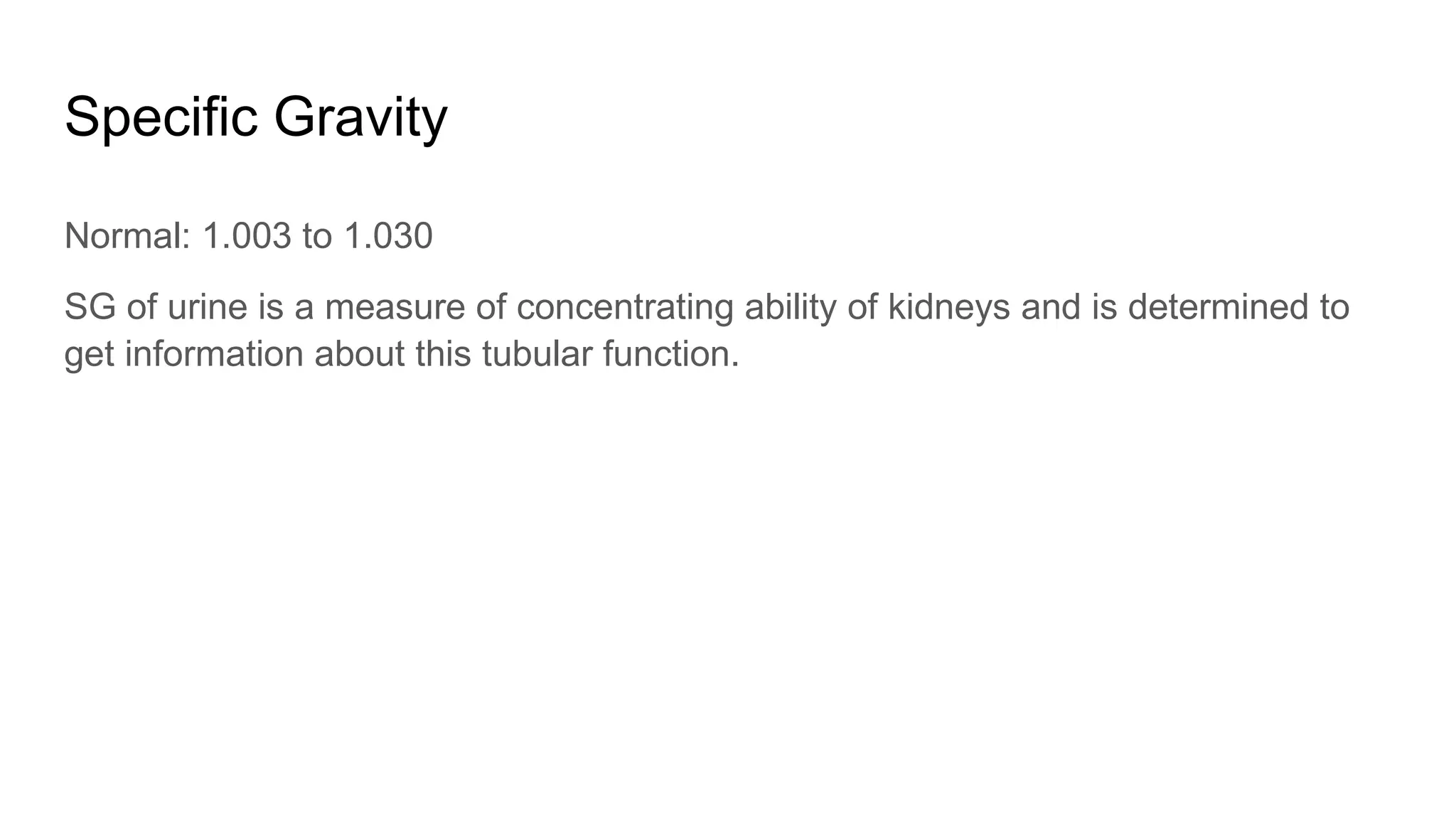
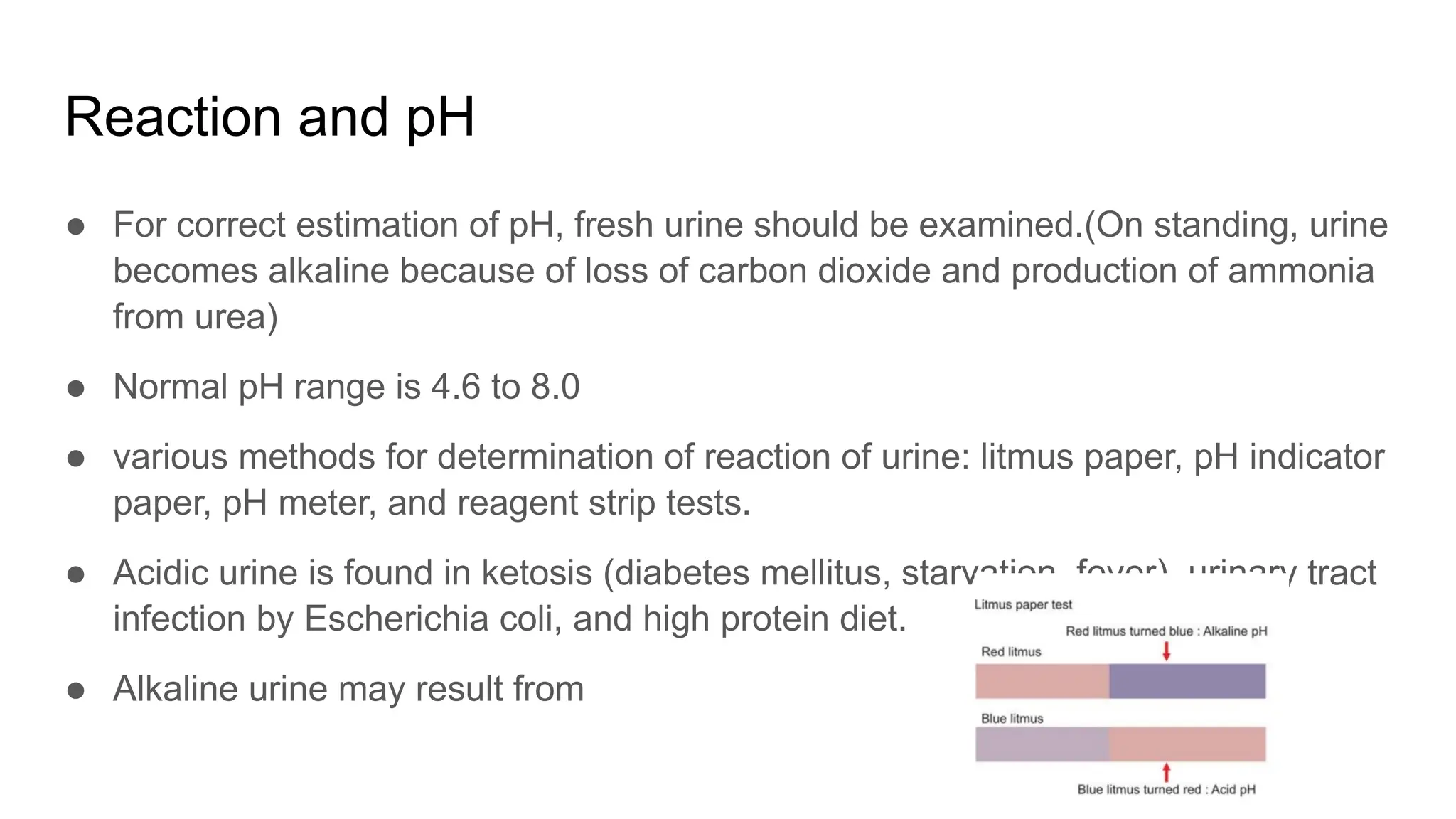
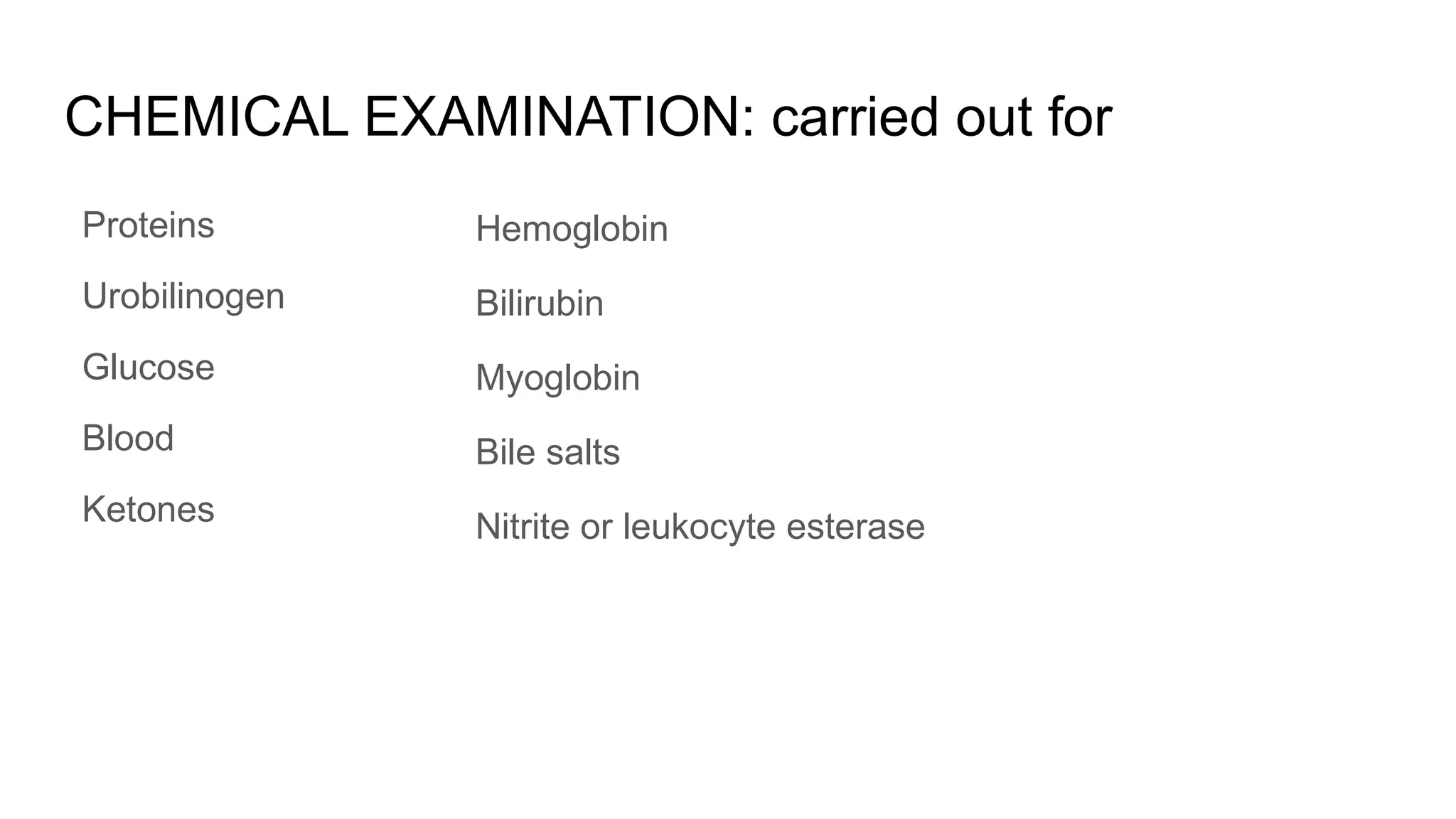
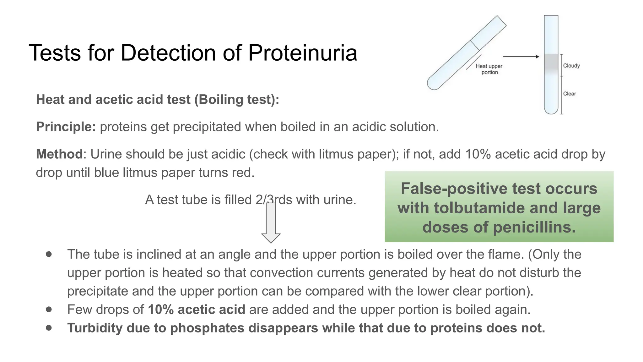
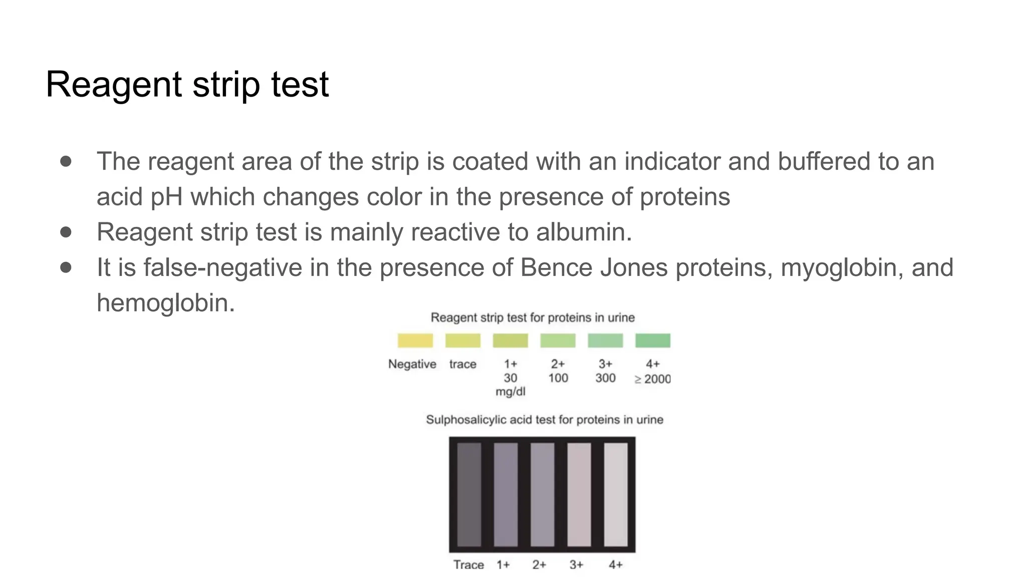
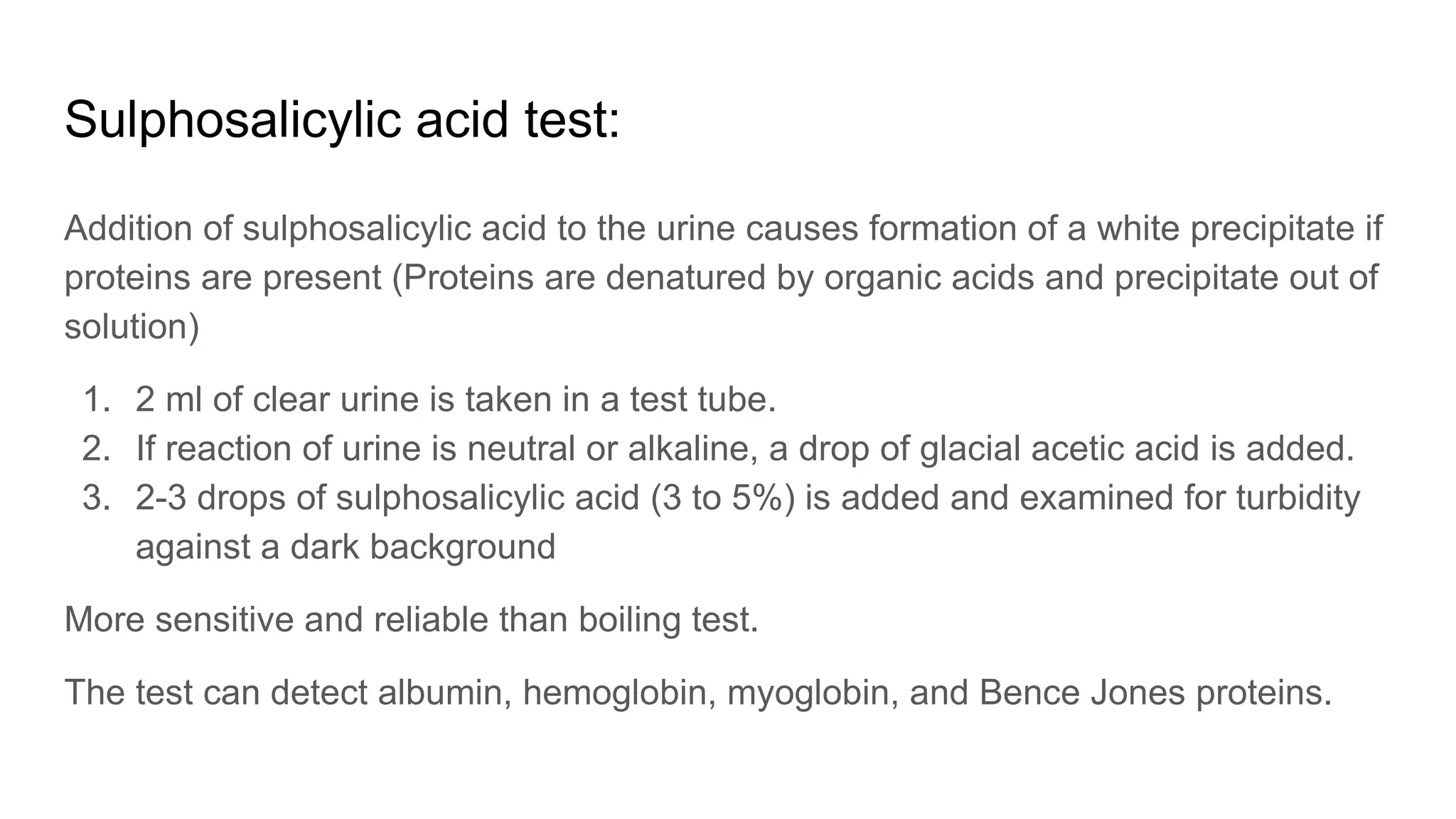
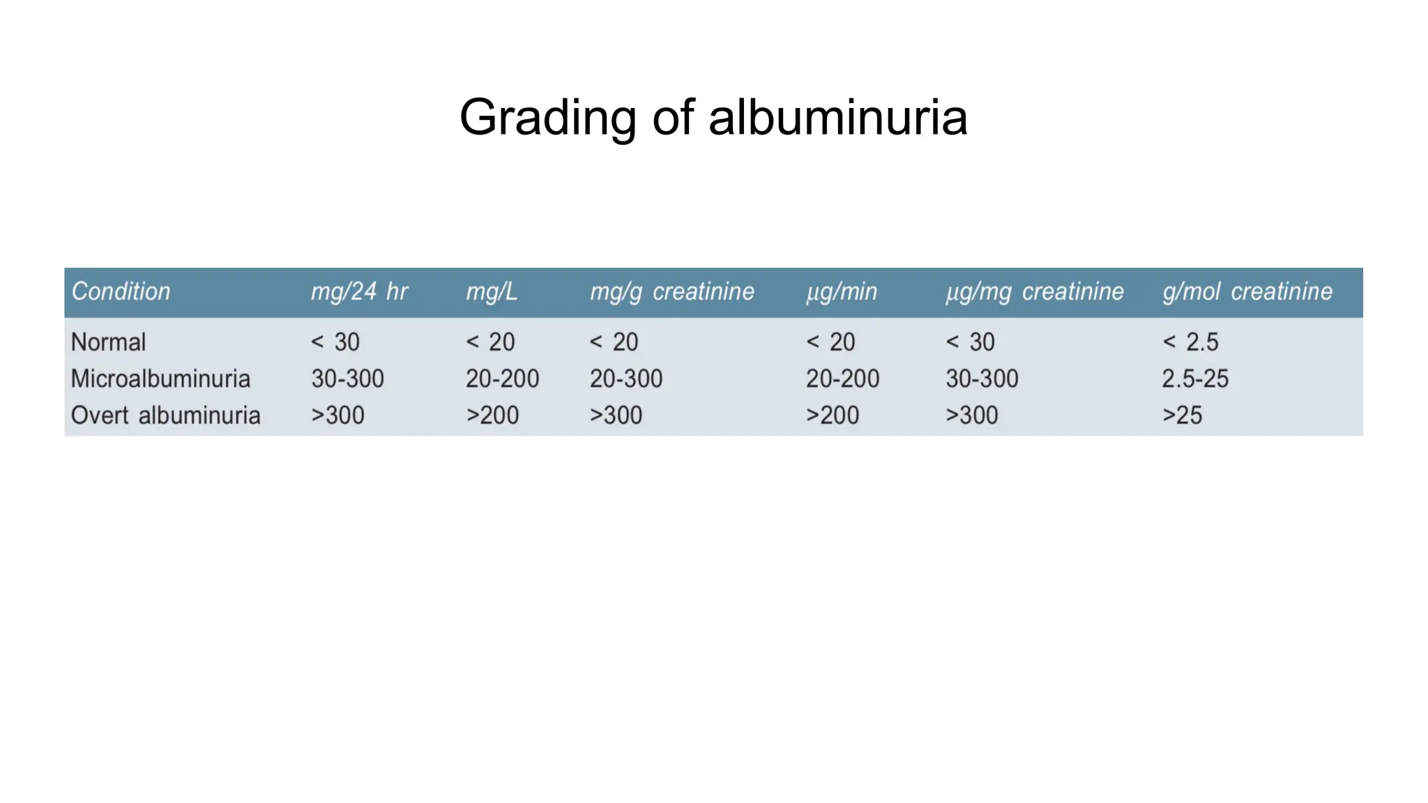
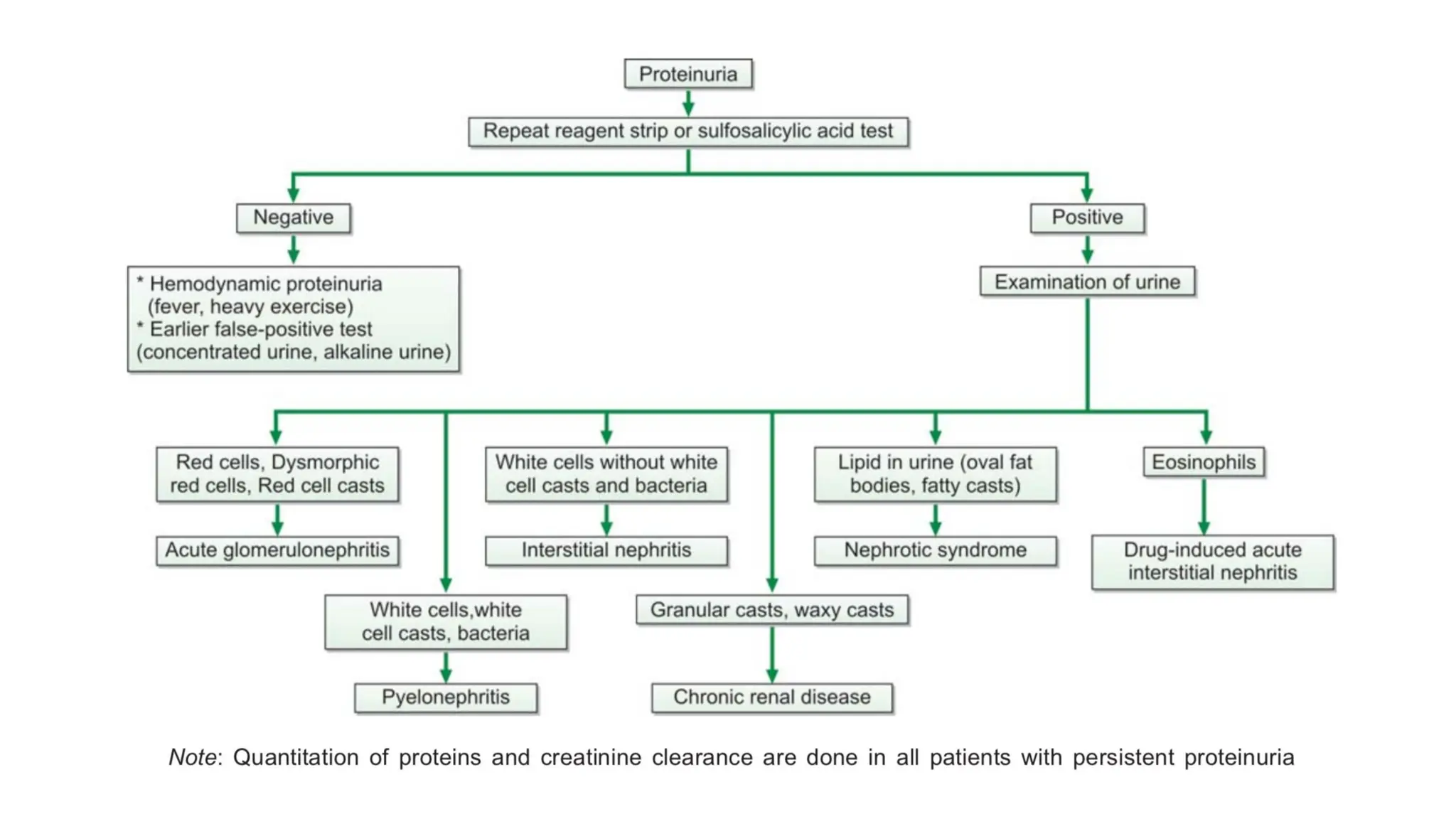
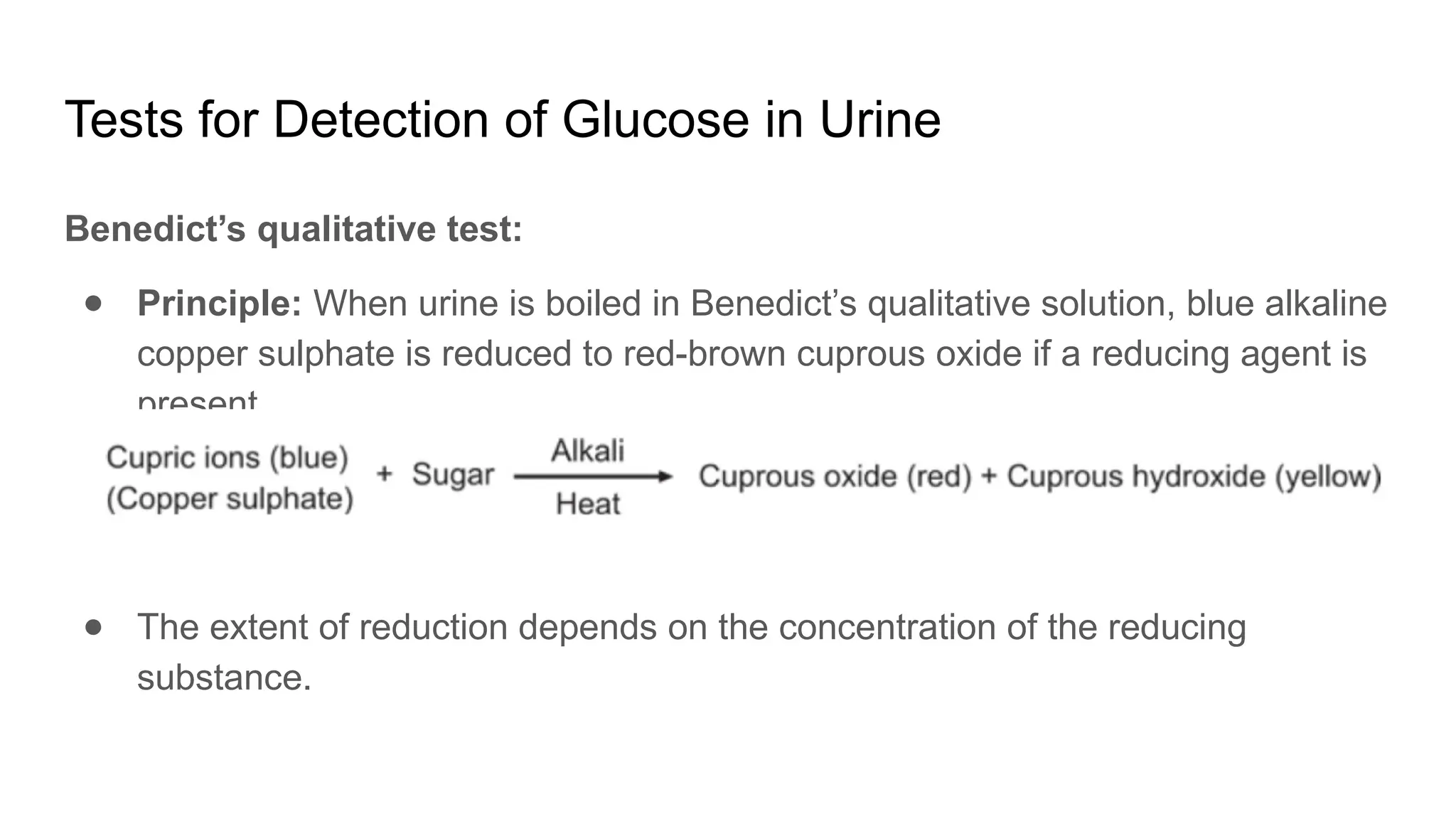
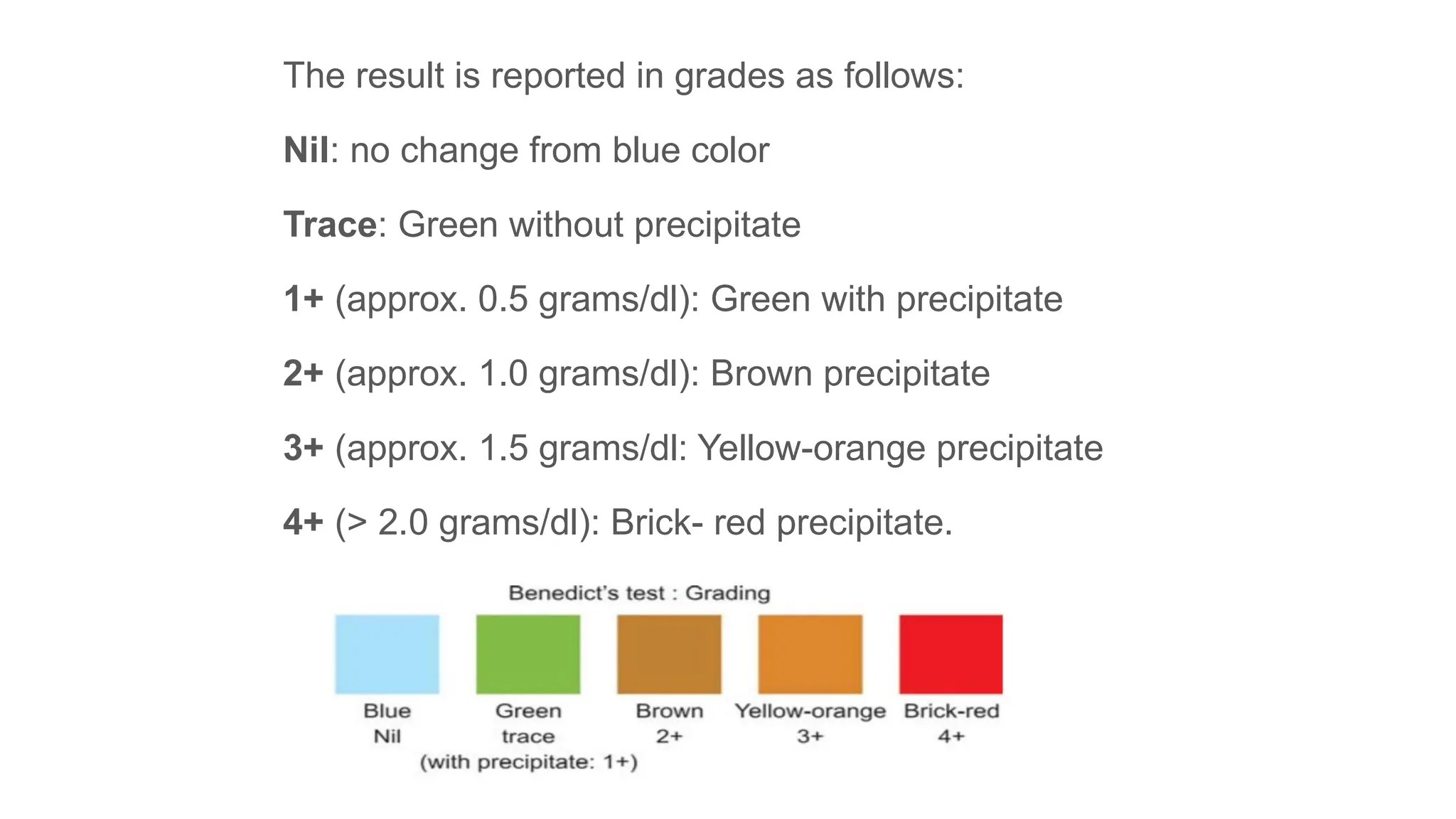
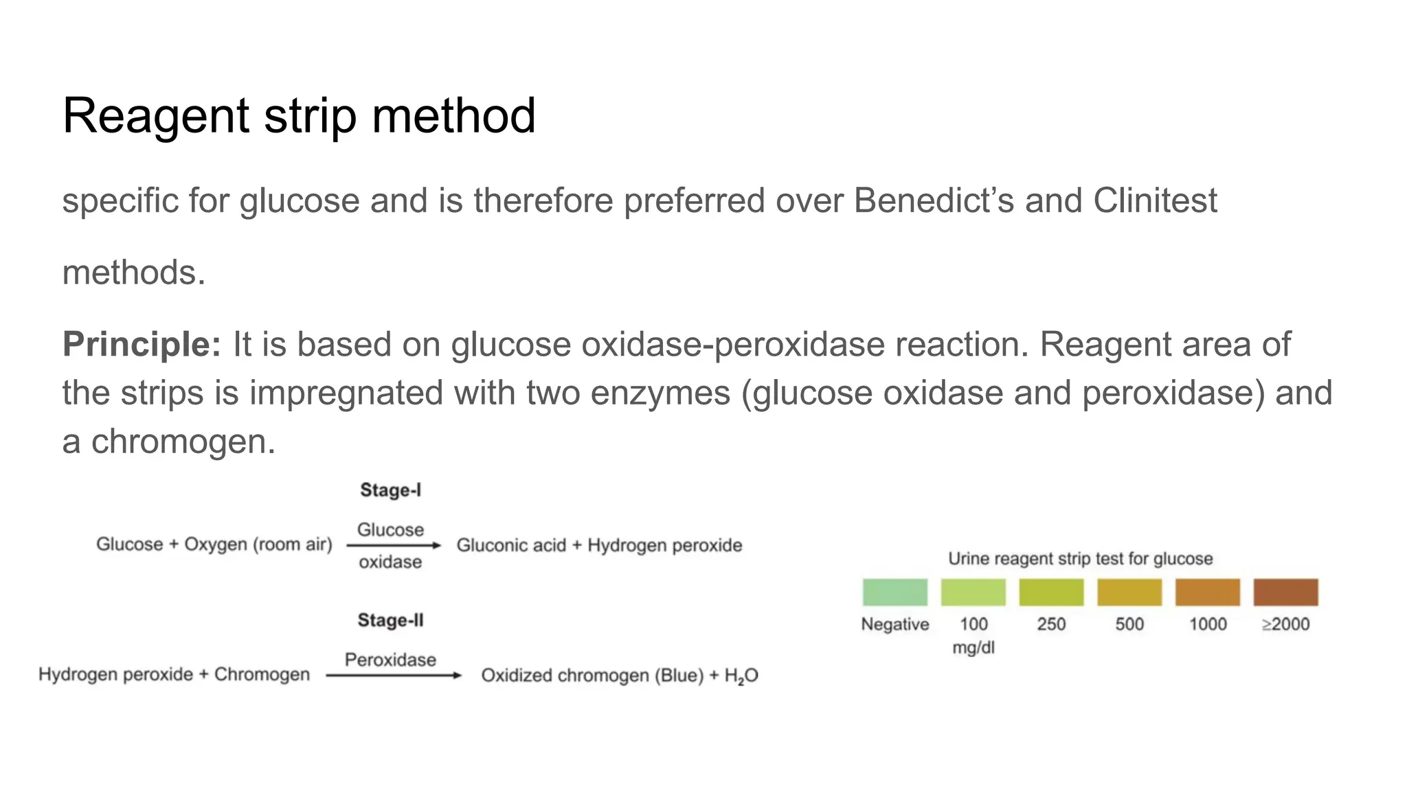
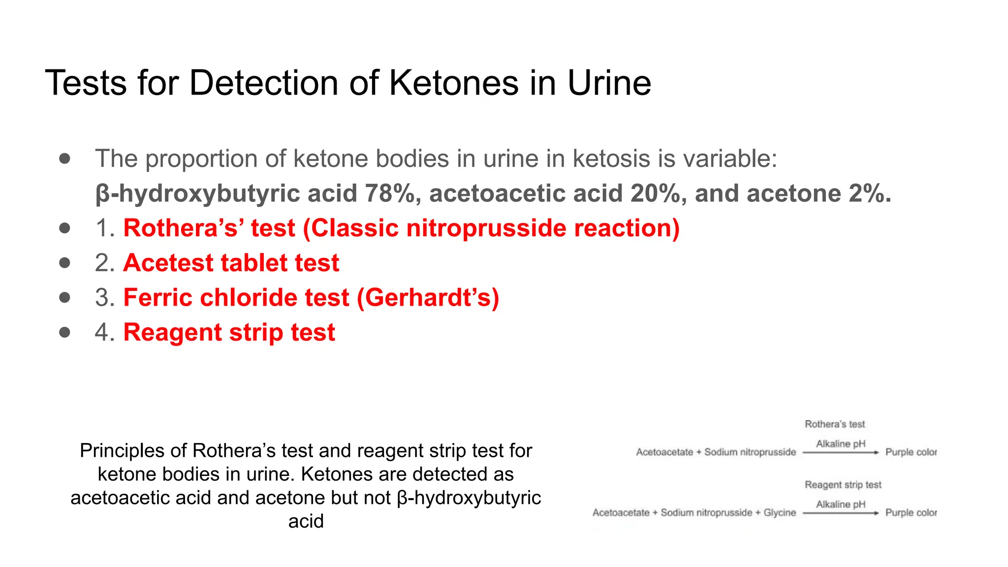
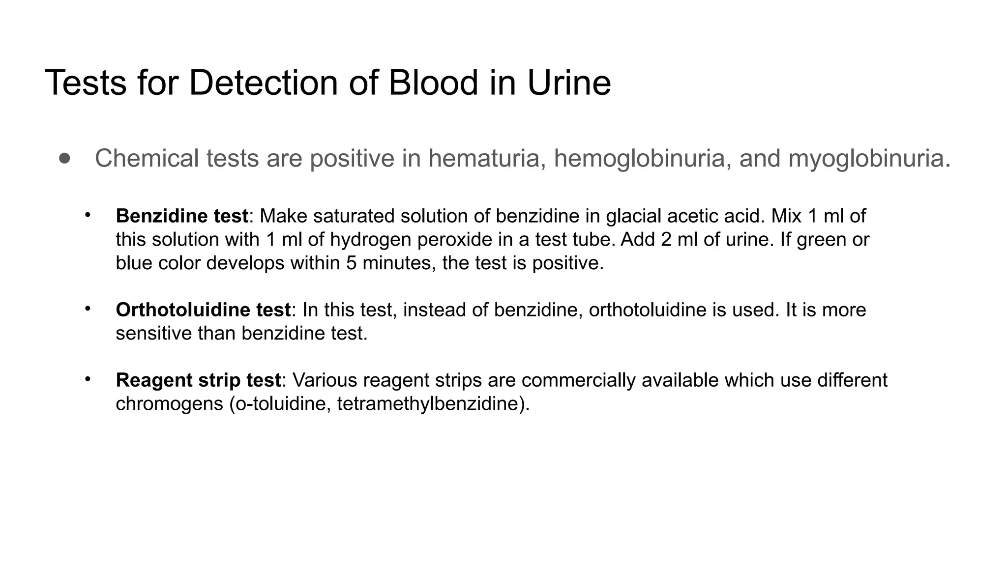
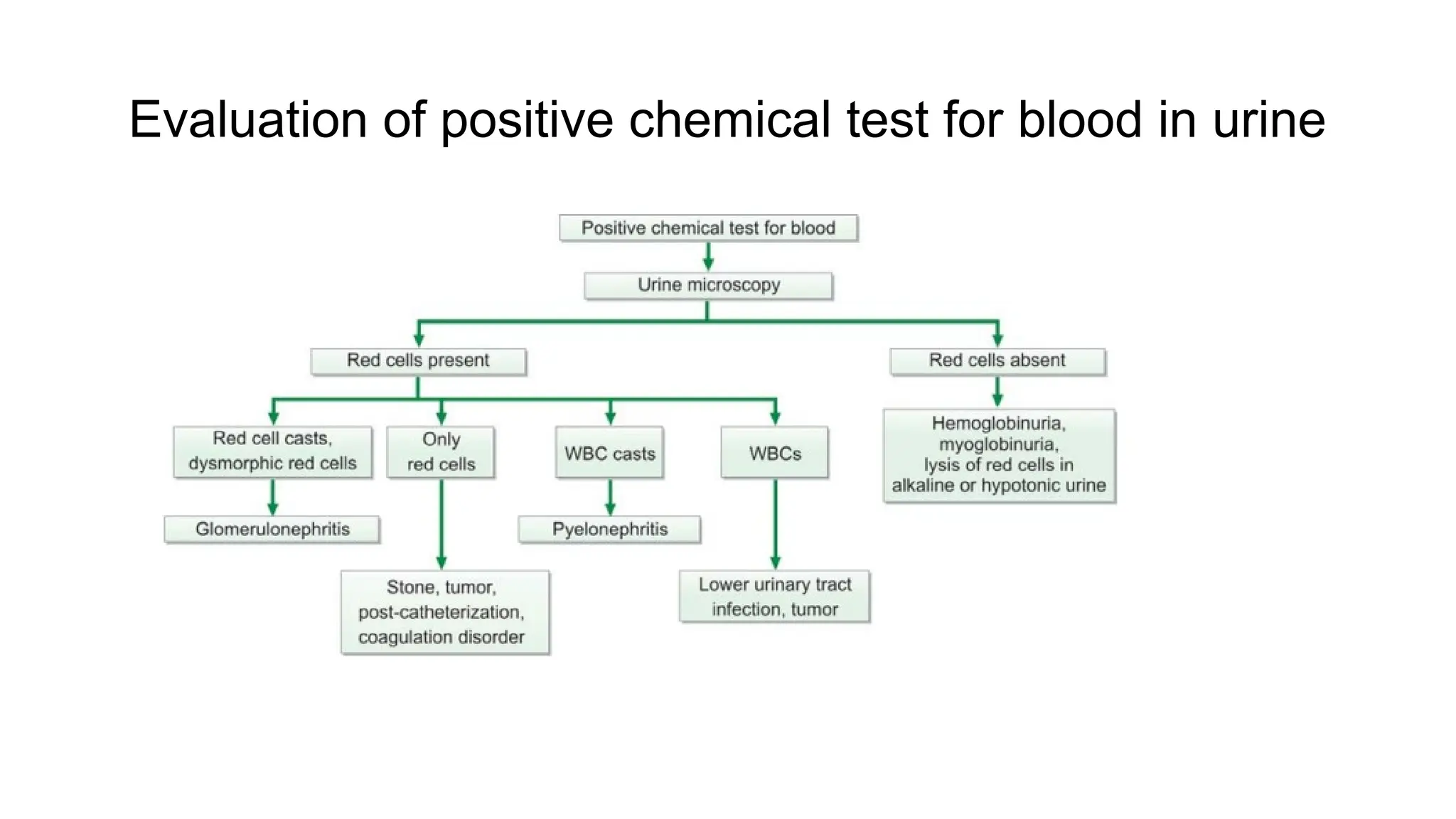
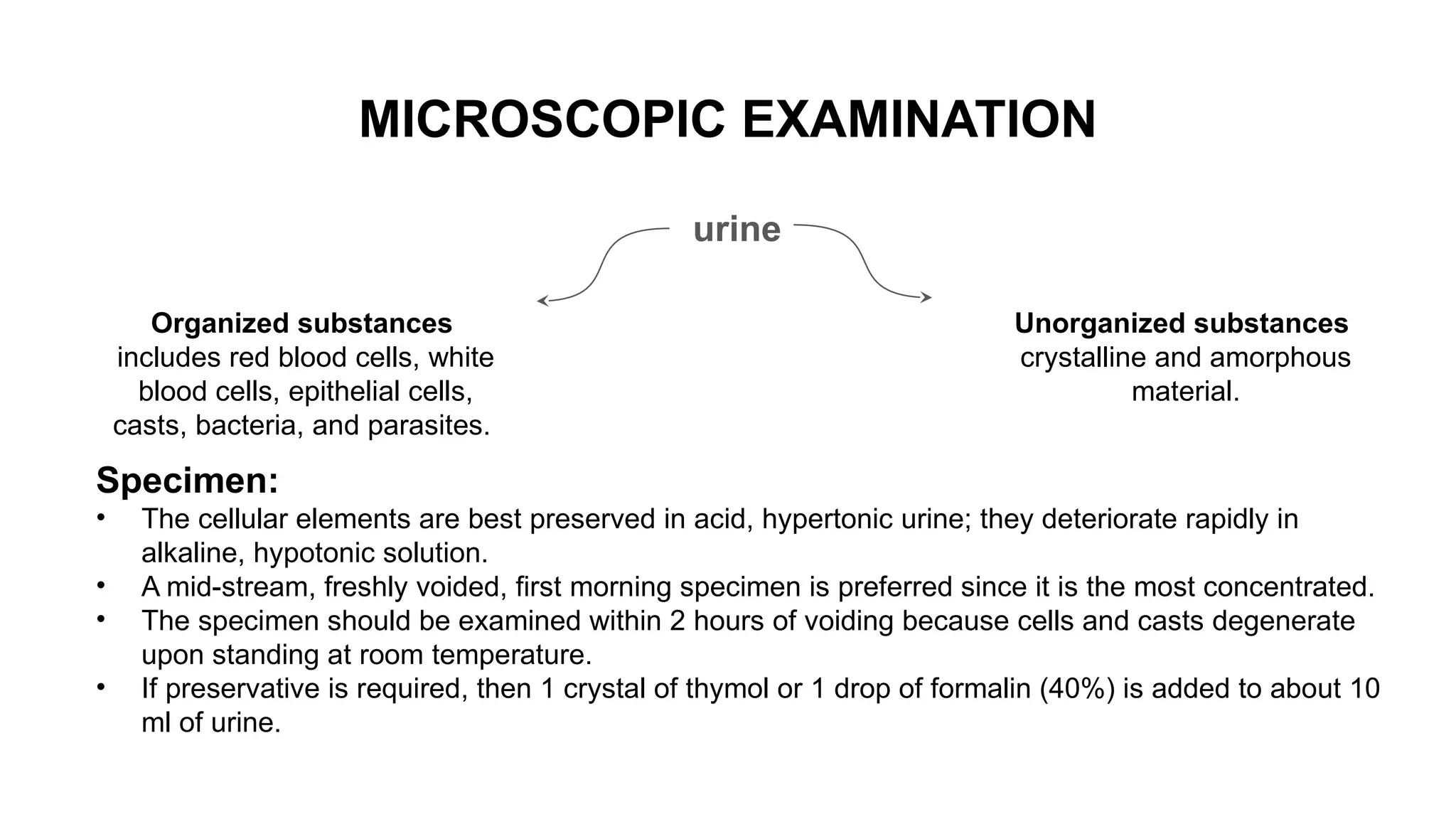
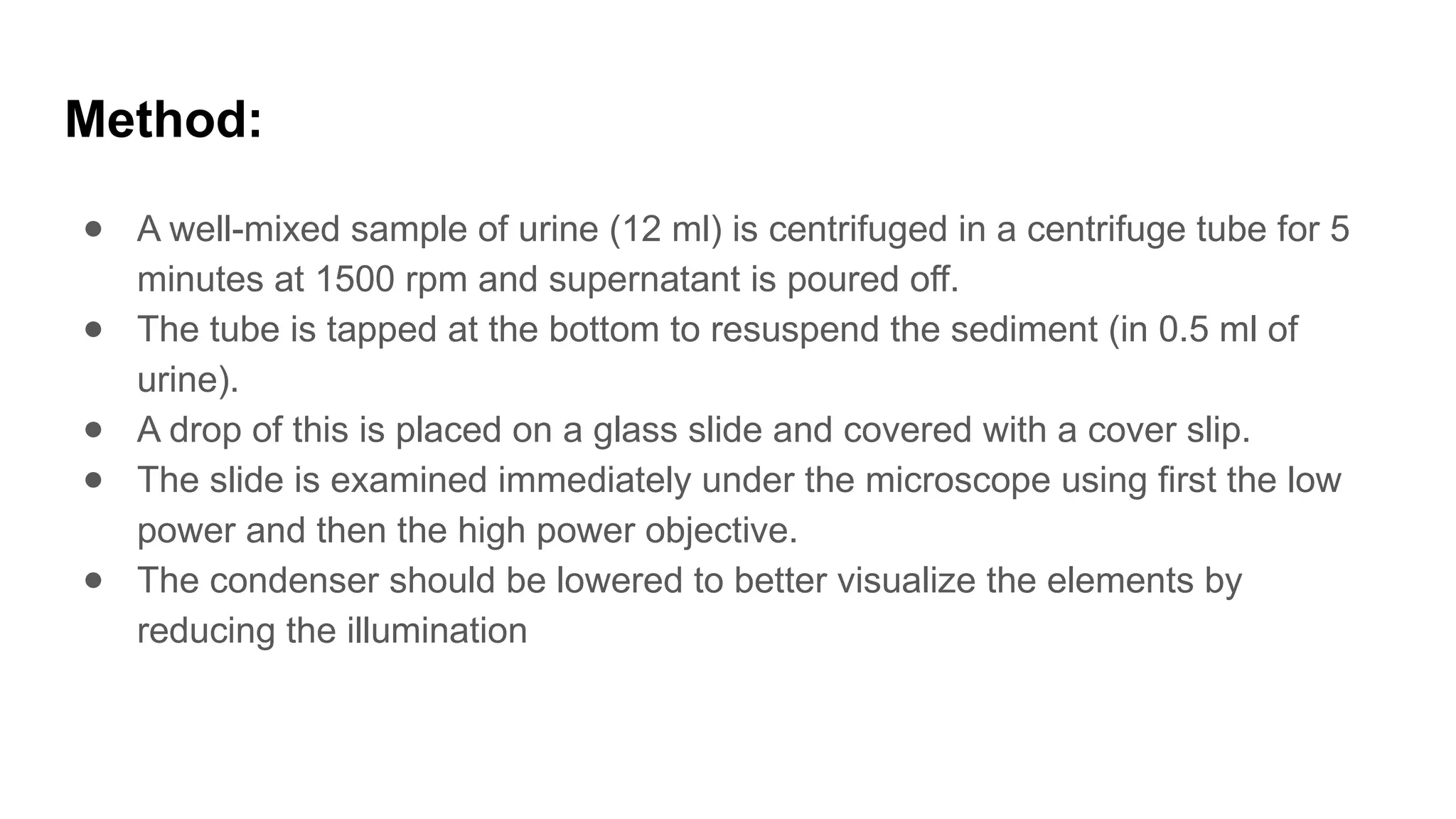
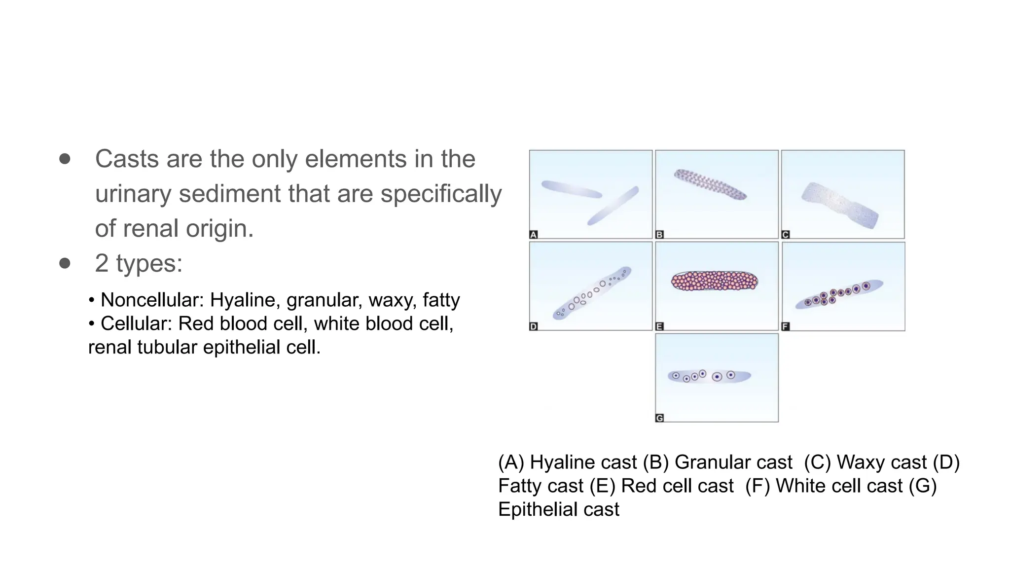
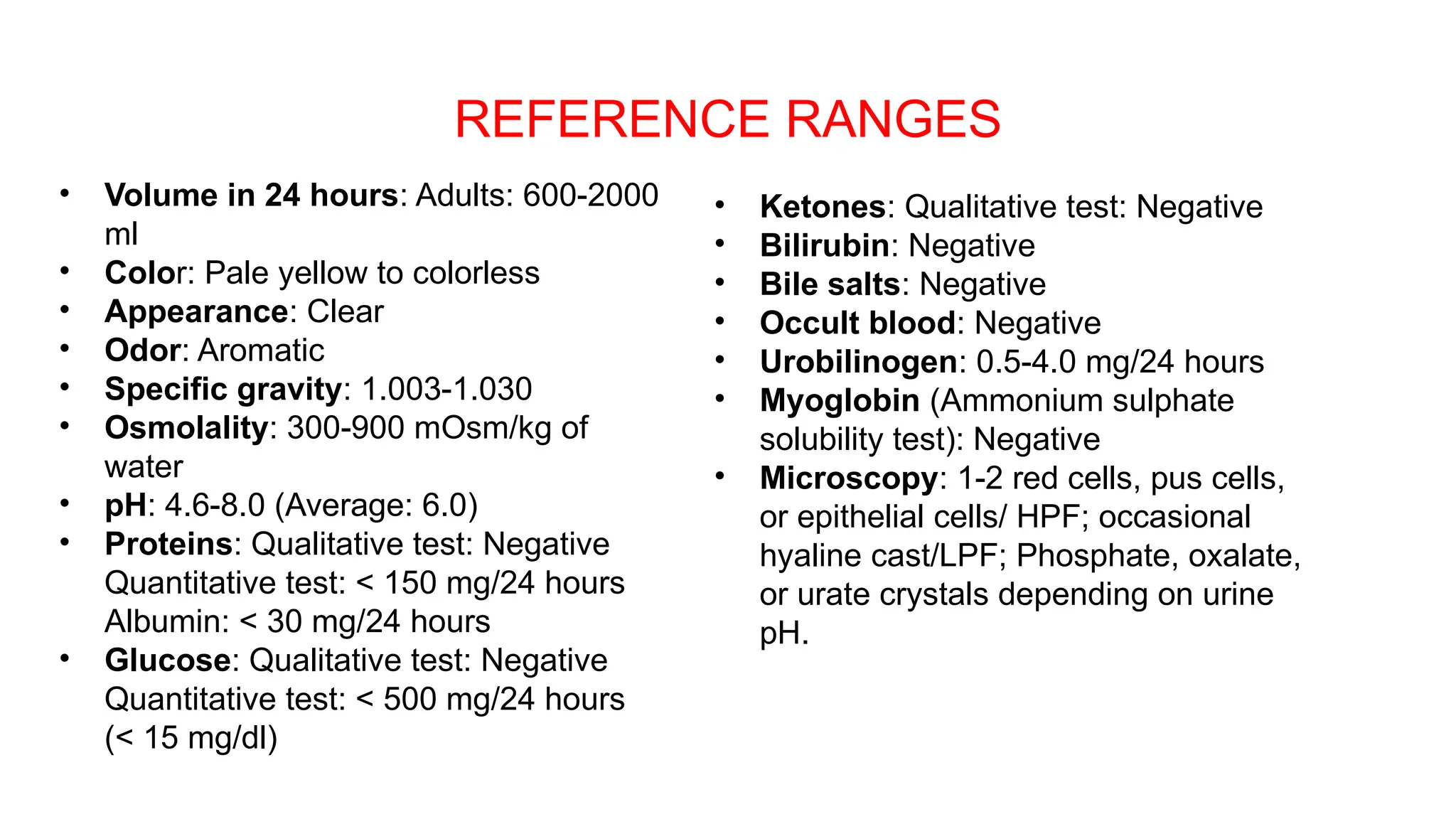
Routine urine examination including physical
chemical and microscopy
Principle and interpretation
Presenter- Dr. Simran Suman (JR-3)
Moderator- Dr. Guddi Rani Singh (Assistant Professor)
Collection of urine
● First morning, midstream: Preferred for routine urine examination. (Most
concentrated and has acidic pH, cells and casts well preserved)
● Random, midstream: Routine urine examination.
● First morning, midstream, clean catch: Bacteriological examination.
● Postprandial: Estimation of glucose, urobilinogen
● 24-hour: Quantitative estimation of proteins or hormones.
● Catheterised: Bacteriological examination in infants, bedridden patients, and
in obstruction of urinary tract.
● Plastic bag (e.g. colostomy bag) tied around genitals: Infants; incontinent
adults.
Urine sample must be tested in the laboratory within 2 hours of
collection to get the correct results.
PHYSICAL EXAMINATION
Volume: The average 24-hr urinary output in adults is 600-2000.
Mean Urine output Causes
Polyuria > 2000 ml/24 hours. diabetes mellitus
diabetes insipidus
chronic renal failure
diuretic therapy.
Oliguria < 400 ml/24 hours. febrile states, acute glomerulo-
nephritis (decreased glomerular
filtration), congestive cardiac
failure or dehydration
Anuria < 100 ml/24 hours or complete
cessation of urine output
acute tubular necrosis (e.g. in
shock, hemolytic transfusion
reaction),
acute glomerulonephritis, and
complete urinary tract
obstruction.
Colour : Normal urine color in a fresh state is pale yellow or amber
Odor
Freshly voided urine has a typical aromatic odor due to volatile organic acids.
• Fruity: Ketoacidosis, starvation
• Mousy or musty: Phenylketonuria
• Fishy: Urinary tract infection with Proteus, tyrosinaemia.
• Ammoniacal: Urinary tract infection with Escherichia coli, old standing urine.
• Foul: Urinary tract infection
• Sulfurous: Cystinuria.
Appearance (Normal: clear)
Specific Gravity
Normal: 1.003 to 1.030
SG of urine is a measure of concentrating ability of kidneys and is determined to
get information about this tubular function.
Reaction and pH
● For correct estimation of pH, fresh urine should be examined.(On standing, urine
becomes alkaline because of loss of carbon dioxide and production of ammonia
from urea)
● Normal pH range is 4.6 to 8.0
● various methods for determination of reaction of urine: litmus paper, pH indicator
paper, pH meter, and reagent strip tests.
● Acidic urine is found in ketosis (diabetes mellitus, starvation, fever), urinary tract
infection by Escherichia coli, and high protein diet.
● Alkaline urine may result from
CHEMICAL EXAMINATION: carried out for
Proteins
Urobilinogen
Glucose
Blood
Ketones
Hemoglobin
Bilirubin
Myoglobin
Bile salts
Nitrite or leukocyte esterase
Proteins
Normally, kidneys excrete scant amount of protein in urine (up to 150 mg/24
hours).
Albumin and proteins derived from urinary tract (Tamm-Horsfall protein, secretory
IgA, and proteins from tubular epithelial cells, leucocytes, and other desquamated
cells)
Proteinuria refers to protein excretion in urine greater than 150 mg/24 hours
in adults.
Causes of Proteinuria
Nephrotic syndrome
Tests for Detection of Proteinuria
Heat and acetic acid test (Boiling test):
Principle: proteins get precipitated when boiled in an acidic solution.
Method: Urine should be just acidic (check with litmus paper); if not, add 10% acetic acid drop by
drop until blue litmus paper turns red.
A test tube is filled 2/3rds with urine.
● The tube is inclined at an angle and the upper portion is boiled over the flame. (Only the
upper portion is heated so that convection currents generated by heat do not disturb the
precipitate and the upper portion can be compared with the lower clear portion).
● Few drops of 10% acetic acid are added and the upper portion is boiled again.
● Turbidity due to phosphates disappears while that due to proteins does not.
False-positive test occurs
with tolbutamide and large
doses of penicillins.
Reagent strip test
● The reagent area of the strip is coated with an indicator and buffered to an
acid pH which changes color in the presence of proteins
● Reagent strip test is mainly reactive to albumin.
● It is false-negative in the presence of Bence Jones proteins, myoglobin, and
hemoglobin.
Sulphosalicylic acid test:
Addition of sulphosalicylic acid to the urine causes formation of a white precipitate if
proteins are present (Proteins are denatured by organic acids and precipitate out of
solution)
1. 2 ml of clear urine is taken in a test tube.
2. If reaction of urine is neutral or alkaline, a drop of glacial acetic acid is added.
3. 2-3 drops of sulphosalicylic acid (3 to 5%) is added and examined for turbidity
against a dark background
More sensitive and reliable than boiling test.
The test can detect albumin, hemoglobin, myoglobin, and Bence Jones proteins.
Grading of albuminuria
Glucose
Normally a very small amount of glucose is excreted in urine (< 500 mg/24 hours
or <15 mg/dl) that cannot be detected by the routine tests
Causes of Glycosuria
1.Glycosuria with hyperglycemia:
● Endocrine diseases: diabetes mellitus, acromegaly,Cushing’s syndrome,
hyperthyroidism, pancreatic disease.
● Non-endocrine disease: CNS diseases,liver diseases
● Drugs: adrenocorticotrophic hormone, corticosteroids, thiazides.
2. Glycosuria without hyperglycemia
● Renal glycosuria: This accounts for 5% of cases of glycosuria in general
population.
● The normal renal threshold for glucose is 180 mg/dl.
● Renal glycosuria is a benign condition in which renal threshold is set below
180 mgs/dl but glucose tolerance is normal; the disorder is transmitted as
autosomal dominant.
Tests for Detection of Glucose in Urine
Benedict’s qualitative test:
● Principle: When urine is boiled in Benedict’s qualitative solution, blue alkaline
copper sulphate is reduced to red-brown cuprous oxide if a reducing agent is
present
● The extent of reduction depends on the concentration of the reducing
substance.
Method
1. Take 5 ml of Benedict’s qualitative reagent in a test tube (composition of
Benedict’s qualitative reagent: copper sulphate 17.3 gram, sodium carbonate 100
gram, sodium citrate 173 gram, distilled water 1000 ml).
2. Add 0.5 ml (or 8 drops) of urine. Mix well.
3. Boil over a flame for 2 minutes.
4. Allow to cool at room temperature.
5. Note the color change, if any.
Benedict’s test gives positive reaction with carbohydrates other than glucose, it is also
used as a screening test (for detection of galactose, lactose, fructose, maltose, and
pentoses in urine) for inborn errors of carbohydrate metabolism in infants and children.
The result is reported in grades as follows:
Nil: no change from blue color
Trace: Green without precipitate
1+ (approx. 0.5 grams/dl): Green with precipitate
2+ (approx. 1.0 grams/dl): Brown precipitate
3+ (approx. 1.5 grams/dl: Yellow-orange precipitate
4+ (> 2.0 grams/dl): Brick- red precipitate.
Reagent strip method
specific for glucose and is therefore preferred over Benedict’s and Clinitest
methods.
Principle: It is based on glucose oxidase-peroxidase reaction. Reagent area of
the strips is impregnated with two enzymes (glucose oxidase and peroxidase) and
a chromogen.
Ketones
● Normally ketone bodies are not detectable in the urine of healthy persons.
● Causes of ketonuria:
1. Uncontrolled diabetes mellitus with ketoacidosis
2. Glycogen storage disease (von Gierke’s disease)
3. Decreased availability of carbohydrates in the diet:
Starvation
Persistent vomiting in children
Weight reduction program (severe carbohydrate restriction with normal fat
intake)
4. Increased metabolic needs:
Fever in children
Severe thyrotoxicosis
Pregnancy
Protein calorie malnutrition
Tests for Detection of Ketones in Urine
● The proportion of ketone bodies in urine in ketosis is variable:
β-hydroxybutyric acid 78%, acetoacetic acid 20%, and acetone 2%.
● 1. Rothera’s’ test (Classic nitroprusside reaction)
● 2. Acetest tablet test
● 3. Ferric chloride test (Gerhardt’s)
● 4. Reagent strip test
Principles of Rothera’s test and reagent strip test for
ketone bodies in urine. Ketones are detected as
acetoacetic acid and acetone but not β-hydroxybutyric
acid
Rothera’s’ test (Classic nitroprusside reaction)
● Acetoacetic acid or acetone reacts with nitroprusside in alkaline solution to form a
purple-colored complex
● Rothera’s test is sensitive to 1-5 mg/dl of acetoacetate and to 10-25 mg/dl of acetone.
● Method
1. 5 ml of urine taken in a test tube and saturated it with ammonium sulphate.
2. small crystal of sodium nitroprusside added and Mixed well.
3. Slowly run along the side of the test tube liquor ammonia to form a layer.
4. Immediate formation of a purple permanganate colored ring at the junction of the two
fluids indicates a positive test.
● False-positive test can occur in the presence of L-dopa in urine and in phenylketonuria
Bile Pigment (Bilirubin)
● Bilirubin (a breakdown product of hemoglobin) is undetectable in the urine of
normal persons.
● Presence of bilirubin in urine indicates conjugated hyperbilirubinemia
(obstructive or hepatocellular jaundice). This is because only conjugated
bilirubin is water-soluble. Bilirubin in urine is absent in hemolytic jaundice; this
is because unconjugated bilirubin is water-insoluble.
● In acute viral hepatitis, bilirubin appears in urine even before jaundice is
clinically apparent.
● In a fever of unknown origin bilirubinuria suggests hepatitis.
● Methods for detection of bilirubin in urine are foam test, Gmelin’s test,
Lugol iodine test, Fouchet’s test, Ictotest tablet test, and reagent strip
test.
● Clinical and laboratory findings in bilirubinuria
• Jaundice
• Urine color: Dark yellow with yellow foam
• Elevated serum conjugated bilirubin
Blood
● The presence of abnormal number of intact red blood cells in urine is called
as hematuria.
● Bleeding in urine may be noted macroscopically or with naked eye (gross
hematuria).
● If bleeding is noted only by microscopic examination or by chemical tests,
then it is called as occult, microscopic or hidden hematuria
● Causes of Hematuria:
• Diseases of urinary tract
1. Glomerular diseases: Glomerulonephritis, Berger’s disease, lupus nephritis, Henoch-Schonlein
purpura
2. Nonglomerular diseases: Calculus, tumor, infection, tuberculosis, pyelonephritis,
hydronephrosis, polycystic kidney disease, trauma, after strenuous physical exercise, diseases of
prostate (benign hyperplasia of prostate, carcinoma of prostate).
• Hematological conditions: Coagulation disorders, sickle cell disease
Tests for Detection of Blood in Urine
● Chemical tests are positive in hematuria, hemoglobinuria, and myoglobinuria.
• Benzidine test: Make saturated solution of benzidine in glacial acetic acid. Mix 1 ml of
this solution with 1 ml of hydrogen peroxide in a test tube. Add 2 ml of urine. If green or
blue color develops within 5 minutes, the test is positive.
• Orthotoluidine test: In this test, instead of benzidine, orthotoluidine is used. It is more
sensitive than benzidine test.
• Reagent strip test: Various reagent strips are commercially available which use different
chromogens (o-toluidine, tetramethylbenzidine).
Evaluation of positive chemical test for blood in urine
MICROSCOPIC EXAMINATION
urine
Organized substances
includes red blood cells, white
blood cells, epithelial cells,
casts, bacteria, and parasites.
Unorganized substances
crystalline and amorphous
material.
Specimen:
• The cellular elements are best preserved in acid, hypertonic urine; they deteriorate rapidly in
alkaline, hypotonic solution.
• A mid-stream, freshly voided, first morning specimen is preferred since it is the most concentrated.
• The specimen should be examined within 2 hours of voiding because cells and casts degenerate
upon standing at room temperature.
• If preservative is required, then 1 crystal of thymol or 1 drop of formalin (40%) is added to about 10
ml of urine.
Method:
● A well-mixed sample of urine (12 ml) is centrifuged in a centrifuge tube for 5
minutes at 1500 rpm and supernatant is poured off.
● The tube is tapped at the bottom to resuspend the sediment (in 0.5 ml of
urine).
● A drop of this is placed on a glass slide and covered with a cover slip.
● The slide is examined immediately under the microscope using first the low
power and then the high power objective.
● The condenser should be lowered to better visualize the elements by
reducing the illumination
Urinary findings in renal diseases
Different types of urinary sediment
Cells in urine
(1) Isomorphic red blood cells,
(2) Crenated red cells,
(3) Swollen red cells,
(4) Dysmorphic red cells,
(5) White blood cells (pus cells),
(6) Squamous epithelial cell,
(7) Transitional epithelial cells,
(8) Renal tubular epithelial cells,
(9) Oval fat bodies,
(10) Maltese cross pattern of oval fat bodies
(11) spermatozoa
RBC
● In a fresh urine sample, red cells appear as small, smooth, yellowish,
anucleate biconcave disks about 7 μ in diameter (called as isomorphic red
cells).
● It may appear swollen (thin discs of greater diameter, 9-10 μ) in dilute or
hypotonic urine, or may appear crenated (smaller diameter with spikey
surface) in hypertonic urine.
● In glomerulonephritis, red cells are typically described as being dysmorphic.
● > 80% of dysmorphic red cells is strongly suggestive of glomerular pathology.
White Blood Cells (Pus Cells)
● Normally 0-2 white cells may be seen per high power field.
● Pus cells greater than 10/HPF or presence of clumps is suggestive of urinary
tract infection.
● Increased numbers of white cells occur in fever, pyelonephritis, lower
urinary tract infection, tubulo interstitial nephritis, and renal transplant
rejection.
● Simultaneous presence of white cells and white cell casts indicates presence
of renal infection (pyelo nephritis).
● Eosinophils (>1% of urinary leucocytes) are a characteristic feature of acute
interstitial nephritis due to drug reaction
Renal Tubular Epithelial Cells
● Renal tubular epithelial cells are difficult to distin guish from pus cells in
unstained preparations.
● Increased numbers are found in conditions causing tubular damage like acute
tubular necrosis, pyelonephritis, viral infection of kidney, allograft
rejection, and salicylate or heavy metal poisoning.
● These cells are small (about the same size or slightly larger than white blood
cell), polyhedral, columnar, or oval, and have granular cytoplasm. A single,
large, refractile, eccentric nucleus is often seen.
Organisms
● Significant bacteriuria exists when there are >105
bacterial colony forming
units/ml of urine in a clean catch midstream sample, >104
colony forming
units/ml of urine in catheterized sample, and >103
colonyforming units/ml of
urine in a suprapubic aspiration sample.
Casts- Urinary casts are cylindrical, cigar-shaped microscopic structures that form in distal renal tubules and collecting ducts.
● Casts are the only elements in the
urinary sediment that are specifically
of renal origin.
● 2 types:
(A) Hyaline cast (B) Granular cast (C) Waxy cast (D)
Fatty cast (E) Red cell cast (F) White cell cast (G)
Epithelial cast
• Noncellular: Hyaline, granular, waxy, fatty
• Cellular: Red blood cell, white blood cell,
renal tubular epithelial cell.
Crystals
(A) Normal crystals: (1) Calcium oxalate, (2) Triple
phosphates, (3) Uric acid, (4) Amorphous
phosphates, (5) Amorphous urates, (6) Ammonium
urate.
(B) Abnormal crystals: (1) Cysteine, (2) Cholesterol,
(3) Bilirubin, (4) Tyrosine, (5) Sulfonamide, and (6)
Leucine
• Crystals present in acid urine- Uric acid,
amorphous nitrate and calcium oxalate crystals
• Crystals present in alkaline urine- Calcium
carbonate, Phosphates and Ammonium urate
crystals
REFERENCE RANGES
• Volume in 24 hours: Adults: 600-2000
ml
• Color: Pale yellow to colorless
• Appearance: Clear
• Odor: Aromatic
• Specific gravity: 1.003-1.030
• Osmolality: 300-900 mOsm/kg of
water
• pH: 4.6-8.0 (Average: 6.0)
• Proteins: Qualitative test: Negative
Quantitative test: < 150 mg/24 hours
Albumin: < 30 mg/24 hours
• Glucose: Qualitative test: Negative
Quantitative test: < 500 mg/24 hours
(< 15 mg/dl)
• Ketones: Qualitative test: Negative
• Bilirubin: Negative
• Bile salts: Negative
• Occult blood: Negative
• Urobilinogen: 0.5-4.0 mg/24 hours
• Myoglobin (Ammonium sulphate
solubility test): Negative
• Microscopy: 1-2 red cells, pus cells,
or epithelial cells/ HPF; occasional
hyaline cast/LPF; Phosphate, oxalate,
or urate crystals depending on urine
pH.
THANK YOU

Routine urine examination including physical chemical and microscopy Principle and interpretation

  • 1.
    Routine urine examinationincluding physical chemical and microscopy Principle and interpretation Presenter- Dr. Simran Suman (JR-3) Moderator- Dr. Guddi Rani Singh (Assistant Professor)
  • 2.
    Collection of urine ●First morning, midstream: Preferred for routine urine examination. (Most concentrated and has acidic pH, cells and casts well preserved) ● Random, midstream: Routine urine examination. ● First morning, midstream, clean catch: Bacteriological examination. ● Postprandial: Estimation of glucose, urobilinogen ● 24-hour: Quantitative estimation of proteins or hormones. ● Catheterised: Bacteriological examination in infants, bedridden patients, and in obstruction of urinary tract. ● Plastic bag (e.g. colostomy bag) tied around genitals: Infants; incontinent adults. Urine sample must be tested in the laboratory within 2 hours of collection to get the correct results.
  • 3.
    PHYSICAL EXAMINATION Volume: Theaverage 24-hr urinary output in adults is 600-2000. Mean Urine output Causes Polyuria > 2000 ml/24 hours. diabetes mellitus diabetes insipidus chronic renal failure diuretic therapy. Oliguria < 400 ml/24 hours. febrile states, acute glomerulo- nephritis (decreased glomerular filtration), congestive cardiac failure or dehydration Anuria < 100 ml/24 hours or complete cessation of urine output acute tubular necrosis (e.g. in shock, hemolytic transfusion reaction), acute glomerulonephritis, and complete urinary tract obstruction.
  • 4.
    Colour : Normalurine color in a fresh state is pale yellow or amber
  • 5.
    Odor Freshly voided urinehas a typical aromatic odor due to volatile organic acids. • Fruity: Ketoacidosis, starvation • Mousy or musty: Phenylketonuria • Fishy: Urinary tract infection with Proteus, tyrosinaemia. • Ammoniacal: Urinary tract infection with Escherichia coli, old standing urine. • Foul: Urinary tract infection • Sulfurous: Cystinuria.
  • 6.
  • 7.
    Specific Gravity Normal: 1.003to 1.030 SG of urine is a measure of concentrating ability of kidneys and is determined to get information about this tubular function.
  • 8.
    Reaction and pH ●For correct estimation of pH, fresh urine should be examined.(On standing, urine becomes alkaline because of loss of carbon dioxide and production of ammonia from urea) ● Normal pH range is 4.6 to 8.0 ● various methods for determination of reaction of urine: litmus paper, pH indicator paper, pH meter, and reagent strip tests. ● Acidic urine is found in ketosis (diabetes mellitus, starvation, fever), urinary tract infection by Escherichia coli, and high protein diet. ● Alkaline urine may result from
  • 9.
    CHEMICAL EXAMINATION: carriedout for Proteins Urobilinogen Glucose Blood Ketones Hemoglobin Bilirubin Myoglobin Bile salts Nitrite or leukocyte esterase
  • 10.
    Proteins Normally, kidneys excretescant amount of protein in urine (up to 150 mg/24 hours). Albumin and proteins derived from urinary tract (Tamm-Horsfall protein, secretory IgA, and proteins from tubular epithelial cells, leucocytes, and other desquamated cells) Proteinuria refers to protein excretion in urine greater than 150 mg/24 hours in adults.
  • 11.
  • 12.
  • 13.
    Tests for Detectionof Proteinuria Heat and acetic acid test (Boiling test): Principle: proteins get precipitated when boiled in an acidic solution. Method: Urine should be just acidic (check with litmus paper); if not, add 10% acetic acid drop by drop until blue litmus paper turns red. A test tube is filled 2/3rds with urine. ● The tube is inclined at an angle and the upper portion is boiled over the flame. (Only the upper portion is heated so that convection currents generated by heat do not disturb the precipitate and the upper portion can be compared with the lower clear portion). ● Few drops of 10% acetic acid are added and the upper portion is boiled again. ● Turbidity due to phosphates disappears while that due to proteins does not. False-positive test occurs with tolbutamide and large doses of penicillins.
  • 14.
    Reagent strip test ●The reagent area of the strip is coated with an indicator and buffered to an acid pH which changes color in the presence of proteins ● Reagent strip test is mainly reactive to albumin. ● It is false-negative in the presence of Bence Jones proteins, myoglobin, and hemoglobin.
  • 15.
    Sulphosalicylic acid test: Additionof sulphosalicylic acid to the urine causes formation of a white precipitate if proteins are present (Proteins are denatured by organic acids and precipitate out of solution) 1. 2 ml of clear urine is taken in a test tube. 2. If reaction of urine is neutral or alkaline, a drop of glacial acetic acid is added. 3. 2-3 drops of sulphosalicylic acid (3 to 5%) is added and examined for turbidity against a dark background More sensitive and reliable than boiling test. The test can detect albumin, hemoglobin, myoglobin, and Bence Jones proteins.
  • 16.
  • 18.
    Glucose Normally a verysmall amount of glucose is excreted in urine (< 500 mg/24 hours or <15 mg/dl) that cannot be detected by the routine tests Causes of Glycosuria 1.Glycosuria with hyperglycemia: ● Endocrine diseases: diabetes mellitus, acromegaly,Cushing’s syndrome, hyperthyroidism, pancreatic disease. ● Non-endocrine disease: CNS diseases,liver diseases ● Drugs: adrenocorticotrophic hormone, corticosteroids, thiazides.
  • 19.
    2. Glycosuria withouthyperglycemia ● Renal glycosuria: This accounts for 5% of cases of glycosuria in general population. ● The normal renal threshold for glucose is 180 mg/dl. ● Renal glycosuria is a benign condition in which renal threshold is set below 180 mgs/dl but glucose tolerance is normal; the disorder is transmitted as autosomal dominant.
  • 20.
    Tests for Detectionof Glucose in Urine Benedict’s qualitative test: ● Principle: When urine is boiled in Benedict’s qualitative solution, blue alkaline copper sulphate is reduced to red-brown cuprous oxide if a reducing agent is present ● The extent of reduction depends on the concentration of the reducing substance.
  • 21.
    Method 1. Take 5ml of Benedict’s qualitative reagent in a test tube (composition of Benedict’s qualitative reagent: copper sulphate 17.3 gram, sodium carbonate 100 gram, sodium citrate 173 gram, distilled water 1000 ml). 2. Add 0.5 ml (or 8 drops) of urine. Mix well. 3. Boil over a flame for 2 minutes. 4. Allow to cool at room temperature. 5. Note the color change, if any. Benedict’s test gives positive reaction with carbohydrates other than glucose, it is also used as a screening test (for detection of galactose, lactose, fructose, maltose, and pentoses in urine) for inborn errors of carbohydrate metabolism in infants and children.
  • 22.
    The result isreported in grades as follows: Nil: no change from blue color Trace: Green without precipitate 1+ (approx. 0.5 grams/dl): Green with precipitate 2+ (approx. 1.0 grams/dl): Brown precipitate 3+ (approx. 1.5 grams/dl: Yellow-orange precipitate 4+ (> 2.0 grams/dl): Brick- red precipitate.
  • 23.
    Reagent strip method specificfor glucose and is therefore preferred over Benedict’s and Clinitest methods. Principle: It is based on glucose oxidase-peroxidase reaction. Reagent area of the strips is impregnated with two enzymes (glucose oxidase and peroxidase) and a chromogen.
  • 24.
    Ketones ● Normally ketonebodies are not detectable in the urine of healthy persons. ● Causes of ketonuria: 1. Uncontrolled diabetes mellitus with ketoacidosis 2. Glycogen storage disease (von Gierke’s disease) 3. Decreased availability of carbohydrates in the diet: Starvation Persistent vomiting in children Weight reduction program (severe carbohydrate restriction with normal fat intake) 4. Increased metabolic needs: Fever in children Severe thyrotoxicosis Pregnancy Protein calorie malnutrition
  • 25.
    Tests for Detectionof Ketones in Urine ● The proportion of ketone bodies in urine in ketosis is variable: β-hydroxybutyric acid 78%, acetoacetic acid 20%, and acetone 2%. ● 1. Rothera’s’ test (Classic nitroprusside reaction) ● 2. Acetest tablet test ● 3. Ferric chloride test (Gerhardt’s) ● 4. Reagent strip test Principles of Rothera’s test and reagent strip test for ketone bodies in urine. Ketones are detected as acetoacetic acid and acetone but not β-hydroxybutyric acid
  • 26.
    Rothera’s’ test (Classicnitroprusside reaction) ● Acetoacetic acid or acetone reacts with nitroprusside in alkaline solution to form a purple-colored complex ● Rothera’s test is sensitive to 1-5 mg/dl of acetoacetate and to 10-25 mg/dl of acetone. ● Method 1. 5 ml of urine taken in a test tube and saturated it with ammonium sulphate. 2. small crystal of sodium nitroprusside added and Mixed well. 3. Slowly run along the side of the test tube liquor ammonia to form a layer. 4. Immediate formation of a purple permanganate colored ring at the junction of the two fluids indicates a positive test. ● False-positive test can occur in the presence of L-dopa in urine and in phenylketonuria
  • 27.
    Bile Pigment (Bilirubin) ●Bilirubin (a breakdown product of hemoglobin) is undetectable in the urine of normal persons. ● Presence of bilirubin in urine indicates conjugated hyperbilirubinemia (obstructive or hepatocellular jaundice). This is because only conjugated bilirubin is water-soluble. Bilirubin in urine is absent in hemolytic jaundice; this is because unconjugated bilirubin is water-insoluble. ● In acute viral hepatitis, bilirubin appears in urine even before jaundice is clinically apparent. ● In a fever of unknown origin bilirubinuria suggests hepatitis.
  • 28.
    ● Methods fordetection of bilirubin in urine are foam test, Gmelin’s test, Lugol iodine test, Fouchet’s test, Ictotest tablet test, and reagent strip test. ● Clinical and laboratory findings in bilirubinuria • Jaundice • Urine color: Dark yellow with yellow foam • Elevated serum conjugated bilirubin
  • 29.
    Blood ● The presenceof abnormal number of intact red blood cells in urine is called as hematuria. ● Bleeding in urine may be noted macroscopically or with naked eye (gross hematuria). ● If bleeding is noted only by microscopic examination or by chemical tests, then it is called as occult, microscopic or hidden hematuria ● Causes of Hematuria: • Diseases of urinary tract 1. Glomerular diseases: Glomerulonephritis, Berger’s disease, lupus nephritis, Henoch-Schonlein purpura 2. Nonglomerular diseases: Calculus, tumor, infection, tuberculosis, pyelonephritis, hydronephrosis, polycystic kidney disease, trauma, after strenuous physical exercise, diseases of prostate (benign hyperplasia of prostate, carcinoma of prostate). • Hematological conditions: Coagulation disorders, sickle cell disease
  • 30.
    Tests for Detectionof Blood in Urine ● Chemical tests are positive in hematuria, hemoglobinuria, and myoglobinuria. • Benzidine test: Make saturated solution of benzidine in glacial acetic acid. Mix 1 ml of this solution with 1 ml of hydrogen peroxide in a test tube. Add 2 ml of urine. If green or blue color develops within 5 minutes, the test is positive. • Orthotoluidine test: In this test, instead of benzidine, orthotoluidine is used. It is more sensitive than benzidine test. • Reagent strip test: Various reagent strips are commercially available which use different chromogens (o-toluidine, tetramethylbenzidine).
  • 31.
    Evaluation of positivechemical test for blood in urine
  • 32.
    MICROSCOPIC EXAMINATION urine Organized substances includesred blood cells, white blood cells, epithelial cells, casts, bacteria, and parasites. Unorganized substances crystalline and amorphous material. Specimen: • The cellular elements are best preserved in acid, hypertonic urine; they deteriorate rapidly in alkaline, hypotonic solution. • A mid-stream, freshly voided, first morning specimen is preferred since it is the most concentrated. • The specimen should be examined within 2 hours of voiding because cells and casts degenerate upon standing at room temperature. • If preservative is required, then 1 crystal of thymol or 1 drop of formalin (40%) is added to about 10 ml of urine.
  • 33.
    Method: ● A well-mixedsample of urine (12 ml) is centrifuged in a centrifuge tube for 5 minutes at 1500 rpm and supernatant is poured off. ● The tube is tapped at the bottom to resuspend the sediment (in 0.5 ml of urine). ● A drop of this is placed on a glass slide and covered with a cover slip. ● The slide is examined immediately under the microscope using first the low power and then the high power objective. ● The condenser should be lowered to better visualize the elements by reducing the illumination
  • 34.
    Urinary findings inrenal diseases
  • 35.
    Different types ofurinary sediment
  • 36.
    Cells in urine (1)Isomorphic red blood cells, (2) Crenated red cells, (3) Swollen red cells, (4) Dysmorphic red cells, (5) White blood cells (pus cells), (6) Squamous epithelial cell, (7) Transitional epithelial cells, (8) Renal tubular epithelial cells, (9) Oval fat bodies, (10) Maltese cross pattern of oval fat bodies (11) spermatozoa
  • 37.
    RBC ● In afresh urine sample, red cells appear as small, smooth, yellowish, anucleate biconcave disks about 7 μ in diameter (called as isomorphic red cells). ● It may appear swollen (thin discs of greater diameter, 9-10 μ) in dilute or hypotonic urine, or may appear crenated (smaller diameter with spikey surface) in hypertonic urine. ● In glomerulonephritis, red cells are typically described as being dysmorphic. ● > 80% of dysmorphic red cells is strongly suggestive of glomerular pathology.
  • 38.
    White Blood Cells(Pus Cells) ● Normally 0-2 white cells may be seen per high power field. ● Pus cells greater than 10/HPF or presence of clumps is suggestive of urinary tract infection. ● Increased numbers of white cells occur in fever, pyelonephritis, lower urinary tract infection, tubulo interstitial nephritis, and renal transplant rejection. ● Simultaneous presence of white cells and white cell casts indicates presence of renal infection (pyelo nephritis). ● Eosinophils (>1% of urinary leucocytes) are a characteristic feature of acute interstitial nephritis due to drug reaction
  • 39.
    Renal Tubular EpithelialCells ● Renal tubular epithelial cells are difficult to distin guish from pus cells in unstained preparations. ● Increased numbers are found in conditions causing tubular damage like acute tubular necrosis, pyelonephritis, viral infection of kidney, allograft rejection, and salicylate or heavy metal poisoning. ● These cells are small (about the same size or slightly larger than white blood cell), polyhedral, columnar, or oval, and have granular cytoplasm. A single, large, refractile, eccentric nucleus is often seen.
  • 40.
    Organisms ● Significant bacteriuriaexists when there are >105 bacterial colony forming units/ml of urine in a clean catch midstream sample, >104 colony forming units/ml of urine in catheterized sample, and >103 colonyforming units/ml of urine in a suprapubic aspiration sample.
  • 41.
    Casts- Urinary castsare cylindrical, cigar-shaped microscopic structures that form in distal renal tubules and collecting ducts.
  • 42.
    ● Casts arethe only elements in the urinary sediment that are specifically of renal origin. ● 2 types: (A) Hyaline cast (B) Granular cast (C) Waxy cast (D) Fatty cast (E) Red cell cast (F) White cell cast (G) Epithelial cast • Noncellular: Hyaline, granular, waxy, fatty • Cellular: Red blood cell, white blood cell, renal tubular epithelial cell.
  • 43.
    Crystals (A) Normal crystals:(1) Calcium oxalate, (2) Triple phosphates, (3) Uric acid, (4) Amorphous phosphates, (5) Amorphous urates, (6) Ammonium urate. (B) Abnormal crystals: (1) Cysteine, (2) Cholesterol, (3) Bilirubin, (4) Tyrosine, (5) Sulfonamide, and (6) Leucine • Crystals present in acid urine- Uric acid, amorphous nitrate and calcium oxalate crystals • Crystals present in alkaline urine- Calcium carbonate, Phosphates and Ammonium urate crystals
  • 44.
    REFERENCE RANGES • Volumein 24 hours: Adults: 600-2000 ml • Color: Pale yellow to colorless • Appearance: Clear • Odor: Aromatic • Specific gravity: 1.003-1.030 • Osmolality: 300-900 mOsm/kg of water • pH: 4.6-8.0 (Average: 6.0) • Proteins: Qualitative test: Negative Quantitative test: < 150 mg/24 hours Albumin: < 30 mg/24 hours • Glucose: Qualitative test: Negative Quantitative test: < 500 mg/24 hours (< 15 mg/dl) • Ketones: Qualitative test: Negative • Bilirubin: Negative • Bile salts: Negative • Occult blood: Negative • Urobilinogen: 0.5-4.0 mg/24 hours • Myoglobin (Ammonium sulphate solubility test): Negative • Microscopy: 1-2 red cells, pus cells, or epithelial cells/ HPF; occasional hyaline cast/LPF; Phosphate, oxalate, or urate crystals depending on urine pH.
  • 45.

Editor's Notes

  • #28 1. Foam test: About 5 ml of urine in a test tube is shaken and observed for development of yellowish foam. Similar result is also obtained with proteins and highly concentrated urine. In normal urine, foam is white. 2. Gmelin’s test: Take 3 ml of concentrated nitric acid in a test tube and slowly place equal quantity of urine over it. The tube is shaken gently; play of colors (yellow, red, violet, blue, and green) indicates positive test 3. Lugol iodine test: Take 4 ml of Lugol iodine solution (Iodine 1 gm, potassium iodide 2 gm, and distilled water to make 100 ml) in a test tube and add 4 drops of urine. Mix by shaking. Development of green color indicates positive test
  • #42 Hyaline cast- homogenous, colorless, transparent, and refractile. Granular cast- strenuous muscle exercise and in fever, acute glomerulonephritis, and pyelonephritis. Waxy cast- homogenous, smooth glassy appearance, cracked or serrated margins and irregular broken-off ends. The ends are straight and sharp and not rounded as in other casts. They are light yellow in color. They are most commonly seen in end-stage renal failure Fatty cast- highly refractile fat globules (nephrotic syndrome) Broad casts- Broad casts form in dilated distal tubules and are seen in chronic renal failure and severe renal tubular obstruction. Both waxy and broad casts are associated with poor prognosis