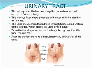
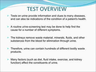
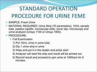
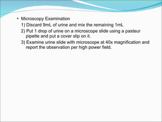
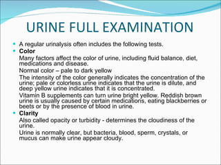
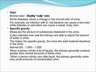
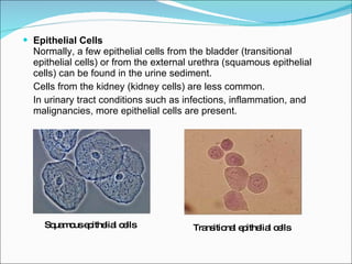
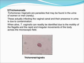
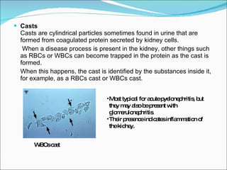
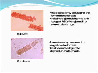
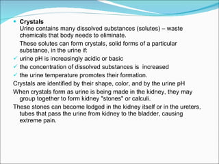
The kidneys and bladder work together to make urine and remove it from our body.  The kidneys filter waste products and water from the blood to form urine.  The urine moves from the kidneys through tubes called ureters to the bladder, which stores the urine until it is full.  From the bladder, urine leaves the body through another thin tube, the urethra.  After the bladder starts to empty, it normally empties all of the urine. URINARY TRACT
TEST OVERVIEW Tests on urine provide information and clues to many diseases, and can also be indications of the condition of a patient's health. A routine urine-screening test may be done to help find the cause for a number of different symptoms. The kidneys remove waste material, minerals, fluids, and other substances from the blood for elimination through urine. Therefore, urine can contain hundreds of different bodily waste products.  Many factors (such as diet, fluid intake, exercise, and kidney function) affect the constituents of urine.
WHY IT IS DONE??? A urine test may be done: To check for a disease or infection of the urinary tract. Symptoms of a urine infection may include colored or bad-smelling urine, pain when urinating, hard to urinate, flank pain, blood in the urine (hematuria), or fever.  To check the treatment of conditions such as diabetes, kidney stones, a urinary tract infection (UTI), or some kidney or liver diseases.  As part of a regular physical examination.
STANDARD OPERATION PROCEDURE FOR URINE FEME SAMPLE: Fresh Urine MATERIAL REQUIRED: Urine Strip (10 parameters), 10mL sample tube, pasteur pipette, microscope slide, cover slip, microscope and urine analyzer (Urisys 1100 or Urisys 1800). PROCEDURE: Full Examination 1) Put 10mL urine in urine tube 2) Dip 1 urine strip in urine 3) Wipe and put it in the reader and press start 4) Analyzer will read the strip and result will be printed out 5) Record result and proceed to spin urine at 1500rpm for 5  minutes.
Microscopy Examination 1) Discard 9mL of urine and mix the remaining 1mL 2) Put 1 drop of urine on a microscope slide using a pasteur  pipette and put a cover slip on it. 3) Examine urine slide with microscope at 40x magnification and  report the observation per high power field.
URINE FULL EXAMINATION A regular urinalysis often includes the following tests. Color Many factors affect the color of urine, including fluid balance, diet, medications and disease. Normal color – pale to dark yellow The intensity of the color generally indicates the concentration of the urine; pale or colorless urine indicates that the urine is dilute, and deep yellow urine indicates that it is concentrated.  Vitamin B supplements can turn urine bright yellow. Reddish brown urine is usually caused by certain medications, eating blackberries or beets or by the presence of blood in urine. Clarity Also called opacity or turbidity - determines the cloudiness of the urine.  Urine is normally clear, but bacteria, blood, sperm, crystals, or mucus can make urine appear cloudy.
Odor Normal odor :  Slightly "nutty" odor. Some diseases cause a change in the normal odor of urine.  For example, an infection with E. coli bacteria can cause a foul odor while diabetes or starvation can cause a sweet, fruity odor. Specific gravity Measures the amount of substances dissolved in the urine.  It also indicates how well the kidneys are able to adjust the amount of water in urine.  The higher the specific gravity, the more the solid material dissolved in the urine.  Normal SG : 1.005 – 1.025 When a person drinks a lot of liquids, the kidneys generally produce greater than normal amounts of dilute urine.   When a person drinks very little liquid, the kidneys generally make only small amounts of concentrated urine.
PH To measure of how acidic or alkaline the urine is.  Normal pH: 4.6 – 8.0 A high (alkaline) pH can be caused by severe vomiting, a kidney  disease, some UTIs, and asthma. A low (acidic)  pH may be caused by severe lung disease (emphysema),  uncontrolled diabetes, aspirin overdose, severe diarrhea,  dehydration, starvation or drinking too much alcohol. Protein Normally there is no protein in the urine (negative). Sometimes a small amount of protein is released into the urine when a person stands up (this condition is called postural proteinuria).  Fever, strenuous exercise, normal pregnancy, and some diseases (especially kidney disease) may also cause protein in the urine.
Glucose Glucose is the type of sugar usually found in blood. Normally there is very little or no glucose in urine. However, when the blood sugar level is very high, as in uncontrolled diabetes, it spills over into the urine.  Glucose can also be present in urine when the kidneys are damaged or diseased, adrenal gland problem, liver damage, brain injury or certain types of poisoning. Ketones Normal : no ketones (negative). When fat is broken down for energy, the body produces by-products called ketones (or ketone bodies) and releases them into the urine.  Large amounts of ketones in the urine may signal a dangerous condition known as Diabetic Ketoacidosis.  A diet low in sugars and starches (carbohydrates), starvation, or prolonged vomiting may also cause ketones in the urine.
Bilirubin Normal : Negative Substance formed by the breakdown of RBCs.   It is passed from the body in stool and should not found in urine.  If it is present, it often means the liver is damaged or that the flow of bile from the gallbladder is blocked. Urobilinogen Normal : Negative Substance formed by the breakdown of bilirubin. It is also passed from the body in stool and only small amounts of urobilinogen are found in urine.  Urobilinogen in urine can be a sign of liver disease (cirrhosis, hepatitis) that the flow of bile from the gallbladder is blocked.
Nitrites Normal : Negative Bacteria that cause a urinary tract infection (UTI) make an enzyme that changes urinary nitrates to nitrites. Nitrites in urine show a UTI is present.  Leukocyte esterase (WBC esterase)   Normal : Negative Leukocyte esterase shows leukocytes (WBCs) in the urine. WBCs in the urine may mean a UTI is present.
URINE MICROSCOPIC EXAMINATION It will typically be done when there are abnormal findings on the physical or chemical examination and also  to verify the results produced from the urine analyzers. It is performed on urine sediment – urine that has been centrifuged to concentrate the substances in it at the bottom of a tube. The fluid at the top of the tube is then discarded and the drops of fluid remaining are examined under a microscope.
In healthy people, the urine contains small numbers of cells and other formed elements from the entire urinary tract, and epithelial cells from the kidney, ureter , bladder, and urethra.  A microscopic examination of urine sediment detects the presence and amounts of red blood cells, white bloods cells, crystals, casts and bacteria or yeast.
Red Blood Cells (RBCs)   Normally, a few RBCs are present in urine sediment (0-5 per hpf). Inflammation, injury, or disease in the kidneys or elsewhere in the urinary tract can cause RBCs to leak out of the blood vessels into the urine.  RBCs can also be a contaminant due to an improper sample collection and blood from hemorrhoids or menstruation.   RBCs may appear normally shaped, swollen by dilute urine or crenated by concentrated urine.    Hematuria is the presence of abnormal numbers of red cells in urine due to: .... glomerular damage .... tumors which erode the urinary tract .... kidney trauma, .... urinary tract stones, .... upper and lower urinary tract infections, .... nephrotoxins, and physical stress
 
White Blood Cells (WBCs)   The number of WBCs in urine sediment is normally low (0-5 per hpf). When the number is high, it indicates an infection or inflammation somewhere in the urinary tract.  WBCs can also be a contaminant, such as those from vaginal secretions. WBCs
Epithelial Cells   Normally, a few epithelial cells from the bladder (transitional epithelial cells) or from the external urethra (squamous epithelial cells) can be found in the urine sediment.  Cells from the kidney (kidney cells) are less common.  In urinary tract conditions such as infections, inflammation, and malignancies, more epithelial cells are present.  Squamous epithelial cells Transitional epithelial cells
Microorganisms  The urinary tract is sterile, so there will be no microorganisms seen in the urine sediment.  1)  Bacteria  from the surrounding skin can enter the urinary tract at the urethra and move up to the bladder, causing a UTI. If the infection is not treated, it can eventually move to the kidneys and cause pyelonephritis.  Less frequently, bacteria from a blood infection (septicemia) may move into the urinary tract and also can cause UTI.   A urine culture and sensitivity may be performed if a UTI is suspected. Bacteria : Cocci Bacteria : Rods
2) Yeast and Fungal (Hyphae)   They are most often present in women who have a vaginal infection, because the urine has been contaminated with vaginal secretions during collection. Hyphae Yeast : Candida
3)Trichomonads   Trchomonas Vaginalis  are parasites that may be found in the urine of women or men (rarely). These actually infecting the vaginal canal and their presence in urine is due to contamination. When alive,  T. vaginalis  can readily be identified due to the motility of the flagella and the rapid and irregular movements of the body across the microscopic field. Trichomonas Vaginalis
Casts   Casts are cylindrical particles sometimes found in urine that are formed from coagulated protein secreted by kidney cells.   When a disease process is present in the kidney, other things such as RBCs or WBCs can become trapped in the protein as the cast is formed.  When this happens, the cast is identified by the substances inside it, for example, as a RBCs cast or WBCs cast. WBCs cast Most typical for acute pyelonephritis, but they may also be present with glomerulonephritis.  Their presence indicates inflammation of the kidney.
RBCs cast Granular cast Have a textured appearance which ranges from fine to coarse.  Usually form as a stage in the degeneration of cellular casts . Red blood cells may stick together and form red blood cell casts.  Indicative of glomerulonephritis, with leakage of RBC's from glomeruli, or severe tubular damage.
Hyaline cast Have a smooth texture. Are not always indicative clinically significant disease. Greater numbers may be seen associated with proteinuria of renal (such as glomerular disease). Waxy cast Have a smooth consistency, squared off ends, as if brittle and easily broken. Found especially in chronic renal disease, diabetic nephropathy, malignant hypertension and glomerulonephritis.
Fatty cast Identified by the presence of refractile lipid droplets.  Often, they are seen in urines in which free lipid droplets are present as well.
Crystals  Urine contains many dissolved substances (solutes) – waste chemicals that body needs to eliminate.  These solutes can form crystals, solid forms of a particular substance, in the urine if: urine pH is increasingly acidic or basic the concentration of dissolved substances is  increased the urine temperature promotes their formation.  Crystals are identified by their shape, color, and by the urine pH When crystals form as urine is being made in the kidney, they may group together to form kidney "stones" or calculi.  These stones can become lodged in the kidney itself or in the ureters, tubes that pass the urine from kidney to the bladder, causing extreme pain.
Calcium Oxalate  Dihydrate Crystals Calcium Oxalate  Monohydrate Crystals In acid urine. Have different shapes like colorless squares whose corner are connected by intersecting lines, spindle, oval or dumbbell shape. Most common constituent of kidney stones.
Uric Acid Crystals Ammonium Urate (or Biurate) In acid urine. Looked like leave shapes. Often yellow to orange-brown in color In alkaline urine. Golden brown color with thorn apple shape. The only urate crystals appear in alkaline urine
Calcium Phosphate Triple Phosphate In alkaline urine. Also called struvite crystals Appear as colorless, prism-like crystal or coffin lids UTI with urease producing bacteria can promote struvite crystalluria by raising urine pH and increasing free ammonia. In alkaline urine. Usually found as rosette and pointed finger forms.
Cystine Crystals Amorphous Phosphate In acid urine. Is an amino acids Seen as flat colorless hexagonal plates. Often aggregate in layers. In alkaline or neutral urine Looked very scattered and bright around the field. As fine, colorless or slightly brown granules. White precipitate is observed on centrifugation.
Amorphous urate In acid urine. Appear as fine pink or brownish granules. Salts of uric acid. Pink precipitate is observed on centrifugation. Cholesterol Crystal Found as transparent plates. Usually one or more corners are cut off or notched Also called ‘stair step crystals’. Usually accompanied by proteinuria, but they are only rarely seen.
Leucine Crystals Tyrosine Crystals In acid urine. Found as colorless to yellowish brown, needle shaped crystals Usually appear together with leucine crystals. Products of protein metabolism and appear in urine people with tissue degeneration or necrosis (such as severe liver disease). In acid urine. Same clinical conditions like tyrosine. Found in form of spheroids and appear as yellowish brown bodies.
What Affects the Test foods that can color the urine, such as blackberries, beets, and rhubarb. exercise strenuously before the test. menstruating or close to starting menstrual period. taking certain medicines that color the urine include vitamin B, phenazopyridine (Pyridium), rifampin, and phenytoin (Dilantin).  Having an X-ray test with contrast material in the past 3 days.
THANK YOU

Urine FEME

  • 1.
  • 2.
    The kidneys andbladder work together to make urine and remove it from our body. The kidneys filter waste products and water from the blood to form urine. The urine moves from the kidneys through tubes called ureters to the bladder, which stores the urine until it is full. From the bladder, urine leaves the body through another thin tube, the urethra. After the bladder starts to empty, it normally empties all of the urine. URINARY TRACT
  • 3.
    TEST OVERVIEW Testson urine provide information and clues to many diseases, and can also be indications of the condition of a patient's health. A routine urine-screening test may be done to help find the cause for a number of different symptoms. The kidneys remove waste material, minerals, fluids, and other substances from the blood for elimination through urine. Therefore, urine can contain hundreds of different bodily waste products. Many factors (such as diet, fluid intake, exercise, and kidney function) affect the constituents of urine.
  • 4.
    WHY IT ISDONE??? A urine test may be done: To check for a disease or infection of the urinary tract. Symptoms of a urine infection may include colored or bad-smelling urine, pain when urinating, hard to urinate, flank pain, blood in the urine (hematuria), or fever. To check the treatment of conditions such as diabetes, kidney stones, a urinary tract infection (UTI), or some kidney or liver diseases. As part of a regular physical examination.
  • 5.
    STANDARD OPERATION PROCEDUREFOR URINE FEME SAMPLE: Fresh Urine MATERIAL REQUIRED: Urine Strip (10 parameters), 10mL sample tube, pasteur pipette, microscope slide, cover slip, microscope and urine analyzer (Urisys 1100 or Urisys 1800). PROCEDURE: Full Examination 1) Put 10mL urine in urine tube 2) Dip 1 urine strip in urine 3) Wipe and put it in the reader and press start 4) Analyzer will read the strip and result will be printed out 5) Record result and proceed to spin urine at 1500rpm for 5 minutes.
  • 6.
    Microscopy Examination 1)Discard 9mL of urine and mix the remaining 1mL 2) Put 1 drop of urine on a microscope slide using a pasteur pipette and put a cover slip on it. 3) Examine urine slide with microscope at 40x magnification and report the observation per high power field.
  • 7.
    URINE FULL EXAMINATIONA regular urinalysis often includes the following tests. Color Many factors affect the color of urine, including fluid balance, diet, medications and disease. Normal color – pale to dark yellow The intensity of the color generally indicates the concentration of the urine; pale or colorless urine indicates that the urine is dilute, and deep yellow urine indicates that it is concentrated. Vitamin B supplements can turn urine bright yellow. Reddish brown urine is usually caused by certain medications, eating blackberries or beets or by the presence of blood in urine. Clarity Also called opacity or turbidity - determines the cloudiness of the urine. Urine is normally clear, but bacteria, blood, sperm, crystals, or mucus can make urine appear cloudy.
  • 8.
    Odor Normal odor: Slightly "nutty" odor. Some diseases cause a change in the normal odor of urine. For example, an infection with E. coli bacteria can cause a foul odor while diabetes or starvation can cause a sweet, fruity odor. Specific gravity Measures the amount of substances dissolved in the urine. It also indicates how well the kidneys are able to adjust the amount of water in urine. The higher the specific gravity, the more the solid material dissolved in the urine. Normal SG : 1.005 – 1.025 When a person drinks a lot of liquids, the kidneys generally produce greater than normal amounts of dilute urine. When a person drinks very little liquid, the kidneys generally make only small amounts of concentrated urine.
  • 9.
    PH To measureof how acidic or alkaline the urine is. Normal pH: 4.6 – 8.0 A high (alkaline) pH can be caused by severe vomiting, a kidney disease, some UTIs, and asthma. A low (acidic) pH may be caused by severe lung disease (emphysema), uncontrolled diabetes, aspirin overdose, severe diarrhea, dehydration, starvation or drinking too much alcohol. Protein Normally there is no protein in the urine (negative). Sometimes a small amount of protein is released into the urine when a person stands up (this condition is called postural proteinuria). Fever, strenuous exercise, normal pregnancy, and some diseases (especially kidney disease) may also cause protein in the urine.
  • 10.
    Glucose Glucose isthe type of sugar usually found in blood. Normally there is very little or no glucose in urine. However, when the blood sugar level is very high, as in uncontrolled diabetes, it spills over into the urine. Glucose can also be present in urine when the kidneys are damaged or diseased, adrenal gland problem, liver damage, brain injury or certain types of poisoning. Ketones Normal : no ketones (negative). When fat is broken down for energy, the body produces by-products called ketones (or ketone bodies) and releases them into the urine. Large amounts of ketones in the urine may signal a dangerous condition known as Diabetic Ketoacidosis. A diet low in sugars and starches (carbohydrates), starvation, or prolonged vomiting may also cause ketones in the urine.
  • 11.
    Bilirubin Normal :Negative Substance formed by the breakdown of RBCs. It is passed from the body in stool and should not found in urine. If it is present, it often means the liver is damaged or that the flow of bile from the gallbladder is blocked. Urobilinogen Normal : Negative Substance formed by the breakdown of bilirubin. It is also passed from the body in stool and only small amounts of urobilinogen are found in urine. Urobilinogen in urine can be a sign of liver disease (cirrhosis, hepatitis) that the flow of bile from the gallbladder is blocked.
  • 12.
    Nitrites Normal :Negative Bacteria that cause a urinary tract infection (UTI) make an enzyme that changes urinary nitrates to nitrites. Nitrites in urine show a UTI is present. Leukocyte esterase (WBC esterase) Normal : Negative Leukocyte esterase shows leukocytes (WBCs) in the urine. WBCs in the urine may mean a UTI is present.
  • 13.
    URINE MICROSCOPIC EXAMINATIONIt will typically be done when there are abnormal findings on the physical or chemical examination and also to verify the results produced from the urine analyzers. It is performed on urine sediment – urine that has been centrifuged to concentrate the substances in it at the bottom of a tube. The fluid at the top of the tube is then discarded and the drops of fluid remaining are examined under a microscope.
  • 14.
    In healthy people,the urine contains small numbers of cells and other formed elements from the entire urinary tract, and epithelial cells from the kidney, ureter , bladder, and urethra. A microscopic examination of urine sediment detects the presence and amounts of red blood cells, white bloods cells, crystals, casts and bacteria or yeast.
  • 15.
    Red Blood Cells(RBCs) Normally, a few RBCs are present in urine sediment (0-5 per hpf). Inflammation, injury, or disease in the kidneys or elsewhere in the urinary tract can cause RBCs to leak out of the blood vessels into the urine. RBCs can also be a contaminant due to an improper sample collection and blood from hemorrhoids or menstruation. RBCs may appear normally shaped, swollen by dilute urine or crenated by concentrated urine. Hematuria is the presence of abnormal numbers of red cells in urine due to: .... glomerular damage .... tumors which erode the urinary tract .... kidney trauma, .... urinary tract stones, .... upper and lower urinary tract infections, .... nephrotoxins, and physical stress
  • 16.
  • 17.
    White Blood Cells(WBCs) The number of WBCs in urine sediment is normally low (0-5 per hpf). When the number is high, it indicates an infection or inflammation somewhere in the urinary tract. WBCs can also be a contaminant, such as those from vaginal secretions. WBCs
  • 18.
    Epithelial Cells Normally, a few epithelial cells from the bladder (transitional epithelial cells) or from the external urethra (squamous epithelial cells) can be found in the urine sediment. Cells from the kidney (kidney cells) are less common. In urinary tract conditions such as infections, inflammation, and malignancies, more epithelial cells are present. Squamous epithelial cells Transitional epithelial cells
  • 19.
    Microorganisms Theurinary tract is sterile, so there will be no microorganisms seen in the urine sediment. 1) Bacteria from the surrounding skin can enter the urinary tract at the urethra and move up to the bladder, causing a UTI. If the infection is not treated, it can eventually move to the kidneys and cause pyelonephritis. Less frequently, bacteria from a blood infection (septicemia) may move into the urinary tract and also can cause UTI. A urine culture and sensitivity may be performed if a UTI is suspected. Bacteria : Cocci Bacteria : Rods
  • 20.
    2) Yeast andFungal (Hyphae) They are most often present in women who have a vaginal infection, because the urine has been contaminated with vaginal secretions during collection. Hyphae Yeast : Candida
  • 21.
    3)Trichomonads Trchomonas Vaginalis are parasites that may be found in the urine of women or men (rarely). These actually infecting the vaginal canal and their presence in urine is due to contamination. When alive, T. vaginalis can readily be identified due to the motility of the flagella and the rapid and irregular movements of the body across the microscopic field. Trichomonas Vaginalis
  • 22.
    Casts Casts are cylindrical particles sometimes found in urine that are formed from coagulated protein secreted by kidney cells. When a disease process is present in the kidney, other things such as RBCs or WBCs can become trapped in the protein as the cast is formed. When this happens, the cast is identified by the substances inside it, for example, as a RBCs cast or WBCs cast. WBCs cast Most typical for acute pyelonephritis, but they may also be present with glomerulonephritis. Their presence indicates inflammation of the kidney.
  • 23.
    RBCs cast Granularcast Have a textured appearance which ranges from fine to coarse. Usually form as a stage in the degeneration of cellular casts . Red blood cells may stick together and form red blood cell casts. Indicative of glomerulonephritis, with leakage of RBC's from glomeruli, or severe tubular damage.
  • 24.
    Hyaline cast Havea smooth texture. Are not always indicative clinically significant disease. Greater numbers may be seen associated with proteinuria of renal (such as glomerular disease). Waxy cast Have a smooth consistency, squared off ends, as if brittle and easily broken. Found especially in chronic renal disease, diabetic nephropathy, malignant hypertension and glomerulonephritis.
  • 25.
    Fatty cast Identifiedby the presence of refractile lipid droplets. Often, they are seen in urines in which free lipid droplets are present as well.
  • 26.
    Crystals Urinecontains many dissolved substances (solutes) – waste chemicals that body needs to eliminate. These solutes can form crystals, solid forms of a particular substance, in the urine if: urine pH is increasingly acidic or basic the concentration of dissolved substances is increased the urine temperature promotes their formation. Crystals are identified by their shape, color, and by the urine pH When crystals form as urine is being made in the kidney, they may group together to form kidney "stones" or calculi. These stones can become lodged in the kidney itself or in the ureters, tubes that pass the urine from kidney to the bladder, causing extreme pain.
  • 27.
    Calcium Oxalate Dihydrate Crystals Calcium Oxalate Monohydrate Crystals In acid urine. Have different shapes like colorless squares whose corner are connected by intersecting lines, spindle, oval or dumbbell shape. Most common constituent of kidney stones.
  • 28.
    Uric Acid CrystalsAmmonium Urate (or Biurate) In acid urine. Looked like leave shapes. Often yellow to orange-brown in color In alkaline urine. Golden brown color with thorn apple shape. The only urate crystals appear in alkaline urine
  • 29.
    Calcium Phosphate TriplePhosphate In alkaline urine. Also called struvite crystals Appear as colorless, prism-like crystal or coffin lids UTI with urease producing bacteria can promote struvite crystalluria by raising urine pH and increasing free ammonia. In alkaline urine. Usually found as rosette and pointed finger forms.
  • 30.
    Cystine Crystals AmorphousPhosphate In acid urine. Is an amino acids Seen as flat colorless hexagonal plates. Often aggregate in layers. In alkaline or neutral urine Looked very scattered and bright around the field. As fine, colorless or slightly brown granules. White precipitate is observed on centrifugation.
  • 31.
    Amorphous urate Inacid urine. Appear as fine pink or brownish granules. Salts of uric acid. Pink precipitate is observed on centrifugation. Cholesterol Crystal Found as transparent plates. Usually one or more corners are cut off or notched Also called ‘stair step crystals’. Usually accompanied by proteinuria, but they are only rarely seen.
  • 32.
    Leucine Crystals TyrosineCrystals In acid urine. Found as colorless to yellowish brown, needle shaped crystals Usually appear together with leucine crystals. Products of protein metabolism and appear in urine people with tissue degeneration or necrosis (such as severe liver disease). In acid urine. Same clinical conditions like tyrosine. Found in form of spheroids and appear as yellowish brown bodies.
  • 33.
    What Affects theTest foods that can color the urine, such as blackberries, beets, and rhubarb. exercise strenuously before the test. menstruating or close to starting menstrual period. taking certain medicines that color the urine include vitamin B, phenazopyridine (Pyridium), rifampin, and phenytoin (Dilantin). Having an X-ray test with contrast material in the past 3 days.
  • 34.