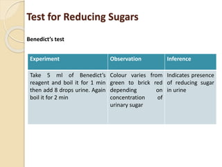
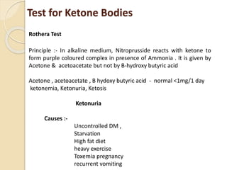
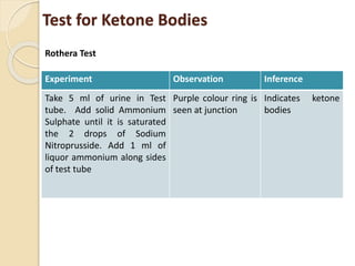
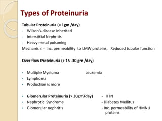
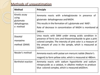
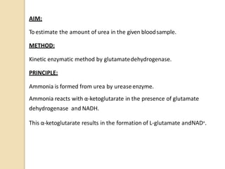
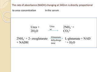
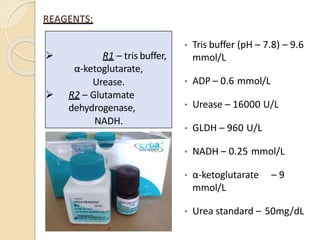
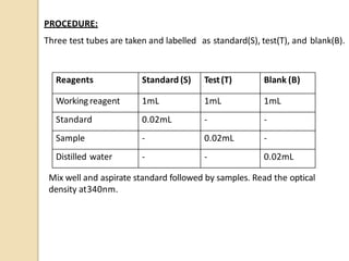
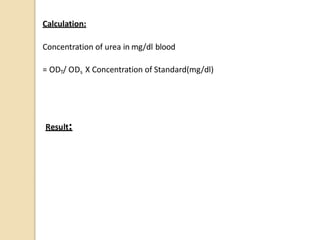
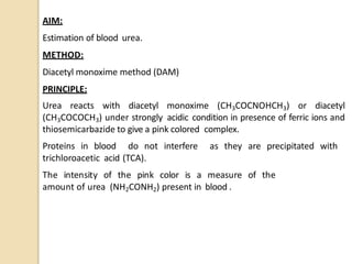
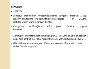
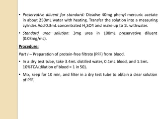
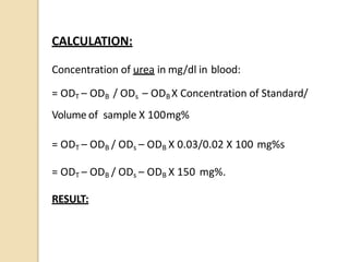
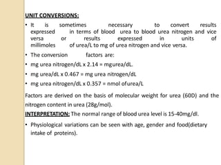
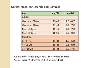
By
Dr .Y. Venkatamani
2nd Year PG
MD Biochemistry
Gandhi Medical College
Secunderabad
ABNORMAL CONSTITUENTS OF URINE
Composition of Normal Urine
Indications For Urinalysis
• Suspected renal diseases like glomerulonephritis, nephrotic
syndrome, pyelonephritis, and renal failure.
• Detection of urinary tract infection.
• Detection and management of metabolic disorders like diabetes
mellitus.
• Differential diagnosis of jaundice.
• Detection and management of plasma cell dyscrasias.
• Diagnosis of pregnancy
• For certain hormones like Catecholamines, 17-Hydroxy steroids,
urinary free cortisol
• Urinary VMA to detect excess of epinephrine and nor epinephrine
 To detect various conditions like lipuria, chyluria, choluria,
Haemoglobinuria, Myoglobinuria,
 Detection of drugs, poisoning
The chemical examination is carried out for the following substances :-
• Proteins
• Glucose
• Ketones
• Bilirubin
• Bile salts
• Urobilinogen
• Blood
• Hemoglobin
• Myoglobin
• Nitrite or leukocyte esterase
Abnormal constituents of
Urine
New Born Screening for Inborn Errors of
Metabolism
• Phenylketonuria
• Tyrosyluria
• Alcaptonuria
• Melanuria
• Branched Chain Amino Acid Disorders
 Maple Syrup urine disese
 Organic Acidemias
• Tryptophan disorders – hartnup’s disease
• 5-Hydroxyindoleacetic acid - (5-HIAA)
• Cystine disorders
• Homocystinuria
• Porphyrin disorders
• Mucopolysaccharide disorders
• Purine disorders
• Carbohydrate disorders
Abnormal Constituents of Urine
Substances which are not present in easily detectable quantities in
urine of healthy individuals
Solution A
Physical Characteristics
• Appearance – Transparent , Clear
• Colour – colour less,
• Odour –fruity odour
Test for Reducing Sugars
Benedict’s test
In alkaline medium, sugars undergo tautomerization to give endiols
forms which are powerful reducing agents. They reduce cupric ions to
red cuprous oxide
Benedict’s Reagent :- CuSO4, NO2CO3, Sodium Chloride
Various colors of the precipitate depends upon the concentration of
sugar in urine
Color Sugar content (Grams %)
Blue Nil
Green 0.1 -0.5 g%
Yellow 0.5 - 1.0 g%
Orange 1 -1.5 g%
Red 1.5 - 2 g%
Brick Red > 2 g%
Test for Reducing Sugars
Benedict’s test
Interpretation: from carbohydrates
Glycosuria (>180 mg/dl)
Causes : - Renal Glycosuria , pregnancy, heavy metal poisoning , renal
tubular necrosis, alimentary Glycosuria
Diabetes mellitus false + ve
Hyperthyroidism Vit C
Hyperpituitarism Gluconic acid
Hyperadrenalism Homogentisic acid
Stress Drug salicylates gets oxidized
Severe infection
Test for Reducing Sugars
Experiment Observation Inference
Take 5 ml of Benedict’s
reagent and boil it for 1 min
then add 8 drops urine. Again
boil it for 2 min
Colour varies from
green to brick red
depending on
concentration of
urinary sugar
Indicates presence
of reducing sugar
in urine
Benedict’s test
Test for Ketone Bodies
Rothera Test
Principle :- In alkaline medium, Nitroprusside reacts with ketone to
form purple coloured complex in presence of Ammonia . It is given by
Acetone & acetoacetate but not by B-hydroxy butyric acid
Acetone , acetoacetate , B hydoxy butyric acid - normal <1mg/1 day
ketonemia, Ketonuria, Ketosis
Ketonuria
Causes :-
Uncontrolled DM ,
Starvation
High fat diet
heavy exercise
Toxemia pregnancy
recurrent vomiting
Test for Ketone Bodies
Experiment Observation Inference
Take 5 ml of urine in Test
tube. Add solid Ammonium
Sulphate until it is saturated
the 2 drops of Sodium
Nitroprusside. Add 1 ml of
liquor ammonium along sides
of test tube
Purple colour ring is
seen at junction
Indicates ketone
bodies
Rothera Test
Rothera Test
Test for Urine
Heat Coagulation test
Principle: - By heating denaturation of proteins occurs denaturation causes
loss of bioactivity of proteins due to unfolding of Protein tertiary structure
Isoelectric PH (PI) of Albumin – 5.4
Globulin – 5.5
Caesin -4.6
Urine Protein composition
Total Proteins – 150 g/day Albumin – 15 mg
Tamm- Horsfall protein– 70 mg Mucopolysaccharides-15mg
Blood group related antigens – 35 mg Immunoglobulin - 5 mg
Rest hormones and enzymes - 10 mg
Bence Jones Proteins - Light chain IG excreted in urine of multiple myeloma
patients . It coagulates 40-600C, upon further heating turbidity appears on
cooling it reappears
Solution B
Test for Urine
Heat Coagulation test
Experiment Observation Inference
Fill 3/4 of test tube with clear urine. If
urine is turbid filter if before
performing. Heat the upper 1/3rd of
test tube. Add few drops of 1/3 acetic
acid to the solution
White
Coagulum is
formed
Indicates presence of
protein is urine
Solution B
Proteinuria
Presence of protein in urine is called proteinuria
Causes – Increased glomerular permeability
Reduced tubular reabsorption
Increased secretion of proteins
Physiological – - Exercise, Postural, Pregnancy, exposure to cold
Pathological -
Pre-renal Renal
dehydration Nephrotic syndrome
Burns Renal failure
Toxemia pregnancy Chronic / Acute GMN
Fever Pyelonephritis
TB of Kidney
Types of Proteinuria
Tubular Proteinuria (< 1gm /day)
- Wilson’s disease inherited
- Interstitial Nephritis
- Heavy metal poisoning
Mechanism - Inc. permeability to LMW proteins, Reduced tubular function
Over flow Proteinuria (> 15 -30 gm /day)
• Multiple Myeloma Leukemia
• Lymphoma
• Production is more
• Glomerular Proteinuria (> 30gm/day) - HTN
• Nephrotic Syndrome - Diabetes Mellitus
• Glomerular nephritis - Inc. permeability of HMNU
proteins
Tests for Blood
Benzidine Test
Principle : - Peroxides in RBC splits H2O2 to produce nascent O2 which
reacts with Benzidine in acidic medium to give bluish green colour
complex
• Hematuria (blood)
• Hemoglobinuria (RBC)
• Myoglobinuria
Applied Aspect – Hematuria
Causes of
1) Gross Hematuria 2) Microscopic Hematuria
Renal stones sickle cell anemia
Acute glomerulonephritis Pyelonephritis kidney disease
malignancy Coagulation disorders
Malaria Malignant hypertension
Trauma Hemolytic Anemia
TB
Tests for Blood
Benzidine Test
Experiment Observation Inference
Take 2 ml of Benzidine
solution. Add 2 ml of
urine 1 ml of H2O2
Bluish green colour is seen Presence of blood in
urine
Solution C
Physical characteristics
• Appearance – transparent , clear
• Colour –green
• Odour – odourless
Benzidine Test
Tests for Bile Salts
Hay’s Test
Bile salts act as emulsifying agents which reduces the surface tension,
hence sulphur powder sinks to bottom
Interpretation
Primary bile salts (liver) - Cholicacid, Chenodeoxycholicacid coagulation
is by Glycine and Taurine
Secondary Bile Salts (Intestine): Deoxy cholic acid , lithocholic acid
Bile Salts : Na & K salts of taurocholic acid & glycocholate
obstructive jaundices – Presence of Bile salts in urine
Prehepatic – Na salts & pigments in urine
Urine Pre Hepatic Hepatic Post Hepatic
Bile Salts -ve - ve + ve
Bile pigments - ve + ve + ve
Tests for Bile Salts
Experiment Observation Inference
Take 5 ml of urine
Sprinkle pinch of sulfur
powder. Control – take 5
ml of water solution.
Add pinch of sulfur
powder
Powder sinks
Powder floats
Presence of bile slats
in urine
Absence of bile salts
in urine
Hay’s Test
Tests for Bile Pigments
Fouchet’s test
Principle : Bile pigments are absorbed on BaSO4 precipitate which are
oxidized by Fouchet’s reagent to biliverdin, & bili cyanin which are blue
and green respectively . Variation of colour due to exposure to O2
Bile pigments : - Bilirubin, Biliverdin , Bilicyanin
Diseases :-
• Hepatic Causes : - Hepatitis (Viral, toxic)
Cholesterol (intrahepatic)
• Post Hepatic :- Obstruction gall stones, tumor of bile duct carcinoma
of pancreas
Tests for Bile Pigments
Experiment Observation Inference
Take 5 ml of urine. Add 1 ml of
MgSO4 Add l ml of BaSO4 Until
white ppl. Is obtained. Filter the
solution dry the residue and add
1-2 drops of Fouchet’s reagent
to filter paper
Series of yellow, blue,
green colour are
obtained
Indicates presence
of bile pigments
Fouchet’s test
Fouchet’s test
ESTIMATION OF UREA(NH2CONH2)
 Urea is the highest(75%) non-protein nitrogen compound
in the blood.
 Urea is the major excretory product of protein
metabolism.
 It is formed in the liver from free ammonia generated
during protein catabolism.
 The most important catabolic pathway for eliminating
excess nitrogen in the human body.
UREA (NH2CONH2):
Methods of ureaestimation
Method Principle
Kinetic assay
using
glutamate
dehydrogenas
e method
Ammonia reacts with α-ketoglutarate in presence of
glutamate dehydrogenase and NADH.
This results in the formation of L-glutamate and NAD+.
Rate of decrease in concentration of NADH is monitored at
340nm
Diacetyl
monoxime
method (DAM)
Urea reacts with DAM under strong acidic condition in
presence of ferric ions and thiosemicarbazide to give a pink
colored complex. The intensity of pink color is a measure of
the amount of urea in the sample, which is measured at
520nm.
Nessler’s method Ammonia reacts with potas ium mercuric iodide (Nes ler’s
reagent) to form yellow color, which is measured at 480nm.
Berthelot reaction Ammonia reacts with sodium hypochlorite and sodium
nitroprusside as a catalyst, in alkaline medium to produce
blue colored complex, which is measured at620nm.
AIM:
Toestimate the amount of urea in the given bloodsample.
METHOD:
Kinetic enzymatic method by glutamatedehydrogenase.
PRINCIPLE:
Ammonia is formed from urea by ureaseenzyme.
Ammonia reacts with α-ketoglutarate in the presence of glutamate
dehydrogenase and NADH.
This α-ketoglutarate results in the formation of L-glutamate andNAD+.
The rate of absorbance (NADH) changing at 340nm is directly proportional
to urea concentration in the serum
Urea +
2H2O
2NH4
+ +
CO3
2-
2NH4
+ + 2- oxoglutarate
+ NADH
L-glutamate + NAD
+ + H2O
Urea
se
Glutamate
dehydroge
nase
REAGENTS:
• Tris buffer (pH – 7.8) – 9.6
mmol/L
• ADP – 0.6 mmol/L
• Urease – 16000 U/L
• GLDH – 960 U/L
• NADH – 0.25 mmol/L
• α-ketoglutarate – 9
mmol/L
• Urea standard – 50mg/dL
 R1 – tris buffer,
α-ketoglutarate,
Urease.
 R2 – Glutamate
dehydrogenase,
NADH.
WORKING REAGENTPREPARATION:
The reagents (R1 and R2) are ready to use.
The working reagent is prepared by gently mixing 4 parts of R1 with 1 part of
R2.
REAGENT STORAGE: Stored at 2-8oC.
REAGENT STABILITY: The unopened reagents are stable till the expiry
date stated on the bottle andkit label.
The working reagent (4R1:1R2) is stable for 30 days at 2-8oC, when
protected from contamination andlight.
It is recommended to prepare fresh working solution before assay is
performed.
SIGNS OF REAGENTDETERIORATION:
• Presence of particles andturbidity.
• Workingreagent absorbance<0.8 at 340nm.
• Failure to recover control values within the acceptable range.
SPECIMEN: Use unheamolytic serum, plasma orurine.
• Do not use samples preserved with fluoride and heparinammonium salt as
anti- coagulant.
• Urine: dilute urine 1+100with distilled water and multiply results by 101.
PROCEDURE:
Three test tubes are taken and labelled as standard(S), test(T), and blank(B).
Mix well and aspirate standard followed by samples. Read the optical
density at340nm.
Reagents Standard (S) Test(T) Blank (B)
Working reagent 1mL 1mL 1mL
Standard 0.02mL - -
Sample - 0.02mL -
Distilled water - - 0.02mL
Calculation:
Concentration of urea in mg/dl blood
= ODT/ ODs X Concentration of Standard(mg/dl)
Result:
AIM:
Estimation of blood urea.
METHOD:
Diacetyl monoxime method (DAM)
PRINCIPLE:
Urea reacts with diacetyl monoxime (CH3COCNOHCH3) or diacetyl
(CH3COCOCH3) under strongly acidic condition in presence of ferric ions and
thiosemicarbazide to give a pink colored complex.
Proteins in blood do not interfere as they are precipitated with
trichloroacetic acid (TCA).
The intensity of the pink color is a measure of the
amount of urea (NH2CONH2) present in blood .
REAGENTS:
• 10% TCA
• Diacetyl monoxime/ thiosemicarbazide reagent: Dissolve 1.56g
diacetyl monoxime and41mg thiosemicarbazide in 250mL
distilled water, store in brown bottle.
• Phosphoric acid-sulfuric acid- ferric chloride reagent:
Dissolve
324mg of anhydrous ferric chloride (FeCl3) in 10mL of 56% phosphoric
acid. Add 1mL of this FeCl3 reagent to 1L of 20% sulfuric acid(H2SO4).
• Diacetyl monoxime reagent: Mix equal volume of b and c. this is
to be freshly prepared.
• Preservative diluent for standard: Dissolve 40mg phenyl mercuric acetate
in about 250mL water with heating. Transfer the solution into a measuring
cylinder. Add 0.3mL concentrated H2SO4 and make up to 1L withwater.
• Standard urea solution: 3mg urea in 100mL preservative diluent
(0.03mg/mL).
Procedure:
Part I – Preparation of protein-free filtrate (PFF) from blood.
• In a dry test tube, take 3.4mL distilled water, 0.1mL blood, and 1.5mL
10%TCA(dilution of blood = 1 in 50).
• Mix, keep for 10 min, and filter in a dry test tube to obtain a clear solution
of PFF.
Part II – Use PFF for blood urea estimation.
• Label three test tubes as T(test), B(blank), and S(standard)
Mix and keep the tubes in the boiling water bath for 15 min, cool, and read
the optical density(OD) using colorimeter with a green filter of wavelength
520nm.
Reagents T B S
Protein-free filtrate 1mL - -
Standard ureasolution - - 1mL
Distilled water - 1mL -
Diacetyl monoxime
reagent
5mL 5mL 5mL
CALCULATION:
Concentration of urea in mg/dl in blood:
= ODT – ODB / ODs – ODB X Concentration of Standard/
Volume of sample X 100mg%
= ODT – ODB / ODs – ODB X 0.03/0.02 X 100 mg%s
= ODT – ODB / ODs – ODB X 150 mg%.
RESULT:
UNIT CONVERSIONS:
• It is sometimes necessary to convert results
expressed in terms of blood urea to blood urea nitrogen and vice
versa or results expressed in units of
millimoles of urea/L to mg of urea nitrogen and vice versa.
• The conversion factors are:
• mg urea nitrogen/dL x 2.14 = mgurea/dL.
• mg urea/dL x 0.467 = mg urea nitrogen/dL
• mg urea nitrogen/dL x 0.357 = nmol ofurea/L
Factors are derived on the basis of molecular weight for urea (60D) and the
nitrogen content in urea (28g/mol).
INTERPRETATION: The normal range of blood urea level is 15-40mg/dl.
• Physiological variations can be seen with age, gender and food(dietary
intake of proteins).
Normal ranges for serum(blood) samples
For diluted urine sample, urea is calculated for 24 hours.
Normal range: 26-43g/day (0.43-0.72mol/24hrs)
Age mg/dl mmol/l
Adults
Women <50yrs 13-40 2.6 – 6.7
Women >50yrs 21-43 3.5 – 7.2
Men <50yrs 19-45 3.2 – 7.3
Men >50yrs 18-55 3.0 – 9.2
Children
1 –3 yrs 11- 36 1.8 – 6.0
4 –13 yrs 15- 36 2.5 – 6.0
14 –19 yrs 18- 45 2.9 – 7.5
An abnormal urea level may indicate the following tabulatedconditions
Type Cause Note
High urea (High
urea
concentrationin
plasma is called
(azotemia)
Pre-renal Congestive heart
failure
•Dehydration
•High protein diet
•Increased protein
catabolism
Caused by
reduced renal
blood flow, less
blood is
delivered to
kidney, then less
urea is filtered.
Renal Renal failure
Post-renal Urinary tract
obstruction.
Low urea Low protein intake (Starvation, anorexia)
•Liver disease
•Pregnancy
BUN (Blood urea nitrogen):
• It is the measure of amount of urea nitrogen in blood.
• BUN is an indicator of renal function.
• The normal range is 6-20mg/dL.
Causes
High urea in urine • too much protein in thediet
• too much protein breakdown
in
the body
Low urea in urine • malnutrition
• too little protein in thediet
• kidney issues
Clinical Application:
Measurement of urea is used to :
• Evaluate renalfunction
• Assess hydration status
• Determine nitrogenbalance
• Aid inthe diagnosis of renal diseases
• Verify adequacy of dialysis
• Check a person's protein balance
CLINICAL SIGNIFICANCE:
• The blood urea concentration in normal individual is 15-40mg%.
• While urea concentration increases in the blood with the loss of renal
function, it is neither a specific nor sensitive indicator of renal
impairment.
• Serum urea is also dependent on nonrenal factors, such as protein
intake, hydration state, and renal perfusion.
• Severe diarrhea, vomiting, and excessive fluid loss decreases rate
of glomerular filtration, thereby increasing blood urealevels.
• Lower urinary tract obstruction and pathology resulting in decreased
glomerular filtration also leads to elevated blood urea levels.
• In renal pathology like chronic acute glomerulonephritis, nephrosis,
malignant hypertension, chronic pyelonephritis and damage to the
kidney tissues due to mercury poisoning or calcium deposition due to
hyperthyroidism and hypervitaminosis (vit-D), blood urea levels are
higher than normal.
• Postrenal conditions like enlargement of prostate gland, stones in the
urinary tract, or tumor of the bladder also leads to increased urealevels.

Abnormal Constituents Urine.pptx

  • 1.
    By Dr .Y. Venkatamani 2ndYear PG MD Biochemistry Gandhi Medical College Secunderabad ABNORMAL CONSTITUENTS OF URINE
  • 2.
  • 3.
    Indications For Urinalysis •Suspected renal diseases like glomerulonephritis, nephrotic syndrome, pyelonephritis, and renal failure. • Detection of urinary tract infection. • Detection and management of metabolic disorders like diabetes mellitus. • Differential diagnosis of jaundice. • Detection and management of plasma cell dyscrasias. • Diagnosis of pregnancy • For certain hormones like Catecholamines, 17-Hydroxy steroids, urinary free cortisol • Urinary VMA to detect excess of epinephrine and nor epinephrine  To detect various conditions like lipuria, chyluria, choluria, Haemoglobinuria, Myoglobinuria,  Detection of drugs, poisoning
  • 4.
    The chemical examinationis carried out for the following substances :- • Proteins • Glucose • Ketones • Bilirubin • Bile salts • Urobilinogen • Blood • Hemoglobin • Myoglobin • Nitrite or leukocyte esterase Abnormal constituents of Urine
  • 5.
    New Born Screeningfor Inborn Errors of Metabolism • Phenylketonuria • Tyrosyluria • Alcaptonuria • Melanuria • Branched Chain Amino Acid Disorders  Maple Syrup urine disese  Organic Acidemias • Tryptophan disorders – hartnup’s disease • 5-Hydroxyindoleacetic acid - (5-HIAA) • Cystine disorders • Homocystinuria • Porphyrin disorders • Mucopolysaccharide disorders • Purine disorders • Carbohydrate disorders
  • 6.
    Abnormal Constituents ofUrine Substances which are not present in easily detectable quantities in urine of healthy individuals Solution A Physical Characteristics • Appearance – Transparent , Clear • Colour – colour less, • Odour –fruity odour
  • 7.
    Test for ReducingSugars Benedict’s test In alkaline medium, sugars undergo tautomerization to give endiols forms which are powerful reducing agents. They reduce cupric ions to red cuprous oxide Benedict’s Reagent :- CuSO4, NO2CO3, Sodium Chloride Various colors of the precipitate depends upon the concentration of sugar in urine Color Sugar content (Grams %) Blue Nil Green 0.1 -0.5 g% Yellow 0.5 - 1.0 g% Orange 1 -1.5 g% Red 1.5 - 2 g% Brick Red > 2 g%
  • 8.
    Test for ReducingSugars Benedict’s test Interpretation: from carbohydrates Glycosuria (>180 mg/dl) Causes : - Renal Glycosuria , pregnancy, heavy metal poisoning , renal tubular necrosis, alimentary Glycosuria Diabetes mellitus false + ve Hyperthyroidism Vit C Hyperpituitarism Gluconic acid Hyperadrenalism Homogentisic acid Stress Drug salicylates gets oxidized Severe infection
  • 9.
    Test for ReducingSugars Experiment Observation Inference Take 5 ml of Benedict’s reagent and boil it for 1 min then add 8 drops urine. Again boil it for 2 min Colour varies from green to brick red depending on concentration of urinary sugar Indicates presence of reducing sugar in urine Benedict’s test
  • 11.
    Test for KetoneBodies Rothera Test Principle :- In alkaline medium, Nitroprusside reacts with ketone to form purple coloured complex in presence of Ammonia . It is given by Acetone & acetoacetate but not by B-hydroxy butyric acid Acetone , acetoacetate , B hydoxy butyric acid - normal <1mg/1 day ketonemia, Ketonuria, Ketosis Ketonuria Causes :- Uncontrolled DM , Starvation High fat diet heavy exercise Toxemia pregnancy recurrent vomiting
  • 12.
    Test for KetoneBodies Experiment Observation Inference Take 5 ml of urine in Test tube. Add solid Ammonium Sulphate until it is saturated the 2 drops of Sodium Nitroprusside. Add 1 ml of liquor ammonium along sides of test tube Purple colour ring is seen at junction Indicates ketone bodies Rothera Test
  • 13.
  • 14.
    Test for Urine HeatCoagulation test Principle: - By heating denaturation of proteins occurs denaturation causes loss of bioactivity of proteins due to unfolding of Protein tertiary structure Isoelectric PH (PI) of Albumin – 5.4 Globulin – 5.5 Caesin -4.6 Urine Protein composition Total Proteins – 150 g/day Albumin – 15 mg Tamm- Horsfall protein– 70 mg Mucopolysaccharides-15mg Blood group related antigens – 35 mg Immunoglobulin - 5 mg Rest hormones and enzymes - 10 mg Bence Jones Proteins - Light chain IG excreted in urine of multiple myeloma patients . It coagulates 40-600C, upon further heating turbidity appears on cooling it reappears Solution B
  • 15.
    Test for Urine HeatCoagulation test Experiment Observation Inference Fill 3/4 of test tube with clear urine. If urine is turbid filter if before performing. Heat the upper 1/3rd of test tube. Add few drops of 1/3 acetic acid to the solution White Coagulum is formed Indicates presence of protein is urine Solution B
  • 17.
    Proteinuria Presence of proteinin urine is called proteinuria Causes – Increased glomerular permeability Reduced tubular reabsorption Increased secretion of proteins Physiological – - Exercise, Postural, Pregnancy, exposure to cold Pathological - Pre-renal Renal dehydration Nephrotic syndrome Burns Renal failure Toxemia pregnancy Chronic / Acute GMN Fever Pyelonephritis TB of Kidney
  • 18.
    Types of Proteinuria TubularProteinuria (< 1gm /day) - Wilson’s disease inherited - Interstitial Nephritis - Heavy metal poisoning Mechanism - Inc. permeability to LMW proteins, Reduced tubular function Over flow Proteinuria (> 15 -30 gm /day) • Multiple Myeloma Leukemia • Lymphoma • Production is more • Glomerular Proteinuria (> 30gm/day) - HTN • Nephrotic Syndrome - Diabetes Mellitus • Glomerular nephritis - Inc. permeability of HMNU proteins
  • 19.
    Tests for Blood BenzidineTest Principle : - Peroxides in RBC splits H2O2 to produce nascent O2 which reacts with Benzidine in acidic medium to give bluish green colour complex • Hematuria (blood) • Hemoglobinuria (RBC) • Myoglobinuria Applied Aspect – Hematuria Causes of 1) Gross Hematuria 2) Microscopic Hematuria Renal stones sickle cell anemia Acute glomerulonephritis Pyelonephritis kidney disease malignancy Coagulation disorders Malaria Malignant hypertension Trauma Hemolytic Anemia TB
  • 20.
    Tests for Blood BenzidineTest Experiment Observation Inference Take 2 ml of Benzidine solution. Add 2 ml of urine 1 ml of H2O2 Bluish green colour is seen Presence of blood in urine Solution C Physical characteristics • Appearance – transparent , clear • Colour –green • Odour – odourless
  • 21.
  • 22.
    Tests for BileSalts Hay’s Test Bile salts act as emulsifying agents which reduces the surface tension, hence sulphur powder sinks to bottom Interpretation Primary bile salts (liver) - Cholicacid, Chenodeoxycholicacid coagulation is by Glycine and Taurine Secondary Bile Salts (Intestine): Deoxy cholic acid , lithocholic acid Bile Salts : Na & K salts of taurocholic acid & glycocholate obstructive jaundices – Presence of Bile salts in urine Prehepatic – Na salts & pigments in urine Urine Pre Hepatic Hepatic Post Hepatic Bile Salts -ve - ve + ve Bile pigments - ve + ve + ve
  • 23.
    Tests for BileSalts Experiment Observation Inference Take 5 ml of urine Sprinkle pinch of sulfur powder. Control – take 5 ml of water solution. Add pinch of sulfur powder Powder sinks Powder floats Presence of bile slats in urine Absence of bile salts in urine Hay’s Test
  • 25.
    Tests for BilePigments Fouchet’s test Principle : Bile pigments are absorbed on BaSO4 precipitate which are oxidized by Fouchet’s reagent to biliverdin, & bili cyanin which are blue and green respectively . Variation of colour due to exposure to O2 Bile pigments : - Bilirubin, Biliverdin , Bilicyanin Diseases :- • Hepatic Causes : - Hepatitis (Viral, toxic) Cholesterol (intrahepatic) • Post Hepatic :- Obstruction gall stones, tumor of bile duct carcinoma of pancreas
  • 26.
    Tests for BilePigments Experiment Observation Inference Take 5 ml of urine. Add 1 ml of MgSO4 Add l ml of BaSO4 Until white ppl. Is obtained. Filter the solution dry the residue and add 1-2 drops of Fouchet’s reagent to filter paper Series of yellow, blue, green colour are obtained Indicates presence of bile pigments Fouchet’s test
  • 27.
  • 28.
  • 29.
     Urea isthe highest(75%) non-protein nitrogen compound in the blood.  Urea is the major excretory product of protein metabolism.  It is formed in the liver from free ammonia generated during protein catabolism.  The most important catabolic pathway for eliminating excess nitrogen in the human body. UREA (NH2CONH2):
  • 30.
    Methods of ureaestimation MethodPrinciple Kinetic assay using glutamate dehydrogenas e method Ammonia reacts with α-ketoglutarate in presence of glutamate dehydrogenase and NADH. This results in the formation of L-glutamate and NAD+. Rate of decrease in concentration of NADH is monitored at 340nm Diacetyl monoxime method (DAM) Urea reacts with DAM under strong acidic condition in presence of ferric ions and thiosemicarbazide to give a pink colored complex. The intensity of pink color is a measure of the amount of urea in the sample, which is measured at 520nm. Nessler’s method Ammonia reacts with potas ium mercuric iodide (Nes ler’s reagent) to form yellow color, which is measured at 480nm. Berthelot reaction Ammonia reacts with sodium hypochlorite and sodium nitroprusside as a catalyst, in alkaline medium to produce blue colored complex, which is measured at620nm.
  • 31.
    AIM: Toestimate the amountof urea in the given bloodsample. METHOD: Kinetic enzymatic method by glutamatedehydrogenase. PRINCIPLE: Ammonia is formed from urea by ureaseenzyme. Ammonia reacts with α-ketoglutarate in the presence of glutamate dehydrogenase and NADH. This α-ketoglutarate results in the formation of L-glutamate andNAD+.
  • 32.
    The rate ofabsorbance (NADH) changing at 340nm is directly proportional to urea concentration in the serum Urea + 2H2O 2NH4 + + CO3 2- 2NH4 + + 2- oxoglutarate + NADH L-glutamate + NAD + + H2O Urea se Glutamate dehydroge nase
  • 33.
    REAGENTS: • Tris buffer(pH – 7.8) – 9.6 mmol/L • ADP – 0.6 mmol/L • Urease – 16000 U/L • GLDH – 960 U/L • NADH – 0.25 mmol/L • α-ketoglutarate – 9 mmol/L • Urea standard – 50mg/dL  R1 – tris buffer, α-ketoglutarate, Urease.  R2 – Glutamate dehydrogenase, NADH.
  • 34.
    WORKING REAGENTPREPARATION: The reagents(R1 and R2) are ready to use. The working reagent is prepared by gently mixing 4 parts of R1 with 1 part of R2. REAGENT STORAGE: Stored at 2-8oC. REAGENT STABILITY: The unopened reagents are stable till the expiry date stated on the bottle andkit label. The working reagent (4R1:1R2) is stable for 30 days at 2-8oC, when protected from contamination andlight. It is recommended to prepare fresh working solution before assay is performed.
  • 35.
    SIGNS OF REAGENTDETERIORATION: •Presence of particles andturbidity. • Workingreagent absorbance<0.8 at 340nm. • Failure to recover control values within the acceptable range. SPECIMEN: Use unheamolytic serum, plasma orurine. • Do not use samples preserved with fluoride and heparinammonium salt as anti- coagulant. • Urine: dilute urine 1+100with distilled water and multiply results by 101.
  • 36.
    PROCEDURE: Three test tubesare taken and labelled as standard(S), test(T), and blank(B). Mix well and aspirate standard followed by samples. Read the optical density at340nm. Reagents Standard (S) Test(T) Blank (B) Working reagent 1mL 1mL 1mL Standard 0.02mL - - Sample - 0.02mL - Distilled water - - 0.02mL
  • 37.
    Calculation: Concentration of ureain mg/dl blood = ODT/ ODs X Concentration of Standard(mg/dl) Result:
  • 38.
    AIM: Estimation of bloodurea. METHOD: Diacetyl monoxime method (DAM) PRINCIPLE: Urea reacts with diacetyl monoxime (CH3COCNOHCH3) or diacetyl (CH3COCOCH3) under strongly acidic condition in presence of ferric ions and thiosemicarbazide to give a pink colored complex. Proteins in blood do not interfere as they are precipitated with trichloroacetic acid (TCA). The intensity of the pink color is a measure of the amount of urea (NH2CONH2) present in blood .
  • 39.
    REAGENTS: • 10% TCA •Diacetyl monoxime/ thiosemicarbazide reagent: Dissolve 1.56g diacetyl monoxime and41mg thiosemicarbazide in 250mL distilled water, store in brown bottle. • Phosphoric acid-sulfuric acid- ferric chloride reagent: Dissolve 324mg of anhydrous ferric chloride (FeCl3) in 10mL of 56% phosphoric acid. Add 1mL of this FeCl3 reagent to 1L of 20% sulfuric acid(H2SO4). • Diacetyl monoxime reagent: Mix equal volume of b and c. this is to be freshly prepared.
  • 40.
    • Preservative diluentfor standard: Dissolve 40mg phenyl mercuric acetate in about 250mL water with heating. Transfer the solution into a measuring cylinder. Add 0.3mL concentrated H2SO4 and make up to 1L withwater. • Standard urea solution: 3mg urea in 100mL preservative diluent (0.03mg/mL). Procedure: Part I – Preparation of protein-free filtrate (PFF) from blood. • In a dry test tube, take 3.4mL distilled water, 0.1mL blood, and 1.5mL 10%TCA(dilution of blood = 1 in 50). • Mix, keep for 10 min, and filter in a dry test tube to obtain a clear solution of PFF.
  • 41.
    Part II –Use PFF for blood urea estimation. • Label three test tubes as T(test), B(blank), and S(standard) Mix and keep the tubes in the boiling water bath for 15 min, cool, and read the optical density(OD) using colorimeter with a green filter of wavelength 520nm. Reagents T B S Protein-free filtrate 1mL - - Standard ureasolution - - 1mL Distilled water - 1mL - Diacetyl monoxime reagent 5mL 5mL 5mL
  • 42.
    CALCULATION: Concentration of ureain mg/dl in blood: = ODT – ODB / ODs – ODB X Concentration of Standard/ Volume of sample X 100mg% = ODT – ODB / ODs – ODB X 0.03/0.02 X 100 mg%s = ODT – ODB / ODs – ODB X 150 mg%. RESULT:
  • 43.
    UNIT CONVERSIONS: • Itis sometimes necessary to convert results expressed in terms of blood urea to blood urea nitrogen and vice versa or results expressed in units of millimoles of urea/L to mg of urea nitrogen and vice versa. • The conversion factors are: • mg urea nitrogen/dL x 2.14 = mgurea/dL. • mg urea/dL x 0.467 = mg urea nitrogen/dL • mg urea nitrogen/dL x 0.357 = nmol ofurea/L Factors are derived on the basis of molecular weight for urea (60D) and the nitrogen content in urea (28g/mol). INTERPRETATION: The normal range of blood urea level is 15-40mg/dl. • Physiological variations can be seen with age, gender and food(dietary intake of proteins).
  • 44.
    Normal ranges forserum(blood) samples For diluted urine sample, urea is calculated for 24 hours. Normal range: 26-43g/day (0.43-0.72mol/24hrs) Age mg/dl mmol/l Adults Women <50yrs 13-40 2.6 – 6.7 Women >50yrs 21-43 3.5 – 7.2 Men <50yrs 19-45 3.2 – 7.3 Men >50yrs 18-55 3.0 – 9.2 Children 1 –3 yrs 11- 36 1.8 – 6.0 4 –13 yrs 15- 36 2.5 – 6.0 14 –19 yrs 18- 45 2.9 – 7.5
  • 45.
    An abnormal urealevel may indicate the following tabulatedconditions Type Cause Note High urea (High urea concentrationin plasma is called (azotemia) Pre-renal Congestive heart failure •Dehydration •High protein diet •Increased protein catabolism Caused by reduced renal blood flow, less blood is delivered to kidney, then less urea is filtered. Renal Renal failure Post-renal Urinary tract obstruction. Low urea Low protein intake (Starvation, anorexia) •Liver disease •Pregnancy
  • 46.
    BUN (Blood ureanitrogen): • It is the measure of amount of urea nitrogen in blood. • BUN is an indicator of renal function. • The normal range is 6-20mg/dL. Causes High urea in urine • too much protein in thediet • too much protein breakdown in the body Low urea in urine • malnutrition • too little protein in thediet • kidney issues
  • 47.
    Clinical Application: Measurement ofurea is used to : • Evaluate renalfunction • Assess hydration status • Determine nitrogenbalance • Aid inthe diagnosis of renal diseases • Verify adequacy of dialysis • Check a person's protein balance
  • 48.
    CLINICAL SIGNIFICANCE: • Theblood urea concentration in normal individual is 15-40mg%. • While urea concentration increases in the blood with the loss of renal function, it is neither a specific nor sensitive indicator of renal impairment. • Serum urea is also dependent on nonrenal factors, such as protein intake, hydration state, and renal perfusion. • Severe diarrhea, vomiting, and excessive fluid loss decreases rate of glomerular filtration, thereby increasing blood urealevels.
  • 49.
    • Lower urinarytract obstruction and pathology resulting in decreased glomerular filtration also leads to elevated blood urea levels. • In renal pathology like chronic acute glomerulonephritis, nephrosis, malignant hypertension, chronic pyelonephritis and damage to the kidney tissues due to mercury poisoning or calcium deposition due to hyperthyroidism and hypervitaminosis (vit-D), blood urea levels are higher than normal. • Postrenal conditions like enlargement of prostate gland, stones in the urinary tract, or tumor of the bladder also leads to increased urealevels.