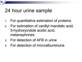
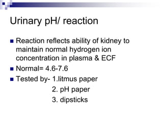
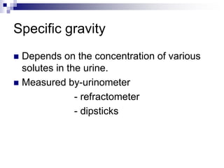
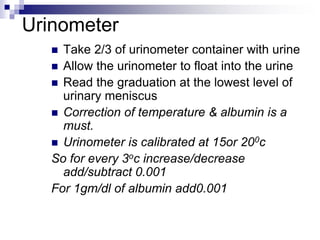
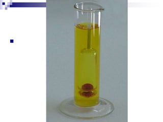
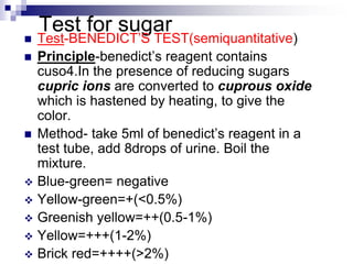
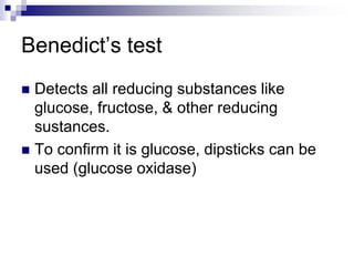
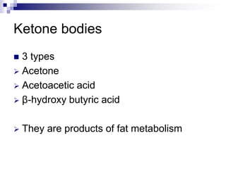
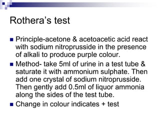
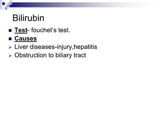
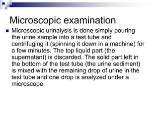
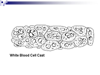
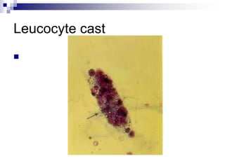
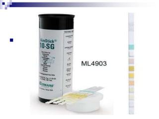
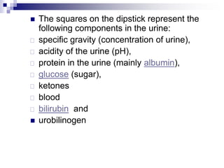
Urine analysis
Introduction
 Urine is formed in the kidneys, is a product
of ultrafiltration of plasma by the renal
glomeruli.
Collection of urine
 Early morning sample-qualitative
 Random sample- routine
 24hrs sample- quantitative
 Midstream sample-UTI
 Post prandial sample-D.M
24 hour urine sample
1. For quantitative estimation of proteins
2. For estimation of vanillyl mandelic acid,
5-hydroxyindole acetic acid,
metanephrines
3. For detection of AFB in urine
4. For detection of microalbuminuria

Urine examination
 Macroscopic examination
 Chemical examination
 Microscopic examination
Macroscopic examination
 Volume
 Color
 Odour
 Reaction or urinary pH
 Specific gravity
Urinary volume
 Normal = 600-1550ml
 Polyuria- >2000ml
 Oliguria-<400ml
 Anuria-complete cessation of urine(<200ml)
 Nocturia-excretion of urine by a adult of >500ml
with a specific gravity of <1.018 at night
(characteristic of chronic glomerulonephritis)
Causes of polyuria
 Diabetes mellitus
 Diabetes insipidus
 Polycystic kidney
 Chronic renal failure
 Diuretics
 Intravenous saline/glucose
oliguria
 Dehydration-vomiting, diarrhoea,
excessive sweating
 Renal ischemia
 Acute tubular necrosis
 Obstruction to the urinary tract
 Acute renal failure
Color & appearance
 Normal= clear & pale yellow
1. Colourless- dilution, diabetes mellitus,
diabetes insipidus, diuretics
2. Milky-purulent genitourinary tract
infection, chyluria
3. Orange-fever, excessive sweating
4. Red-beetroot ingestion,haematuria
5. Brown/ black- alkaptunuria, melanin
Urinary pH/ reaction
 Reaction reflects ability of kidney to
maintain normal hydrogen ion
concentration in plasma & ECF
 Normal= 4.6-7.6
 Tested by- 1.litmus paper
2. pH paper
3. dipsticks
Acidic urine
 Ketosis-diabetes, starvation, fever
 Systemic acidosis
 UTI- E.coli
 Acidification therapy
Alkaline urine
 Strict vegetarian
 Systemic alkalosis
 UTI- Proteus
 Alkalization therapy
Odour
 Normal= aromatic due to the volatile fatty
acids
 Ammonical – bacterial action
 Fruity- ketonuria
Specific gravity
 Depends on the concentration of various
solutes in the urine.
 Measured by-urinometer
- refractometer
- dipsticks
Urinometer
 Take 2/3 of urinometer container with urine
 Allow the urinometer to float into the urine
 Read the graduation at the lowest level of
urinary meniscus
 Correction of temperature & albumin is a
must.
 Urinometer is calibrated at 15or 200c
So for every 3oc increase/decrease
add/subtract 0.001
For 1gm/dl of albumin add0.001

High specific
gravity(hyperosthenuria)
 Normal-1.016-1.022
 Causes
All causes of oliguria
Gycosuria
Low specific
gravity(hyposthenuria)
 All causes of polyuria except gycosuria
 Fixed specific gravity (isosthenuria)=1.010
Seen in chronic renal disease when kidney
has lost the ability to concentrate or dilute
Chemical examination
 Proteins
 Sugars
 Ketone bodies
 Bilirubin
 Bile salts
 Urobilinogen
 Blood
Tests for proteins
 Test – HEAT & ACETIC ACID TEST
 Principle-proteins are denatured & coagulated
on heating to give white cloud precipitate.
 Method-take 2/3 of test tube with urine, heat only
the upper part keeping lower part as control.
 Presence of phosphates, carbonates, proteins
gives a white cloud formation. Add acetic acid 1-
2 drops, if the cloud persists it indicates it is
protein(acetic acid dissolves the
carbonates/phosphates)
Other tests
 Sulphosalicylic acid test
 Dipsticks
 Esbach’s albuminometer- for quantitative
estimation of proteins
Causes of proteinuria
 Prerenal causes-Heavy
exercise,Fever,hypertension, multiple myeloma,
ecalmpsia
 Renal –acute & chronic
glomerulonephritis,Renal tubular
dysfunction,Polycystic kidney, nephrotic
syndrome
 Post renal- acute & chronic cystitis, tuberculosis
cystitis
 Selective proteinuria
 Nonselective proteinuria
microalbuminuria
 The level of albumin protein produced by
microalbuminuria cannot be detected by
urine dipstick methods. In a properly
functioning body, albumin is not normally
present in urine because it is retained in
the bloodstream by the kidneys.
Microalbuminuria is diagnosed either from
a 24-hour urine collection
Significance of microalbuminuria
 an indicator of subclinical cardiovascular
disease
 an important prognostic marker for kidney
disease
 in diabetes mellitus
 in hypertension
 increasing microalbuminuria during the first 48
hours after admission to an intensive care unit
predicts elevated risk for acute respiratory failure
, multiple organ failure , and overall mortality
Bence Jones proteins
 These are light chain globulins seen in multiple
myeloma, macroglobulimias, lymphoma.
 Test- Thermal method(waterbath):
Proteins has unusual property of
precipitating at 400 -600c & then dissolving
when the urine is brought to boiling(1000c) &
reappears when the urine is cooled.
Test for sugar
 Test-BENEDICT’S TEST(semiquantitative)
 Principle-benedict’s reagent contains
cuso4.In the presence of reducing sugars
cupric ions are converted to cuprous oxide
which is hastened by heating, to give the
color.
 Method- take 5ml of benedict’s reagent in a
test tube, add 8drops of urine. Boil the
mixture.
 Blue-green= negative
 Yellow-green=+(<0.5%)
 Greenish yellow=++(0.5-1%)
 Yellow=+++(1-2%)
 Brick red=++++(>2%)
Benedict’s test
 Detects all reducing substances like
glucose, fructose, & other reducing
sustances.
 To confirm it is glucose, dipsticks can be
used (glucose oxidase)
Causes of glycosuria
 Glycosuria with hyperglycaemia-
diabetes,acromegaly, cushing’s disease,
hyperthyroidism, drugs like corticosteroids.
 Glycosuria without hyperglycaemia-
renal tubular dysfunction
Ketone bodies
 3 types
 Acetone
 Acetoacetic acid
 β-hydroxy butyric acid
 They are products of fat metabolism
Rothera’s test
 Principle-acetone & acetoacetic acid react
with sodium nitroprusside in the presence
of alkali to produce purple colour.
 Method- take 5ml of urine in a test tube &
saturate it with ammonium sulphate. Then
add one crystal of sodium nitroprusside.
Then gently add 0.5ml of liquor ammonia
along the sides of the test tube.
 Change in colour indicates + test
Causes of ketonuria
 Diabetes
 Non-diabetic causes- high fever,
starvation, severe vomiting/diarrhoea
Bilirubin
 Test- fouchet’s test.
 Causes
 Liver diseases-injury,hepatitis
 Obstruction to biliary tract
Urobilinogen
 Test- ehrlich test
 Causes-hemolytic anemia's
 Bile salts-
Hay’s test
Cause- obstruction to bile flow (obstructive
jaundice)
Blood in urine
 Test- BENZIDINE TEST
 Principle-The peroxidase activity of hemoglobin
decomposes hydrogen peroxide releasing
nascent oxygen which in turn oxidizes benzidine
to give blue color.
 Method- mix 2ml of benzidine solution with 2ml
of hydrogen peroxide in a test tube. Take 2ml of
urine & add 2ml of above mixture. A blue color
indicates + reaction.
Causes of hematuria
 Pre renal- bleeding diathesis,
hemoglobinopathies, malignant
hypertension.
 Renal- trauma, calculi, acute & chronic
glomerulonephritis, renal TB, renal
tumors
 Post renal – severe UTI, calculi,
trauma, tumors of urinary tract
Type Plasma color Urine color
Hematuria normal Smoky red
m/s-plenty of
RBC’s
hemoglobunuria Pink,hepatoglob
in reduced
Red ,
occasional
RBC’s
Myoglobunuria Pink, normal
hepatoglobin
Red, occasional
RBC’s
Microscopic examination
 Microscopic urinalysis is done simply pouring
the urine sample into a test tube and
centrifuging it (spinning it down in a machine) for
a few minutes. The top liquid part (the
supernatant) is discarded. The solid part left in
the bottom of the test tube (the urine sediment)
is mixed with the remaining drop of urine in the
test tube and one drop is analyzed under a
microscope
Contents of normal urine m/s
 Contains few epithelial cells, occasional
RBC’s, few crystals.
Crystals in urine
Crystals in acidic
urine
 Uric acid
 Calcium oxalate
 Cystine
 Leucine
Crystals in alkaline
urine
 Ammonium
magnesium
phosphates(triple
phosphate crystals)
 Calcium carbonate
crystals

casts
 Urinary casts are cylindrical aggregations
of particles that form in the distal nephron,
dislodge, and pass into the urine. In
urinalysis they indicate kidney disease.
They form via precipitation of Tamm-
Horsfall mucoprotein which is secreted by
renal tubule cells.
Types of casts
 Acellular casts
Hyaline casts
Granular casts
Waxy casts
Fatty casts
Pigment casts
Crystal casts
 Cellular casts
Red cell casts
White cell casts
Epithelial cell cast
Hyaline casts
 The most common type of cast, hyaline
casts are solidified Tamm-Horsfall
mucoprotein secreted from the tubular
epithelial cells
 Seen in fever, strenuous exercise,
damage to the glomerular capillary
Granular casts
 Granular casts can result either from the
breakdown of cellular casts or the
inclusion of aggregates of plasma proteins
(e.g., albumin) or immunoglobulin light
chains
 indicative of chronic renal disease
Waxy casts
 waxy casts suggest severe,
longstanding kidney disease such as
renal failure(end stage renal disease).
Waxy casts


Fatty casts
 Formed by the breakdown of lipid-rich
epithelial cells, these are hyaline casts
with fat globule inclusions
They can be present in various disorders,
including
 nephrotic syndrome,
 diabetic or lupus nephropathy,
 Acute tubular necrosis
Fatty casts

Pigment casts
 Formed by the adhesion of metabolic
breakdown products or drug pigments
 Pigments include those produced
endogenously, such as
 hemoglobin in hemolytic anemia,
 myoglobin in rhabdomyolysis, and
 bilirubin in liver disease.
Crystal casts
 Though crystallized urinary solutes, such
as oxalates, urates, or sulfonamides, may
become enmeshed within a hyaline cast
during its formation.
 The clinical significance of this occurrence
is not felt to be great.
Red cell casts
 The presence of red blood cells within the
cast is always pathologic, and is strongly
indicative of glomerular damage.
 They are usually associated with nephritic
syndromes.
Erythrocyte cast

White blood cell casts
 Indicative of inflammation or infection,
 pyelonephritis
 acute allergic interstitial nephritis,
 nephrotic syndrome, or
 post-streptococcal acute
glomerulonephritis
Leucocyte cast

Epithelial casts
 This cast is formed by inclusion or
adhesion of desquamated epithelial cells
of the tubule lining.
These can be seen in
 acute tubular necrosis and
 toxic ingestion, such as from mercury,
diethylene glycol, or salicylate.
Urine dipsticks
 Urine dipstick is a narrow plastic strip which
has several squares of different colors
attached to it. Each small square represents
a component of the test used to interpret
urinalysis. The entire strip is dipped in the
urine sample and color changes in each
square are noted. The color change takes
place after several seconds to a few minutes
from dipping the strip. If read too early or
too long after the strip is dipped, the results
may not be accurate.

 The squares on the dipstick represent the
following components in the urine:
 specific gravity (concentration of urine),
 acidity of the urine (pH),
 protein in the urine (mainly albumin),
 glucose (sugar),
 ketones
 blood
 bilirubin and
 urobilinogen
 The main advantage of dipsticks is that
they are
1. convenient,
2. easy to interpret,
3. and cost-effective
The main disadvantage is that the
1. Information may not be very accurate as
the test is time-sensitive.
2. It also provides limited information about
the urine as it is qualitative test and not a
quantitative test (for example, it does not
give a precise measure of the quantity of
abnormality).
urine analysis sem ಬೈ  ಗೌತಮ್ ಕನ್ನಡಿಗ

urine analysis sem ಬೈ ಗೌತಮ್ ಕನ್ನಡಿಗ

  • 1.
  • 2.
    Introduction  Urine isformed in the kidneys, is a product of ultrafiltration of plasma by the renal glomeruli.
  • 3.
    Collection of urine Early morning sample-qualitative  Random sample- routine  24hrs sample- quantitative  Midstream sample-UTI  Post prandial sample-D.M
  • 4.
    24 hour urinesample 1. For quantitative estimation of proteins 2. For estimation of vanillyl mandelic acid, 5-hydroxyindole acetic acid, metanephrines 3. For detection of AFB in urine 4. For detection of microalbuminuria
  • 5.
  • 6.
    Urine examination  Macroscopicexamination  Chemical examination  Microscopic examination
  • 7.
    Macroscopic examination  Volume Color  Odour  Reaction or urinary pH  Specific gravity
  • 8.
    Urinary volume  Normal= 600-1550ml  Polyuria- >2000ml  Oliguria-<400ml  Anuria-complete cessation of urine(<200ml)  Nocturia-excretion of urine by a adult of >500ml with a specific gravity of <1.018 at night (characteristic of chronic glomerulonephritis)
  • 9.
    Causes of polyuria Diabetes mellitus  Diabetes insipidus  Polycystic kidney  Chronic renal failure  Diuretics  Intravenous saline/glucose
  • 10.
    oliguria  Dehydration-vomiting, diarrhoea, excessivesweating  Renal ischemia  Acute tubular necrosis  Obstruction to the urinary tract  Acute renal failure
  • 11.
    Color & appearance Normal= clear & pale yellow 1. Colourless- dilution, diabetes mellitus, diabetes insipidus, diuretics 2. Milky-purulent genitourinary tract infection, chyluria 3. Orange-fever, excessive sweating 4. Red-beetroot ingestion,haematuria 5. Brown/ black- alkaptunuria, melanin
  • 12.
    Urinary pH/ reaction Reaction reflects ability of kidney to maintain normal hydrogen ion concentration in plasma & ECF  Normal= 4.6-7.6  Tested by- 1.litmus paper 2. pH paper 3. dipsticks
  • 13.
    Acidic urine  Ketosis-diabetes,starvation, fever  Systemic acidosis  UTI- E.coli  Acidification therapy
  • 14.
    Alkaline urine  Strictvegetarian  Systemic alkalosis  UTI- Proteus  Alkalization therapy
  • 15.
    Odour  Normal= aromaticdue to the volatile fatty acids  Ammonical – bacterial action  Fruity- ketonuria
  • 16.
    Specific gravity  Dependson the concentration of various solutes in the urine.  Measured by-urinometer - refractometer - dipsticks
  • 17.
    Urinometer  Take 2/3of urinometer container with urine  Allow the urinometer to float into the urine  Read the graduation at the lowest level of urinary meniscus  Correction of temperature & albumin is a must.  Urinometer is calibrated at 15or 200c So for every 3oc increase/decrease add/subtract 0.001 For 1gm/dl of albumin add0.001
  • 18.
  • 19.
  • 20.
    Low specific gravity(hyposthenuria)  Allcauses of polyuria except gycosuria  Fixed specific gravity (isosthenuria)=1.010 Seen in chronic renal disease when kidney has lost the ability to concentrate or dilute
  • 21.
    Chemical examination  Proteins Sugars  Ketone bodies  Bilirubin  Bile salts  Urobilinogen  Blood
  • 22.
    Tests for proteins Test – HEAT & ACETIC ACID TEST  Principle-proteins are denatured & coagulated on heating to give white cloud precipitate.  Method-take 2/3 of test tube with urine, heat only the upper part keeping lower part as control.  Presence of phosphates, carbonates, proteins gives a white cloud formation. Add acetic acid 1- 2 drops, if the cloud persists it indicates it is protein(acetic acid dissolves the carbonates/phosphates)
  • 23.
    Other tests  Sulphosalicylicacid test  Dipsticks  Esbach’s albuminometer- for quantitative estimation of proteins
  • 24.
    Causes of proteinuria Prerenal causes-Heavy exercise,Fever,hypertension, multiple myeloma, ecalmpsia  Renal –acute & chronic glomerulonephritis,Renal tubular dysfunction,Polycystic kidney, nephrotic syndrome  Post renal- acute & chronic cystitis, tuberculosis cystitis
  • 25.
     Selective proteinuria Nonselective proteinuria
  • 26.
    microalbuminuria  The levelof albumin protein produced by microalbuminuria cannot be detected by urine dipstick methods. In a properly functioning body, albumin is not normally present in urine because it is retained in the bloodstream by the kidneys. Microalbuminuria is diagnosed either from a 24-hour urine collection
  • 27.
    Significance of microalbuminuria an indicator of subclinical cardiovascular disease  an important prognostic marker for kidney disease  in diabetes mellitus  in hypertension  increasing microalbuminuria during the first 48 hours after admission to an intensive care unit predicts elevated risk for acute respiratory failure , multiple organ failure , and overall mortality
  • 28.
    Bence Jones proteins These are light chain globulins seen in multiple myeloma, macroglobulimias, lymphoma.  Test- Thermal method(waterbath): Proteins has unusual property of precipitating at 400 -600c & then dissolving when the urine is brought to boiling(1000c) & reappears when the urine is cooled.
  • 29.
    Test for sugar Test-BENEDICT’S TEST(semiquantitative)  Principle-benedict’s reagent contains cuso4.In the presence of reducing sugars cupric ions are converted to cuprous oxide which is hastened by heating, to give the color.  Method- take 5ml of benedict’s reagent in a test tube, add 8drops of urine. Boil the mixture.  Blue-green= negative  Yellow-green=+(<0.5%)  Greenish yellow=++(0.5-1%)  Yellow=+++(1-2%)  Brick red=++++(>2%)
  • 30.
    Benedict’s test  Detectsall reducing substances like glucose, fructose, & other reducing sustances.  To confirm it is glucose, dipsticks can be used (glucose oxidase)
  • 31.
    Causes of glycosuria Glycosuria with hyperglycaemia- diabetes,acromegaly, cushing’s disease, hyperthyroidism, drugs like corticosteroids.  Glycosuria without hyperglycaemia- renal tubular dysfunction
  • 32.
    Ketone bodies  3types  Acetone  Acetoacetic acid  β-hydroxy butyric acid  They are products of fat metabolism
  • 33.
    Rothera’s test  Principle-acetone& acetoacetic acid react with sodium nitroprusside in the presence of alkali to produce purple colour.  Method- take 5ml of urine in a test tube & saturate it with ammonium sulphate. Then add one crystal of sodium nitroprusside. Then gently add 0.5ml of liquor ammonia along the sides of the test tube.  Change in colour indicates + test
  • 34.
    Causes of ketonuria Diabetes  Non-diabetic causes- high fever, starvation, severe vomiting/diarrhoea
  • 35.
    Bilirubin  Test- fouchet’stest.  Causes  Liver diseases-injury,hepatitis  Obstruction to biliary tract
  • 36.
    Urobilinogen  Test- ehrlichtest  Causes-hemolytic anemia's  Bile salts- Hay’s test Cause- obstruction to bile flow (obstructive jaundice)
  • 37.
    Blood in urine Test- BENZIDINE TEST  Principle-The peroxidase activity of hemoglobin decomposes hydrogen peroxide releasing nascent oxygen which in turn oxidizes benzidine to give blue color.  Method- mix 2ml of benzidine solution with 2ml of hydrogen peroxide in a test tube. Take 2ml of urine & add 2ml of above mixture. A blue color indicates + reaction.
  • 38.
    Causes of hematuria Pre renal- bleeding diathesis, hemoglobinopathies, malignant hypertension.  Renal- trauma, calculi, acute & chronic glomerulonephritis, renal TB, renal tumors  Post renal – severe UTI, calculi, trauma, tumors of urinary tract
  • 39.
    Type Plasma colorUrine color Hematuria normal Smoky red m/s-plenty of RBC’s hemoglobunuria Pink,hepatoglob in reduced Red , occasional RBC’s Myoglobunuria Pink, normal hepatoglobin Red, occasional RBC’s
  • 40.
    Microscopic examination  Microscopicurinalysis is done simply pouring the urine sample into a test tube and centrifuging it (spinning it down in a machine) for a few minutes. The top liquid part (the supernatant) is discarded. The solid part left in the bottom of the test tube (the urine sediment) is mixed with the remaining drop of urine in the test tube and one drop is analyzed under a microscope
  • 41.
    Contents of normalurine m/s  Contains few epithelial cells, occasional RBC’s, few crystals.
  • 42.
    Crystals in urine Crystalsin acidic urine  Uric acid  Calcium oxalate  Cystine  Leucine Crystals in alkaline urine  Ammonium magnesium phosphates(triple phosphate crystals)  Calcium carbonate
  • 43.
  • 44.
    casts  Urinary castsare cylindrical aggregations of particles that form in the distal nephron, dislodge, and pass into the urine. In urinalysis they indicate kidney disease. They form via precipitation of Tamm- Horsfall mucoprotein which is secreted by renal tubule cells.
  • 45.
    Types of casts Acellular casts Hyaline casts Granular casts Waxy casts Fatty casts Pigment casts Crystal casts  Cellular casts Red cell casts White cell casts Epithelial cell cast
  • 46.
    Hyaline casts  Themost common type of cast, hyaline casts are solidified Tamm-Horsfall mucoprotein secreted from the tubular epithelial cells  Seen in fever, strenuous exercise, damage to the glomerular capillary
  • 48.
    Granular casts  Granularcasts can result either from the breakdown of cellular casts or the inclusion of aggregates of plasma proteins (e.g., albumin) or immunoglobulin light chains  indicative of chronic renal disease
  • 50.
    Waxy casts  waxycasts suggest severe, longstanding kidney disease such as renal failure(end stage renal disease).
  • 51.
  • 52.
  • 53.
    Fatty casts  Formedby the breakdown of lipid-rich epithelial cells, these are hyaline casts with fat globule inclusions They can be present in various disorders, including  nephrotic syndrome,  diabetic or lupus nephropathy,  Acute tubular necrosis
  • 54.
  • 55.
    Pigment casts  Formedby the adhesion of metabolic breakdown products or drug pigments  Pigments include those produced endogenously, such as  hemoglobin in hemolytic anemia,  myoglobin in rhabdomyolysis, and  bilirubin in liver disease.
  • 56.
    Crystal casts  Thoughcrystallized urinary solutes, such as oxalates, urates, or sulfonamides, may become enmeshed within a hyaline cast during its formation.  The clinical significance of this occurrence is not felt to be great.
  • 57.
    Red cell casts The presence of red blood cells within the cast is always pathologic, and is strongly indicative of glomerular damage.  They are usually associated with nephritic syndromes.
  • 59.
  • 60.
    White blood cellcasts  Indicative of inflammation or infection,  pyelonephritis  acute allergic interstitial nephritis,  nephrotic syndrome, or  post-streptococcal acute glomerulonephritis
  • 62.
  • 63.
    Epithelial casts  Thiscast is formed by inclusion or adhesion of desquamated epithelial cells of the tubule lining. These can be seen in  acute tubular necrosis and  toxic ingestion, such as from mercury, diethylene glycol, or salicylate.
  • 64.
    Urine dipsticks  Urinedipstick is a narrow plastic strip which has several squares of different colors attached to it. Each small square represents a component of the test used to interpret urinalysis. The entire strip is dipped in the urine sample and color changes in each square are noted. The color change takes place after several seconds to a few minutes from dipping the strip. If read too early or too long after the strip is dipped, the results may not be accurate.
  • 65.
  • 66.
     The squareson the dipstick represent the following components in the urine:  specific gravity (concentration of urine),  acidity of the urine (pH),  protein in the urine (mainly albumin),  glucose (sugar),  ketones  blood  bilirubin and  urobilinogen
  • 67.
     The mainadvantage of dipsticks is that they are 1. convenient, 2. easy to interpret, 3. and cost-effective
  • 68.
    The main disadvantageis that the 1. Information may not be very accurate as the test is time-sensitive. 2. It also provides limited information about the urine as it is qualitative test and not a quantitative test (for example, it does not give a precise measure of the quantity of abnormality).