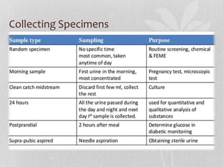
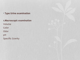
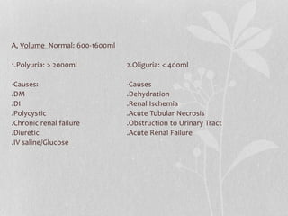
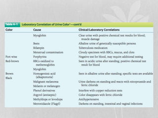
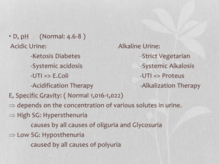
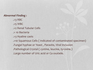
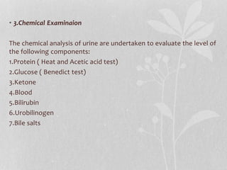
Professor: SENG SEREY
prepared by: NAM MENG LY
URINE ANALYSIS
• Urine Analysis: is an array of tests performed on urine and one of
the most common method of medical diagnosis.
• Why we have to do urine analysis?
• - General evaluation of health
• -Diagnosis of diseases or disorders of kidney and Urinary tract
• -Diagnosis of other systemic disease that affect kidney function
• -Monitoring of patients with diabetes
• -Screening for drug abuse ( Sulfonamide and Aminoglycoside )
Collecting Specimens
• Type Urine examination
1.Macroscopic examination
Volume
Color
Odor
pH
Specific Gravity
A, Volume Normal: 600-1600ml
1.Polyuria: > 2000ml 2.Oliguria: < 400ml
-Causes: -Causes
.DM .Dehydration
.DI .Renal Ischemia
.Polycystic .Acute Tubular Necrosis
.Chronic renal failure .Obstruction to Urinary Tract
.Diuretic .Acute Renal Failure
.IV saline/Glucose
• B, Color
Normal : clear and pale yellow
Colorless: dilution , DM, DI, Diuretic
Milky: Purulent GUT infection, Chyluria
Orange: Fever, excessive sweating
Red: Beetroot ingestion, hematuria
Brown: Melanin, Alkaptunuria.
• C, Odor
• D, pH (Normal: 4.6-8 )
Acidic Urine: Alkaline Urine:
-Ketosis Diabetes -Strict Vegetarian
-Systemic acidosis -Systemic Alkalosis
-UTI => E.Coli -UTI => Proteus
-Acidification Therapy -Alkalization Therapy
E, Specific Gravity: ( Normal 1,016-1,022)
 depends on the concentration of various solutes in urine.
 High SG: Hypersthenuria
causes by all causes of oliguria and Glycosuria
 Low SG: Hyposthenuria
caused by all causes of polyuria
• 2.Microscopic examination
A sample has been taken 10-15ml into a tube for 5-10min which
produces a concentration of sediment at the bottom.
A drop of sediment is poured onto a glass slide with coverslip put
onto observed under microscopes.
• A variety of normal and abnormal cellular elements in urine such as:
-RBC
-WBC
-Mucus
-Various Epithelial Cells
-Various Crystal
-Bacteria
-Casts
Abnormal Finding :
.>3 RBC
.>5 WBC
.>2 Renal Tubular Cells
.> 10 Bacteria
.>3 Hyaline casts
.>10 Squamous Cells ( Indicated of contaminated specimen)
.Fungal hyphae or Yeast , Parasite, Viral Inclusion
.Pathological Crystal ( cystine, leucine, tyrosine.)
.Large number of Uric acid or Ca oxalade.
• Presents of Casts:
Type of Casts:
Acellular casts Cellular casts
Hyaline Casts (fever, exercise) RBC casts( Glomerular damage)
Granular casts (albumin) WBC casts(Infection)
Waxy casts( Renal Failure) Epithelial casts(toxic ingestion
Fatty casts ( NS, DM, SLE,ATN) Hg,ATN)
Pigment casts( Hemolytic anemia, Rhabdomyosis, Liver disease)
Crystal casts( crystallized urinary solute: Oxalate urate,,,)
• 3.Chemical Examinaion
The chemical analysis of urine are undertaken to evaluate the level of
the following components:
1.Protein ( Heat and Acetic acid test)
2.Glucose ( Benedict test)
3.Ketone
4.Blood
5.Bilirubin
6.Urobilinogen
7.Bile salts
The Dipstick method
1.Tests for proteins
•Test – HEAT & ACETIC ACID TEST
•Principle-proteins are denatured & coagulated on
heating to give white cloud precipitate.
•Method-take 2/3 of test tube with urine, heat only
the upper part keeping lower part as control.
•Presence of phosphates, carbonates, proteins
gives a white cloud formation. Add acetic acid 1-2
drops, if the cloud persists it indicates it is
protein(acetic acid dissolves the
carbonates/phosphates)
Other tests
• Sulphosalicylic acid test
• Dipsticks
• Esbach’s albuminometer- for quantitative estimation of proteins
Causes of proteinuria
•Prerenal causes-Heavy
exercise,Fever,hypertension, multiple myeloma,
eclumpsia
•Renal –acute & chronic glomerulonephritis,Renal
tubular dysfunction,Polycystic kidney, nephrotic
syndrome
•Post renal- acute & chronic cystitis, tuberculosis
cystitis
• Selective proteinuria
• Nonselective proteinuria
microalbuminuria
• The level of albumin protein produced by microalbuminuria
cannot be detected by urine dipstick methods. In a properly
functioning body, albumin is not normally present in urine
because it is retained in the bloodstream by the kidneys.
Microalbuminuria is diagnosed either from a 24-hour urine
collection
Significance of microalbuminuria
•an indicator of subclinical cardiovascular disease
•an important prognostic marker for kidney disease
•in diabetes mellitus
•in hypertension
•increasing microalbuminuria during the first 48
hours after admission to an intensive care unit
predicts elevated risk for acute respiratory failure ,
multiple organ failure , and overall mortality
Bence Jones proteins
•These are light chain globulins seen in multiple
myeloma, macroglobulimias, lymphoma.
•Test- Thermal method(waterbath):
Proteins has unusual property of precipitating at
400 -600c & then dissolving when the urine is
brought to boiling(1000c) & reappears when the
urine is cooled.
2.Test for sugar
• Test-BENEDICT’S TEST(semiquantitative)
• Principle-benedict’s reagent contains CuSO4.In the
presence of reducing sugars cupric ions are converted
to cuprous oxide which is hastened by heating, to give
the color.
• Method- take 5ml of benedict’s reagent in a test tube,
add 8drops of urine. Boil the mixture.
Blue-green= negative
Yellow-green=+(<0.5%)
Greenish yellow=++(0.5-1%)
Yellow=+++(1-2%)
Brick red=++++(>2%)
Benedict’s test
• Detects all reducing substances like glucose, fructose, & other
reducing sustances.
• To confirm it is glucose, dipsticks can be used (glucose oxidase)
Causes of glycosuria
• Glycosuria with hyperglycaemia- diabetes,acromegaly, cushing’s
disease, hyperthyroidism, drugs like corticosteroids.
• Glycosuria without hyperglycaemia- renal tubular dysfunction
3.Ketone bodies
• 3 types
Acetone
Acetoacetic acid
β-hydroxy butyric acid
They are products of fat metabolism
Rothera’s test
• Principle-acetone & acetoacetic acid react with sodium
nitroprusside in the presence of alkali to produce purple colour.
• Method- take 5ml of urine in a test tube & saturate it with
ammonium sulphate. Then add one crystal of sodium
nitroprusside. Then gently add 0.5ml of liquor ammonia along
the sides of the test tube.
• Change in colour indicates + test
Causes of ketonuria
• Diabetes
• Non-diabetic causes- high fever, starvation, severe
vomiting/diarrhoea
4.Bilirubin
•Test- fouchet’s test.
•Causes
Liver diseases-injury,hepatitis
Obstruction to biliary tract
5.Urobilinogen
• Test- ehrlich test
• Causes-hemolytic anemia's
6.Bile salts
Hay’s test
Cause- obstruction to bile flow (obstructive jaundice)
7.Blood in urine
•Test- BENZIDINE TEST
•Method- mix 2ml of benzidine solution with 2ml of
hydrogen peroxide in a test tube. Take 2ml of urine
& add 2ml of above mixture. A blue color indicates
+ reaction.
Causes of hematuria
• Pre renal- bleeding diathesis, hemoglobinopathies,
malignant hypertension.
• Renal- trauma, calculi, acute & chronic glomerulonephritis,
renal TB, renal tumors
• Post renal – severe UTI, calculi, trauma, tumors of urinary
tract
Type Plasma color Urine color
Hematuria normal Smoky red
m/s-plenty of
RBC’s
hemoglobunuria Pink,hepatoglob
in reduced
Red ,
occasional
RBC’s
Myoglobunuria Pink, normal
hepatoglobin
Red, occasional
RBC’s
Thanks for pay attention !!!

Urine analysis.ppt

  • 1.
    Professor: SENG SEREY preparedby: NAM MENG LY URINE ANALYSIS
  • 2.
    • Urine Analysis:is an array of tests performed on urine and one of the most common method of medical diagnosis. • Why we have to do urine analysis? • - General evaluation of health • -Diagnosis of diseases or disorders of kidney and Urinary tract • -Diagnosis of other systemic disease that affect kidney function • -Monitoring of patients with diabetes • -Screening for drug abuse ( Sulfonamide and Aminoglycoside )
  • 3.
  • 4.
    • Type Urineexamination 1.Macroscopic examination Volume Color Odor pH Specific Gravity
  • 5.
    A, Volume Normal:600-1600ml 1.Polyuria: > 2000ml 2.Oliguria: < 400ml -Causes: -Causes .DM .Dehydration .DI .Renal Ischemia .Polycystic .Acute Tubular Necrosis .Chronic renal failure .Obstruction to Urinary Tract .Diuretic .Acute Renal Failure .IV saline/Glucose
  • 6.
    • B, Color Normal: clear and pale yellow Colorless: dilution , DM, DI, Diuretic Milky: Purulent GUT infection, Chyluria Orange: Fever, excessive sweating Red: Beetroot ingestion, hematuria Brown: Melanin, Alkaptunuria.
  • 9.
  • 10.
    • D, pH(Normal: 4.6-8 ) Acidic Urine: Alkaline Urine: -Ketosis Diabetes -Strict Vegetarian -Systemic acidosis -Systemic Alkalosis -UTI => E.Coli -UTI => Proteus -Acidification Therapy -Alkalization Therapy E, Specific Gravity: ( Normal 1,016-1,022)  depends on the concentration of various solutes in urine.  High SG: Hypersthenuria causes by all causes of oliguria and Glycosuria  Low SG: Hyposthenuria caused by all causes of polyuria
  • 11.
    • 2.Microscopic examination Asample has been taken 10-15ml into a tube for 5-10min which produces a concentration of sediment at the bottom. A drop of sediment is poured onto a glass slide with coverslip put onto observed under microscopes.
  • 12.
    • A varietyof normal and abnormal cellular elements in urine such as: -RBC -WBC -Mucus -Various Epithelial Cells -Various Crystal -Bacteria -Casts
  • 13.
    Abnormal Finding : .>3RBC .>5 WBC .>2 Renal Tubular Cells .> 10 Bacteria .>3 Hyaline casts .>10 Squamous Cells ( Indicated of contaminated specimen) .Fungal hyphae or Yeast , Parasite, Viral Inclusion .Pathological Crystal ( cystine, leucine, tyrosine.) .Large number of Uric acid or Ca oxalade.
  • 14.
    • Presents ofCasts: Type of Casts: Acellular casts Cellular casts Hyaline Casts (fever, exercise) RBC casts( Glomerular damage) Granular casts (albumin) WBC casts(Infection) Waxy casts( Renal Failure) Epithelial casts(toxic ingestion Fatty casts ( NS, DM, SLE,ATN) Hg,ATN) Pigment casts( Hemolytic anemia, Rhabdomyosis, Liver disease) Crystal casts( crystallized urinary solute: Oxalate urate,,,)
  • 16.
    • 3.Chemical Examinaion Thechemical analysis of urine are undertaken to evaluate the level of the following components: 1.Protein ( Heat and Acetic acid test) 2.Glucose ( Benedict test) 3.Ketone 4.Blood 5.Bilirubin 6.Urobilinogen 7.Bile salts
  • 17.
  • 18.
    1.Tests for proteins •Test– HEAT & ACETIC ACID TEST •Principle-proteins are denatured & coagulated on heating to give white cloud precipitate. •Method-take 2/3 of test tube with urine, heat only the upper part keeping lower part as control. •Presence of phosphates, carbonates, proteins gives a white cloud formation. Add acetic acid 1-2 drops, if the cloud persists it indicates it is protein(acetic acid dissolves the carbonates/phosphates)
  • 19.
    Other tests • Sulphosalicylicacid test • Dipsticks • Esbach’s albuminometer- for quantitative estimation of proteins
  • 20.
    Causes of proteinuria •Prerenalcauses-Heavy exercise,Fever,hypertension, multiple myeloma, eclumpsia •Renal –acute & chronic glomerulonephritis,Renal tubular dysfunction,Polycystic kidney, nephrotic syndrome •Post renal- acute & chronic cystitis, tuberculosis cystitis
  • 21.
    • Selective proteinuria •Nonselective proteinuria
  • 22.
    microalbuminuria • The levelof albumin protein produced by microalbuminuria cannot be detected by urine dipstick methods. In a properly functioning body, albumin is not normally present in urine because it is retained in the bloodstream by the kidneys. Microalbuminuria is diagnosed either from a 24-hour urine collection
  • 23.
    Significance of microalbuminuria •anindicator of subclinical cardiovascular disease •an important prognostic marker for kidney disease •in diabetes mellitus •in hypertension •increasing microalbuminuria during the first 48 hours after admission to an intensive care unit predicts elevated risk for acute respiratory failure , multiple organ failure , and overall mortality
  • 24.
    Bence Jones proteins •Theseare light chain globulins seen in multiple myeloma, macroglobulimias, lymphoma. •Test- Thermal method(waterbath): Proteins has unusual property of precipitating at 400 -600c & then dissolving when the urine is brought to boiling(1000c) & reappears when the urine is cooled.
  • 25.
    2.Test for sugar •Test-BENEDICT’S TEST(semiquantitative) • Principle-benedict’s reagent contains CuSO4.In the presence of reducing sugars cupric ions are converted to cuprous oxide which is hastened by heating, to give the color. • Method- take 5ml of benedict’s reagent in a test tube, add 8drops of urine. Boil the mixture. Blue-green= negative Yellow-green=+(<0.5%) Greenish yellow=++(0.5-1%) Yellow=+++(1-2%) Brick red=++++(>2%)
  • 26.
    Benedict’s test • Detectsall reducing substances like glucose, fructose, & other reducing sustances. • To confirm it is glucose, dipsticks can be used (glucose oxidase)
  • 27.
    Causes of glycosuria •Glycosuria with hyperglycaemia- diabetes,acromegaly, cushing’s disease, hyperthyroidism, drugs like corticosteroids. • Glycosuria without hyperglycaemia- renal tubular dysfunction
  • 28.
    3.Ketone bodies • 3types Acetone Acetoacetic acid β-hydroxy butyric acid They are products of fat metabolism
  • 29.
    Rothera’s test • Principle-acetone& acetoacetic acid react with sodium nitroprusside in the presence of alkali to produce purple colour. • Method- take 5ml of urine in a test tube & saturate it with ammonium sulphate. Then add one crystal of sodium nitroprusside. Then gently add 0.5ml of liquor ammonia along the sides of the test tube. • Change in colour indicates + test
  • 30.
    Causes of ketonuria •Diabetes • Non-diabetic causes- high fever, starvation, severe vomiting/diarrhoea
  • 31.
    4.Bilirubin •Test- fouchet’s test. •Causes Liverdiseases-injury,hepatitis Obstruction to biliary tract
  • 32.
    5.Urobilinogen • Test- ehrlichtest • Causes-hemolytic anemia's 6.Bile salts Hay’s test Cause- obstruction to bile flow (obstructive jaundice)
  • 33.
    7.Blood in urine •Test-BENZIDINE TEST •Method- mix 2ml of benzidine solution with 2ml of hydrogen peroxide in a test tube. Take 2ml of urine & add 2ml of above mixture. A blue color indicates + reaction.
  • 34.
    Causes of hematuria •Pre renal- bleeding diathesis, hemoglobinopathies, malignant hypertension. • Renal- trauma, calculi, acute & chronic glomerulonephritis, renal TB, renal tumors • Post renal – severe UTI, calculi, trauma, tumors of urinary tract
  • 35.
    Type Plasma colorUrine color Hematuria normal Smoky red m/s-plenty of RBC’s hemoglobunuria Pink,hepatoglob in reduced Red , occasional RBC’s Myoglobunuria Pink, normal hepatoglobin Red, occasional RBC’s
  • 36.
    Thanks for payattention !!!

Editor's Notes

  • #7 Alkaptunuria: autosomal recessive , mutation in the HGD gene… accumulation of homogentisic acid ,