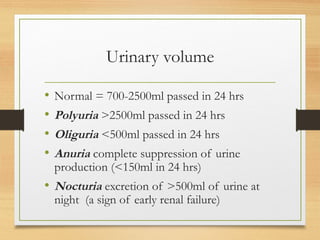
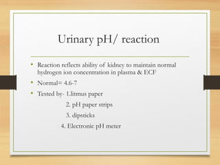
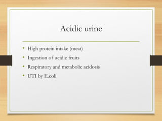
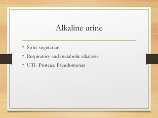
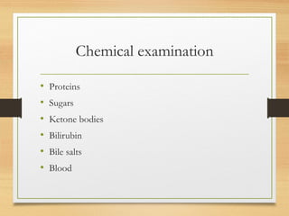
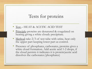
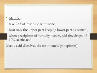
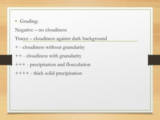
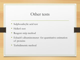
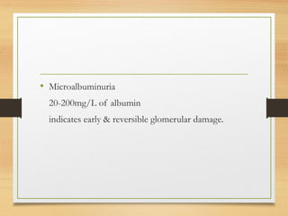
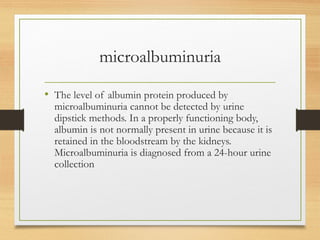
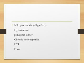
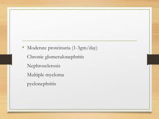
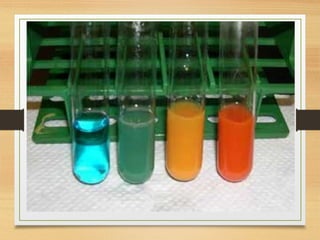
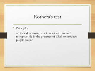
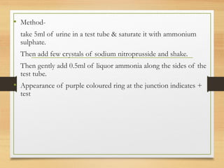
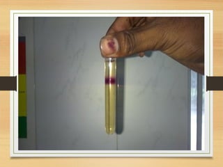
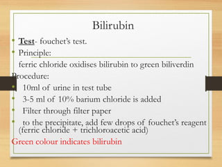
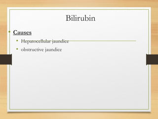
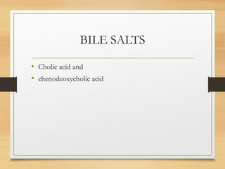
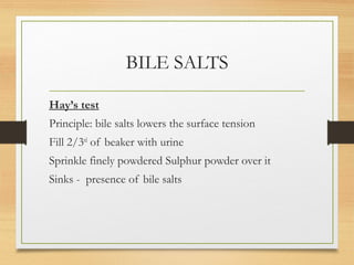
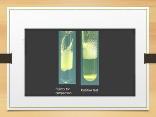
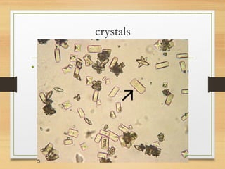
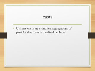
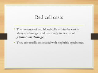
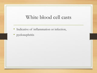
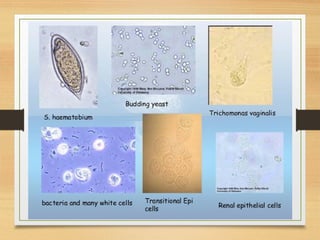
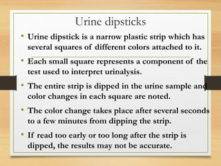
Urine analysis
Dr. Janani Mathialagan
OVERVIEW
• Physical examination
• Chemical examination
• Microscopic examination
Types of collection of urine:
• 24hrs sample – Glycosuria,
microalbuminuria
• Random sample - glucose, protein
• Midstream sample - UTI
PHYSICAL
EXAMINATION
Urine examination
• Physical examination
• Chemical examination
• Microscopic examination
Physical examination
• Volume
• Color
• Odour
• Reaction/pH
• Specific gravity
Urinary volume
• Normal = 700-2500ml passed in 24 hrs
• Polyuria >2500ml passed in 24 hrs
• Oliguria <500ml passed in 24 hrs
• Anuria complete suppression of urine
production (<150ml in 24 hrs)
• Nocturia excretion of >500ml of urine at
night (a sign of early renal failure)
Color
• Normal= clear, pale yellow/ straw colour (Urochrome)
1. Colourless - diabetes mellitus, diabetes insipidus, diuretics,
excessive intake of water.
2. Milky – pus, chyluria(fat)
3. Orange – urobilinogen, conc. Urine, ATT
4. Deep amber colour – high grade fever, muscular exercise
5. Smoky – Vit B12 therapy, aniline dye
6. Red – hematuria, haemoglobinuria
7. Brown – Bile
8. Green – phenol poisoning, putrified sample.
Urinary pH/ reaction
• Reaction reflects ability of kidney to maintain normal
hydrogen ion concentration in plasma & ECF
• Normal= 4.6-7
• Tested by- 1.litmus paper
2. pH paper strips
3. dipsticks
4. Electronic pH meter
Acidic urine
• High protein intake (meat)
• Ingestion of acidic fruits
• Respiratory and metabolic acidosis
• UTI by E.coli
Alkaline urine
• Strict vegetarian
• Respiratory and metabolic alkalosis
• UTI- Proteus, Pseudomonas
Odour
• Normal – faintly aromatic due to the volatile fatty
acids
1.Pungent – ammonia produced by bacterial
contamination
2.Putrid – UTI
3.Fruity – ketoacidosis
Specific gravity
• Ratio of weight of 1ml of urine to 1ml of distilled water.
• Depends on the concentration of various solutes in the urine.
• Purpose – to measure the concentrating and diluting power of
kidneys.
• Measured by-urinometer
- refractometer
- dipsticks
Urinometer
• Take 3/4th of urinometer container with urine
• Allow the urinometer to float in the urine without
touching the walls or bottom of the container.
• Read the graduation at the lowest level of urinary
meniscus
• Urinometer is calibrated at 15 or 200
c
• Correction of temperature is necessary
So for every 3o
c increase/decrease add/subtract 0.001
Normal-1.016-1.022
•
High specific
gravity(hyperosthenuria)
• Causes
Dehydration
Albuminuria
Gycosuria
Low specific gravity
(hypo-osthenuria)
• Diabetes insipidus
• Excess water intake
• Fixed specific gravity
(iso-osthenuria)=1.010
Seen in chronic renal disease when kidney has lost the ability to
concentrate or dilute
CHEMICAL
EXAMINATION
Chemical examination
• Proteins
• Sugars
• Ketone bodies
• Bilirubin
• Bile salts
• Blood
Tests for proteins
• Test – HEAT & ACETIC ACID TEST
• Principle-proteins are denatured & coagulated on
heating giving a white cloudy precipitate.
• Method-take 2/3 of test tube with urine, heat only
the upper part keeping lower part as control.
• Presence of phosphates, carbonates, proteins gives a
white cloud formation. Add acetic acid 1-2 drops, if
the cloud persists it indicates it is protein(acetic acid
dissolves the carbonates/phosphates)
• Method-
take 2/3 of test tube with urine,
heat only the upper part keeping lower part as control.
when precipitate of turbidiy occurs, add few drops of
10% acetic acid
(acetic acid dissolves the carbonates/phosphates)
• Grading:
Negative – no cloudiness
Traces – cloudiness against dark background
+ - cloudiness without granularity
++ - cloudiness with granularity
+++ - precipitation and flocculation
++++ - thick solid precipitation
Other tests
• Sulphosalicylic acid test
• Heller’s test
• Reagent strip method
• Esbach’s albuminometer- for quantitative estimation
of proteins
• Turbidimetric method
Causes of proteinuria
• Normal - <150mg/day
• Prerenal causes-Heavy
exercise,Fever,hypertension, multiple myeloma,
ecalmpsia
• Renal –acute & chronic glomerulonephritis,Renal
tubular dysfunction,Polycystic kidney, nephrotic
syndrome
• Post renal- acute & chronic cystitis, tuberculosis
cystitis
• Microalbuminuria
20-200mg/L of albumin
indicates early & reversible glomerular damage.
microalbuminuria
• The level of albumin protein produced by
microalbuminuria cannot be detected by urine
dipstick methods. In a properly functioning body,
albumin is not normally present in urine because it is
retained in the bloodstream by the kidneys.
Microalbuminuria is diagnosed from a 24-hour urine
collection
• Mild proteinuria: (<1gm/day)
Hypertension
polycystic kidney
Chronic pyelonephritis
UTI
Fever
• Moderate proteinuria (1-3gm/day)
Chronic glomerulonephritis
Nephrosclerosis
Multiple myeloma
pyelonephritis
• Heavy proteinuria (>3gm/day)
Nephrotic syndrome
Renal vein thrombosis
Diabetes mellitus
SLE
Test for sugar
• Test- BENEDICT’S TEST(semiquantitative)
• Principle
Benedict’s reagent contains CuSO4.
In the presence of reducing sugars cupric ions are converted
to cuprous oxide which is hastened by heating, to give the
color.
REDUCING SUBSTANCES PRESENT IN URINE: glucose,
fructose, maltose, lactose (not sucrose)
• Method-
take 5ml of benedict’s reagent in a test tube
add 8 drops of urine.
Boil the mixture.
No change in blue colour = negative
Greenish= Traces (<0.5gm%)
Green/Cloudy green=+(1gm%)
Yellow=++(1-1.5gm%)
Orange=+++(1.5-2gm%)
Brick red=++++(>2gm%)
Benedict’s test
• Detects all reducing substances like glucose, fructose,
& other reducing sustances.
• To confirm it is glucose, dipsticks can be used
(glucose oxidase)
Causes of glycosuria
• Diabetes mellitus
• Renal glucosuria
• Sever burns
• Administration of corticosteroids
• Severe sepsis
• Pregnancy
Ketone bodies
• 3 types
Acetone
Acetoacetic acid
β-hydroxy butyric acid
They are products of fat metabolism
Rothera’s test
• Principle-
acetone & acetoacetic acid react with sodium
nitroprusside in the presence of alkali to produce
purple colour.
• Method-
take 5ml of urine in a test tube & saturate it with ammonium
sulphate.
Then add few crystals of sodium nitroprusside and shake.
Then gently add 0.5ml of liquor ammonia along the sides of the
test tube.
• Appearance of purple coloured ring at the junction indicates +
test
Causes of ketonuria
• Diabetic ketoacidosis
• Dehydration
• Hyperemesis gravidarum
• Fever
• Cachexia
Other tests for ketone bodies
• Gerhardt’s test
• Reagent strip test
Bilirubin
• Test- fouchet’s test.
• Principle:
ferric chloride oxidises bilirubin to green biliverdin
Procedure:
• 10ml of urine in test tube
• 3-5 ml of 10% barium chloride is added
• Filter through filter paper
• to the precipitate, add few drops of fouchet’s reagent
(ferric chloride + trichloroacetic acid)
Green colour indicates bilirubin
Bilirubin
• Causes
• Hepatocellular jaundice
• obstructive jaundice
BILE SALTS
• Cholic acid and
• chenodeoxycholic acid
BILE SALTS
Hay’s test
Principle: bile salts lowers the surface tension
Fill 2/3rd
of beaker with urine
Sprinkle finely powdered Sulphur powder over it
Sinks - presence of bile salts
Blood in urine
• Test- BENZIDINE TEST
• Principle-The peroxidase activity of
hemoglobin decomposes hydrogen peroxide
releasing nascent oxygen which in turn
oxidizes benzidine to give blue color.
Blood in urine
• Method-
mix 2ml of benzidine solution with 2ml of
hydrogen peroxide in a test tube.
Take 2ml of urine & add 2ml of above
mixture.
A blue color indicates + reaction.
Causes of hematuria
• Renal stones
• Renal tumors
• Polycystic kidney
• Bleeding disorders
• trauma
Microscopic
examination
Microscopic examination
• Microscopic urinalysis is done by pouring the urine
sample into a test tube and centrifuging it for a few
minutes.
• The top liquid part (the supernatant) is discarded.
• The solid part left in the bottom of the test tube (the
urine sediment) is mixed with the remaining drop of
urine in the test tube
• one drop of this is analyzed under a microscope
Contents of normal urine m/s
• Contains few epithelial cells, occasional RBC’s, few
crystals.
crystals
•
casts
• Urinary casts are cylindrical aggregations of
particles that form in the distal nephron
Red cell casts
• The presence of red blood cells within the cast is
always pathologic, and is strongly indicative of
glomerular damage.
• They are usually associated with nephritic syndromes.
Erythrocyte cast
•
White blood cell casts
• Indicative of inflammation or infection,
• pyelonephritis
Leucocyte cast
•
Urine dipsticks
• Urine dipstick is a narrow plastic strip which has
several squares of different colors attached to it.
• Each small square represents a component of the
test used to interpret urinalysis.
• The entire strip is dipped in the urine sample and
color changes in each square are noted.
• The color change takes place after several seconds
to a few minutes from dipping the strip.
• If read too early or too long after the strip is
dipped, the results may not be accurate.
Urine dipsticks
• The color change takes place after several seconds
to a few minutes from dipping the strip.
• If read too early or too long after the strip is
dipped, the results may not be accurate.
•
• The main advantage of dipsticks is that they are
1.  convenient,
2.  easy to interpret,
3.  and cost-effective
The main disadvantage is that the
1. Information may not be very accurate as the test is
time-sensitive.
2. It also provides limited information about the urine
as it is qualitative test and not a quantitative test
• Thankyou

Urine analysis

  • 1.
  • 2.
    OVERVIEW • Physical examination •Chemical examination • Microscopic examination
  • 3.
    Types of collectionof urine: • 24hrs sample – Glycosuria, microalbuminuria • Random sample - glucose, protein • Midstream sample - UTI
  • 4.
  • 5.
    Urine examination • Physicalexamination • Chemical examination • Microscopic examination
  • 6.
    Physical examination • Volume •Color • Odour • Reaction/pH • Specific gravity
  • 7.
    Urinary volume • Normal= 700-2500ml passed in 24 hrs • Polyuria >2500ml passed in 24 hrs • Oliguria <500ml passed in 24 hrs • Anuria complete suppression of urine production (<150ml in 24 hrs) • Nocturia excretion of >500ml of urine at night (a sign of early renal failure)
  • 8.
    Color • Normal= clear,pale yellow/ straw colour (Urochrome)
  • 9.
    1. Colourless -diabetes mellitus, diabetes insipidus, diuretics, excessive intake of water. 2. Milky – pus, chyluria(fat) 3. Orange – urobilinogen, conc. Urine, ATT 4. Deep amber colour – high grade fever, muscular exercise 5. Smoky – Vit B12 therapy, aniline dye 6. Red – hematuria, haemoglobinuria 7. Brown – Bile 8. Green – phenol poisoning, putrified sample.
  • 10.
    Urinary pH/ reaction •Reaction reflects ability of kidney to maintain normal hydrogen ion concentration in plasma & ECF • Normal= 4.6-7 • Tested by- 1.litmus paper 2. pH paper strips 3. dipsticks 4. Electronic pH meter
  • 11.
    Acidic urine • Highprotein intake (meat) • Ingestion of acidic fruits • Respiratory and metabolic acidosis • UTI by E.coli
  • 12.
    Alkaline urine • Strictvegetarian • Respiratory and metabolic alkalosis • UTI- Proteus, Pseudomonas
  • 13.
    Odour • Normal –faintly aromatic due to the volatile fatty acids 1.Pungent – ammonia produced by bacterial contamination 2.Putrid – UTI 3.Fruity – ketoacidosis
  • 14.
    Specific gravity • Ratioof weight of 1ml of urine to 1ml of distilled water. • Depends on the concentration of various solutes in the urine. • Purpose – to measure the concentrating and diluting power of kidneys. • Measured by-urinometer - refractometer - dipsticks
  • 15.
    Urinometer • Take 3/4thof urinometer container with urine • Allow the urinometer to float in the urine without touching the walls or bottom of the container. • Read the graduation at the lowest level of urinary meniscus • Urinometer is calibrated at 15 or 200 c • Correction of temperature is necessary So for every 3o c increase/decrease add/subtract 0.001 Normal-1.016-1.022
  • 16.
  • 17.
  • 18.
    Low specific gravity (hypo-osthenuria) •Diabetes insipidus • Excess water intake • Fixed specific gravity (iso-osthenuria)=1.010 Seen in chronic renal disease when kidney has lost the ability to concentrate or dilute
  • 19.
  • 20.
    Chemical examination • Proteins •Sugars • Ketone bodies • Bilirubin • Bile salts • Blood
  • 21.
    Tests for proteins •Test – HEAT & ACETIC ACID TEST • Principle-proteins are denatured & coagulated on heating giving a white cloudy precipitate. • Method-take 2/3 of test tube with urine, heat only the upper part keeping lower part as control. • Presence of phosphates, carbonates, proteins gives a white cloud formation. Add acetic acid 1-2 drops, if the cloud persists it indicates it is protein(acetic acid dissolves the carbonates/phosphates)
  • 22.
    • Method- take 2/3of test tube with urine, heat only the upper part keeping lower part as control. when precipitate of turbidiy occurs, add few drops of 10% acetic acid (acetic acid dissolves the carbonates/phosphates)
  • 24.
    • Grading: Negative –no cloudiness Traces – cloudiness against dark background + - cloudiness without granularity ++ - cloudiness with granularity +++ - precipitation and flocculation ++++ - thick solid precipitation
  • 25.
    Other tests • Sulphosalicylicacid test • Heller’s test • Reagent strip method • Esbach’s albuminometer- for quantitative estimation of proteins • Turbidimetric method
  • 26.
    Causes of proteinuria •Normal - <150mg/day • Prerenal causes-Heavy exercise,Fever,hypertension, multiple myeloma, ecalmpsia • Renal –acute & chronic glomerulonephritis,Renal tubular dysfunction,Polycystic kidney, nephrotic syndrome • Post renal- acute & chronic cystitis, tuberculosis cystitis
  • 27.
    • Microalbuminuria 20-200mg/L ofalbumin indicates early & reversible glomerular damage.
  • 28.
    microalbuminuria • The levelof albumin protein produced by microalbuminuria cannot be detected by urine dipstick methods. In a properly functioning body, albumin is not normally present in urine because it is retained in the bloodstream by the kidneys. Microalbuminuria is diagnosed from a 24-hour urine collection
  • 29.
    • Mild proteinuria:(<1gm/day) Hypertension polycystic kidney Chronic pyelonephritis UTI Fever
  • 30.
    • Moderate proteinuria(1-3gm/day) Chronic glomerulonephritis Nephrosclerosis Multiple myeloma pyelonephritis
  • 31.
    • Heavy proteinuria(>3gm/day) Nephrotic syndrome Renal vein thrombosis Diabetes mellitus SLE
  • 32.
    Test for sugar •Test- BENEDICT’S TEST(semiquantitative) • Principle Benedict’s reagent contains CuSO4. In the presence of reducing sugars cupric ions are converted to cuprous oxide which is hastened by heating, to give the color. REDUCING SUBSTANCES PRESENT IN URINE: glucose, fructose, maltose, lactose (not sucrose)
  • 33.
    • Method- take 5mlof benedict’s reagent in a test tube add 8 drops of urine. Boil the mixture. No change in blue colour = negative Greenish= Traces (<0.5gm%) Green/Cloudy green=+(1gm%) Yellow=++(1-1.5gm%) Orange=+++(1.5-2gm%) Brick red=++++(>2gm%)
  • 34.
    Benedict’s test • Detectsall reducing substances like glucose, fructose, & other reducing sustances. • To confirm it is glucose, dipsticks can be used (glucose oxidase)
  • 36.
    Causes of glycosuria •Diabetes mellitus • Renal glucosuria • Sever burns • Administration of corticosteroids • Severe sepsis • Pregnancy
  • 37.
    Ketone bodies • 3types Acetone Acetoacetic acid β-hydroxy butyric acid They are products of fat metabolism
  • 38.
    Rothera’s test • Principle- acetone& acetoacetic acid react with sodium nitroprusside in the presence of alkali to produce purple colour.
  • 39.
    • Method- take 5mlof urine in a test tube & saturate it with ammonium sulphate. Then add few crystals of sodium nitroprusside and shake. Then gently add 0.5ml of liquor ammonia along the sides of the test tube. • Appearance of purple coloured ring at the junction indicates + test
  • 41.
    Causes of ketonuria •Diabetic ketoacidosis • Dehydration • Hyperemesis gravidarum • Fever • Cachexia
  • 42.
    Other tests forketone bodies • Gerhardt’s test • Reagent strip test
  • 43.
    Bilirubin • Test- fouchet’stest. • Principle: ferric chloride oxidises bilirubin to green biliverdin Procedure: • 10ml of urine in test tube • 3-5 ml of 10% barium chloride is added • Filter through filter paper • to the precipitate, add few drops of fouchet’s reagent (ferric chloride + trichloroacetic acid) Green colour indicates bilirubin
  • 44.
    Bilirubin • Causes • Hepatocellularjaundice • obstructive jaundice
  • 45.
    BILE SALTS • Cholicacid and • chenodeoxycholic acid
  • 46.
    BILE SALTS Hay’s test Principle:bile salts lowers the surface tension Fill 2/3rd of beaker with urine Sprinkle finely powdered Sulphur powder over it Sinks - presence of bile salts
  • 48.
    Blood in urine •Test- BENZIDINE TEST • Principle-The peroxidase activity of hemoglobin decomposes hydrogen peroxide releasing nascent oxygen which in turn oxidizes benzidine to give blue color.
  • 50.
    Blood in urine •Method- mix 2ml of benzidine solution with 2ml of hydrogen peroxide in a test tube. Take 2ml of urine & add 2ml of above mixture. A blue color indicates + reaction.
  • 51.
    Causes of hematuria •Renal stones • Renal tumors • Polycystic kidney • Bleeding disorders • trauma
  • 52.
  • 53.
    Microscopic examination • Microscopicurinalysis is done by pouring the urine sample into a test tube and centrifuging it for a few minutes. • The top liquid part (the supernatant) is discarded. • The solid part left in the bottom of the test tube (the urine sediment) is mixed with the remaining drop of urine in the test tube • one drop of this is analyzed under a microscope
  • 54.
    Contents of normalurine m/s • Contains few epithelial cells, occasional RBC’s, few crystals.
  • 55.
  • 56.
    casts • Urinary castsare cylindrical aggregations of particles that form in the distal nephron
  • 57.
    Red cell casts •The presence of red blood cells within the cast is always pathologic, and is strongly indicative of glomerular damage. • They are usually associated with nephritic syndromes.
  • 58.
  • 59.
    White blood cellcasts • Indicative of inflammation or infection, • pyelonephritis
  • 60.
  • 62.
    Urine dipsticks • Urinedipstick is a narrow plastic strip which has several squares of different colors attached to it. • Each small square represents a component of the test used to interpret urinalysis. • The entire strip is dipped in the urine sample and color changes in each square are noted. • The color change takes place after several seconds to a few minutes from dipping the strip. • If read too early or too long after the strip is dipped, the results may not be accurate.
  • 63.
    Urine dipsticks • Thecolor change takes place after several seconds to a few minutes from dipping the strip. • If read too early or too long after the strip is dipped, the results may not be accurate.
  • 64.
  • 65.
    • The mainadvantage of dipsticks is that they are 1.  convenient, 2.  easy to interpret, 3.  and cost-effective
  • 66.
    The main disadvantageis that the 1. Information may not be very accurate as the test is time-sensitive. 2. It also provides limited information about the urine as it is qualitative test and not a quantitative test
  • 67.