Cerebrum
•Download as PPTX, PDF•
2 likes•491 views

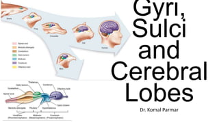
This document provides details on the sulci, gyri, and lobes of the cerebral hemispheres. Some key points: - The cerebral hemispheres are separated by the longitudinal fissure and contain ventricles and white matter. Each hemisphere has frontal, parietal, temporal, occipital, and limbic lobes defined by sulci. - Major sulci include the central sulcus separating frontal and parietal lobes, lateral sulcus outlining the temporal lobe, and parieto-occipital sulcus marking the border of the parietal and occipital lobes. - Gyri are raised ridges of cortex separated by sulci, including precentral and postcentral gy

Recommended
More Related Content
What's hot
What's hot (20)
Similar to Cerebrum
Similar to Cerebrum (20)
More from Komal Parmar
More from Komal Parmar (20)
Recently uploaded
Recently uploaded (20)
Cerebrum
- 2. • The cerebral hemispheres are separated by a deep median cleft, the great longitudinal fissure, which contains a crescent-shaped fold of the dura mater, the falx cerebri. • Each cerebral hemisphere may be considered to have superolateral, medial and inferior (basal) surfaces or aspects. • The tentorial part lies in the middle cranial fossa; posteriorly it lies above the tentorium cerebelli, which separates it from the superior surface of the cerebellum.
- 3. • Each hemisphere consists of an external highly-convoluted cortex, beneath which lies an extensive internal mass of white matter that partly encloses the basal ganglia. • Each hemisphere also contains a lateral ventricle that is continuous with the third ventricle through the interventricular foramen. The two hemispheres are linked by the commissural fibres of the corpus callosum.
- 5. • The area of the adult cerebral cortex is approximately 2200 cm²: its convoluted form increases the cortical volume to three times greater than it would be if the surface were smooth.
- 6. Sulci and related Gyri - The very obvious and easily identifiable ones
- 7. Sulci Types
- 8. Lateral Sulcus • The lateral fissure is a deep cleft on the lateral and inferior surfaces. • It separates the frontal and parietal lobes above from the temporal lobe below and accommodates the middle cerebral vessels. • It commences inferiorly at the anterior perforated substance, extending laterally between the orbital surface of the frontal lobe and the anterior pole of the temporal lobe and accommodating the sphenoparietal venous sinus.
- 12. Central Sulcus of Rolando • The central sulcus is the boundary between the frontal and parietal lobes and demarcates the primary motor and somatosensory areas of the cortex, located in the precentral and postcentral gyri, respectively. • It starts in or near the superomedial border of the hemisphere, a little behind the midpoint between the frontal and occipital poles. • It runs sinuously downwards and forwards to end a little above the posterior ramus of the lateral sulcus.
- 17. • The superior frontal gyrus, above the superior frontal sulcus, is continuous over the superomedial margin with the medial frontal gyrus and may be incompletely divided. • The middle frontal gyrus lies between the superior and inferior frontal sulci. • The inferior frontal gyrus lies below the inferior frontal sulcus which is invaded by the ascending ramus of the lateral fissure. • In the left hemisphere, the cortical areas around this ramus make up the motor speech area (Broca’s area; areas 44 and 45). Frontal Gyri and Sulci
- 18. Cingulate Sulcus • starts below the rostrum and passes first forwards, then up and finally backwards, conforming to the callosal curvature. • Its posterior end turns up to the superomedial margin of the hemisphere approximately 4 cm behind its midpoint, and is posterior to the upper end of the central sulcus. • • Divides the anterior region of medial surface into outer and inner zone. • The outer zone, except for its posterior extremity, is part of the frontal lobe, and is subdivided into anterior and posterior areas by a short sulcus, which ascends from the cingulate sulcus above the midpoint of the corpus callosum. • The larger anterior area is the medial frontal gyrus; the posterior is the paracentral lobule. • The superior end of the central sulcus usually invades the paracentral lobule posteriorly.
- 19. • The zone under the cingulate sulcus is the cingulate gyrus. Starting below the rostrum, this gyrus follows the callosal curve, separated by the callosal sulcus. • It continues round the splenium to the inferior surface, and then into the parahippocampal gyrus, through the narrow isthmus.
- 20. • The parietooccipital and the calcarine sulci • These two deep sulci converge anteriorly to meet a little posterior to the splenium. • The parieto-occipital sulcus marks the boundary between parietal and occipital lobes. • It starts on the superomedial margin of the hemisphere approximately 5 cm anterior to the occipital pole, sloping down and slightly forwards to the calcarine sulcus. • The calcarine sulcus starts near the occipital pole. • Though usually restricted to the medial surface, its posterior end may reach the lateral surface. • Directed anteriorly, it joins the parietooccipital sulcus at an acute angle behind the splenium.
- 21. • Continuing forwards, the calcarine sulcus crosses the inferomedial margin of the hemisphere, and forms the inferior boundary of the isthmus, which connects the cingulate with the parahippocampal gyrus. • The visual cortex lies above and below the posterior part of the calcarine sulcus, behind the junction with the parieto-occipital. • The calcarine sulcus is deep and produces an elevation, the calcar avis, in the wall of the posterior horn of the lateral ventricle.
- 23. Precuneus and Cuneus • The area posterior to the upturned end of the cingulate sulcus, and anterior to the parieto-occipital sulcus, is the precuneus. • It forms the medial surface of the parietal lobe with the part of the paracentral lobule behind the central sulcus. • The medial surface of the occipital lobe is formed by the cuneus, a wedge of cortex bounded in front by the parieto-occipital sulcus, below by the calcarine sulcus, and above by the superomedial margin.
- 24. Orbital Surface • On the orbital part of the basal surface, a rostrocaudal olfactory sulcus traverses it near its medial margin, overlapped by the olfactory bulb and tract. • The medial strip thus demarked is the gyrus rectus. • The rest of this surface bears irregular orbital sulci, generally H- shaped, which divide it into the anterior, medial, posterior and lateral orbital gyri
- 26. Collateral sulcus • The collateral sulcus starts near the occipital pole, and extends anteriorly and parallel to the calcarine sulcus, separated from it by the lingual gyrus. • The lingual gyrus, between the calcarine and collateral sulci, passes into the parahippocampal gyrus, which begins at the isthmus where it is continuous with the cingulate gyrus.
- 29. • Anteriorly, the parahippocampal gyrus continues into the hook- shaped uncus, which lies lateral to the midbrain and posterolateral to the anterior perforated substance. • The uncus is part of the piriform cortex of the olfactory system, phylogenetically one of the oldest parts of the cortex, and is separated from the temporal pole by the rhinal sulcus (fissure) which marks the lateral limit of the entorhinal cortex (area)
- 33. Occipitotemporal sulcus • The occipitotemporal sulcus is parallel and lateral to the collateral sulcus, does not usually reach the occipital pole, and is frequently divided. • The medial occipitotemporal gyrus extends from the occipital to the temporal pole and is limited medially by the collateral and rhinal sulci and laterally by the occipitotemporal sulcus. • The lateral occipitotemporal gyrus is continuous, round the inferolateral margin of the hemisphere, with the inferior temporal gyrus.
- 35. Lobes and Sulci & related Gyri, again
- 37. 1. Frontal Lobe • The frontal lobe is the rostral region of the hemisphere, anterior to the central sulcus and above the lateral fissure. • The precentral gyrus runs parallel to the central sulcus on the superolateral surface and extends onto the medial surface, and is limited anteriorly by the precentral sulcus. • The area of the frontal lobe anterior to the precentral sulcus is divided into the superior, middle and inferior frontal gyri. • The medial surface extends from the frontal pole to the paracentral lobule and consists of the medial side of superior frontal gyrus (medial frontal gyrus) and the anterior half of cingulate gyrus. • The frontal pole lies in front of these gyri. The ventral surface of the frontal lobe, the orbitofrontal cortex, overlies the bony orbit.
- 39. Orbital Surface • On the orbital part of the basal surface, a rostrocaudal olfactory sulcus traverses it near its medial margin, overlapped by the olfactory bulb and tract. • The medial strip thus demarked is the gyrus rectus. • The rest of this surface bears irregular orbital sulci, generally H- shaped, which divide it into the anterior, medial, posterior and lateral orbital gyri
- 40. • straight gyrus,(or gyrus rectus) and is continuous with the superior frontal gyrus on the medial surface.
- 41. Frontal Pole • It does not have easily defined boundaries, but is roughly equivalent to the frontopolar cortex, which in turn is continuous with the anterior margins of the gyri of the lateral, medial and inferior surfaces of the frontal lobe. • Prefrontal Cortex
- 42. Inferior Frontal Gyrus • The inferior frontal gyrus is highly convoluted and has three cytoarchitecturally diverse regions. • The three subdivisions are an opercular part, a triangular part, and an orbital part. These divisions are marked by two rami arising from the lateral sulcus.The ascending ramus separates the opercular and triangular parts.[4] The anterior (horizontal) ramus separates the triangular and orbital parts.
- 44. Cingulate Cortex
- 45. 2. Parietal Lobe
- 46. Boundaries • On Superolateral Surface- • Anteriorly- Central Sulcus • Posteriorly and Inferiorly- • Imaginary Line 1- Indentation of Parieto-occipital Sulcus to Preoccipital Notch • Imaginary Line 2- Extension of Sylvian Fissure • On Medial Surface- • Anteriorly- Central Sulcus • Posteriorly- Parieto-occipital Sulcus
- 48. • The lateral aspect of the parietal lobe is divided into three areas by postcentral and intraparietal sulci. • The postcentral sulcus, often divided into upper and lower parts, is posterior and parallel to the central sulcus. • Inferiorly, it ends above the posterior ramus of the lateral fissure. The postcentral gyrus or primary somatosensory cortex lies between the central and postcentral sulci. • Posterior to the postcentral sulcus there is a large area, subdivided by the intraparietal sulcus. • It usually starts in the postcentral sulcus near its midpoint and extends posteroinferiorly across the parietal lobe, dividing the latter into superior and inferior parietal lobules.
- 49. • The superior parietal lobule lies between the superomedial margin of the hemisphere and the intraparietal sulcus, is continuous anteriorly with the postcentral gyrus round the upper end of the postcentral sulcus. • The inferior parietal lobule, below the intraparietal sulcus and behind the lower part of the postcentral sulcus, is divided into three. • The anterior part is the supramarginal gyrus, which arches over the upturned end of the lateral fissure. It is continuous anteriorly with the lower part of the postcentral gyrus and posteroinferiorly with the superior temporal gyrus. • The middle part of the inferior parietal lobule, called the angular gyrus, arches over the end of the superior temporal sulcus and is continuous posteroinferiorly with the middle temporal gyrus. • The posterior part of the inferior parietal lobule arches over the upturned end of the inferior temporal sulcus on to the occipital lobe
- 52. 3. TEMPORAL LOBE • The temporal lobe is inferior to the lateral fissure. • It is limited behind by an arbitrary line from the preoccipital incisure to the parieto-occipital sulcus (Line No. 1) which meets the superomedial margin of the hemisphere approximately 5 cm from the occipital pole. • Its lateral surface is divided into three parallel gyri by the superior and inferior temporal sulci.
- 53. • The superior temporal sulcus begins near the temporal pole and slopes slightly up and backwards parallel to the posterior ramus of the lateral sulcus. Its end curves up into the parietal lobe. • The inferior temporal sulcus is subjacent and parallel to the superior and is often broken into two or three short sulci. Its posterior end also ascends into the parietal lobe, posterior and parallel to the upturned end of the superior sulcus. Temporal Sulci- Lateral Surfcae
- 54. Temporal Gyri- Lateral Surface • The three parallel gyri on the lateral surface of the temporal lobe are the superior; middle; and inferior temporal gyri. • The temporal pole lies in front of the termination of these gyri.
- 55. • Along its superior margin the superior temporal gyrus is continuous with gyri in the floor of the posterior ramus of the lateral sulcus. • These vary in number, and extend obliquely anterolaterally from the circular sulcus around the insula as the transverse temporal gyri of Heschl • The anterior transverse temporal gyrus and adjoining part of the superior temporal gyrus are auditory in function
- 57. Medial Side of the Temporal Lobe • The cortex of the medial temporal lobe includes major subdivisions of the limbic system, such as the hippocampus and entorhinal cortex. • Areas of neocortex adjacent to these limbic regions are grouped together as medial temporal association cortex.
- 58. Lingual Gyrus • The lingual gyrus aka medial occipitotemporal gyrus of the occipital lobe lies between the calcarine sulcus and the posterior part of the collateral sulcus; behind, it reaches the occipital pole; in front, it is continued on to the tentorial surface of the temporal lobe, and joins the parahippocampal gyrus
- 59. 4. INSULA • The insula lies deep in the fl oor of the lateral fi ssure, almost surrounded by a circular sulcus, and overlapped by adjacent cortical areas, the opercula. • The frontal operculum is between the anterior and ascending rami of the lateral fissure, forming a triangular division of the inferior frontal gyrus. • The frontoparietal operculum, between ascending and posterior rami of the lateral fi ssure, consists of the posterior part of the inferior frontal gyrus, the lower ends of the precentral and postcentral gyri, and the lower end of the anterior part of the inferior parietal lobule. • The temporal operculum, below the posterior ramus of the lateral fissure, is formed by superior temporal and transverse temporal gyri. • Anteriorly, the inferior region of the insula adjoins the orbital part of the inferior frontal gyrus.
- 60. Opercula • The frontal operculum is between the anterior and ascending rami of the lateral fissure, forming a triangular division of the inferior frontal gyrus. • The frontoparietal operculum, between ascending and posterior rami of the lateral fissure, consists of the posterior part of the inferior frontal gyrus, the lower ends of the precentral and postcentral gyri, and the lower end of the anterior part of the inferior parietal lobule. • The temporal operculum, below the posterior ramus of the lateral fissure, is formed by superior temporal and transverse temporal gyri. • Anteriorly, the inferior region of the insula adjoins the orbital part of the inferior frontal gyrus.
- 61. • When the opercula are removed, the insula appears as a pyramidal area, its apex beneath and near the anterior perforated substance, where the circular sulcus is deficient. • The medial part of the apex is termed the limen insulae (gyrus ambiens). • A central insular sulcus, which slants posterosuperiorly from the apex, divides the insular surface into a large anterior and a small posterior part. • The anterior part is divided by shallow sulci into three or four short gyri, whereas the posterior part is one long gyrus, often divided at its upper end. • The cortex of the insula is continuous with that of its opercula in the circular sulcus. The insula is approximately coextensive with the subjacent claustrum and putamen.
- 65. • The claustrum is a thin sheet of grey matter lying deep to the insula. • It is approximately coextensive with the insula, from which it is separated by the extreme capsule. • Medially, the claustrum is separated from the putamen by the external capsule.
- 66. 5. OCCIPITAL LOBE • The occipital lobe lies behind an arbitrary line joining the preoccipital incisure and the parieto-occipital sulcus. • On Lateral Side-
- 68. Lunate Sulcus • In brain anatomy, the lunate sulcus or simian sulcus also known as the sulcus lunatus is a fissure in the occipital lobe variably found in humans and more often larger when present in apes and monkeys.
- 69. Lateral Occipital Sulcus • In the occipital lobe, the lateral occipital sulcus, where present, divides the lateral, or middle occipital gyrus into a superior and an inferior part, which are then continuous in front with the parietal and temporal lobes. • The anterior portion is often incomplete, but in some individuals it may encounter the superior temporal sulcus whilst the posterior portion originates from the middle of the curved lunate sulcus, or from a curved portion of the transverse occipital sulcus if absent.
- 76. 6. LIMBIC LOBE • The limbic lobe includes large parts of the cortex on the medial wall of the hemisphere, principally the subcallosal, cingulate and parahippocampal gyri. It also includes the hippocampal formation • The hippocampal formation consists of the hippocampus proper (Ammon’s horn or ‘cornu ammonis’), the dentate gyrus, the subicular complex (subiculum, presubiculum, parasubiculum) and the entorhinal cortex. • There is a close relationship between these phylogenetically old cortical structures and the termination of the olfactory tract in the frontal and medial temporal lobes.
- 78. Cingulate Gyrus
- 79. Parahippocampal Gyrus • The parahippocampal gyrus includes areas of entorhinal and medial temporal cortical fields. • It has complex interconnections with the cingulate cortex and with the hippocampal formation. • It surrounds the hippocampus
- 80. Entorhinal Cortex • The entorhinal cortex (EC) is an area of the brain located in the medial temporal lobe. • located at the caudal end of the temporal lobe • Divided into Lateral and Medial Entorhinal cortices.
- 81. Hippocampal Gyrus • The hippocampus lies above the subiculum and medial parahippocampal gyrus, forming a curved elevation, approximately 5 cm long, along the floor of the inferior horn of the lateral ventricle
- 86. Subcallosal Gyrus • The subcallosal gyrus (paraterminal gyrus, peduncle of the corpus callosum) is a narrow lamina on the medial surface of the hemisphere in front of the lamina terminalis, behind the parolfactory area, and below the rostrum of the corpus callosum. It is continuous around the genu of the corpus callosum with the indusium griseum.
- 87. Indusiem Griseum • The indusium griseum, (supracallosal gyrus, gyrus epicallosus) consists of a thin membranous layer of grey matter in contact with the upper surface of the corpus callosum and continuous laterally with the grey matter of the cingulate cortex.
- 89. • The amygdala (amygdaloid nuclear complex) consists of lateral, central and basal nuclei that lie in the dorsomedial temporal pole, anterior to the hippocampus, close to the tail of the caudate nucleus and partly deep to the gyrus semilunaris, gyrus ambiens and uncinate gyrus. • Collectively the nuclei form the ventral, superior and medial walls of the tip of the inferior horn of the lateral ventricle. The amygdala is partly continuous above with the inferomedial margin of the claustrum.
- 93. Thank You! See you soon.