Download to read offline
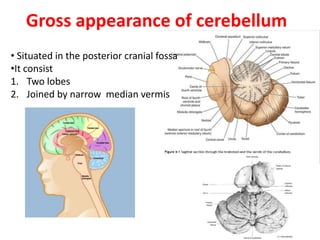
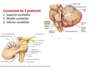
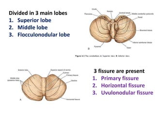
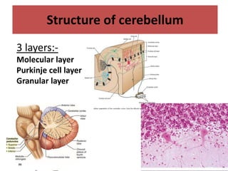
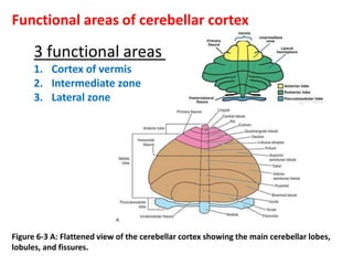
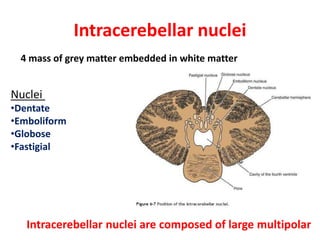
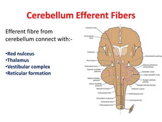

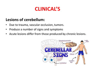

The document provides a detailed overview of the cerebellum, including its gross appearance, structure, functional divisions, and pathways, as well as clinical syndromes associated with cerebellar lesions. It describes the cerebellum's lobes, nuclei, afferent and efferent fibers, and the clinical signs of dysfunction, including hypotonia and ataxia. Additionally, it includes multiple-choice questions and clinical vignettes related to cerebellar anatomy and pathology.


