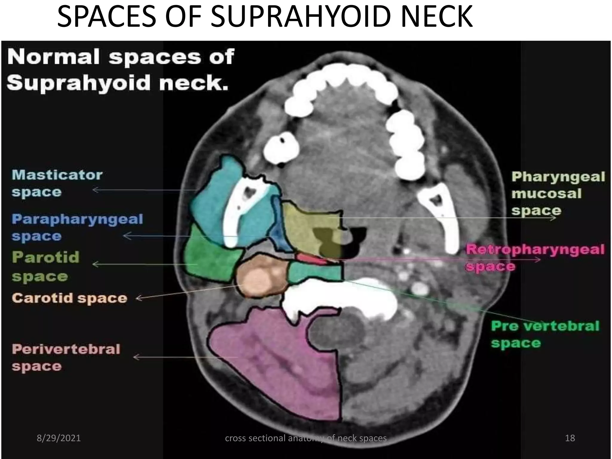
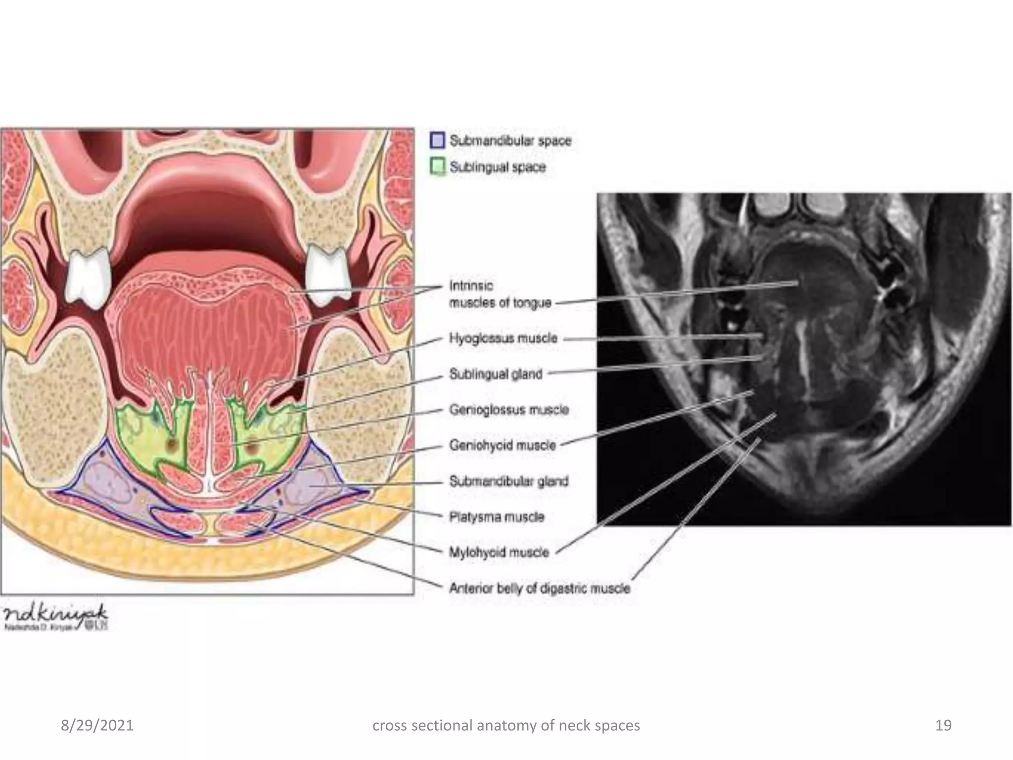
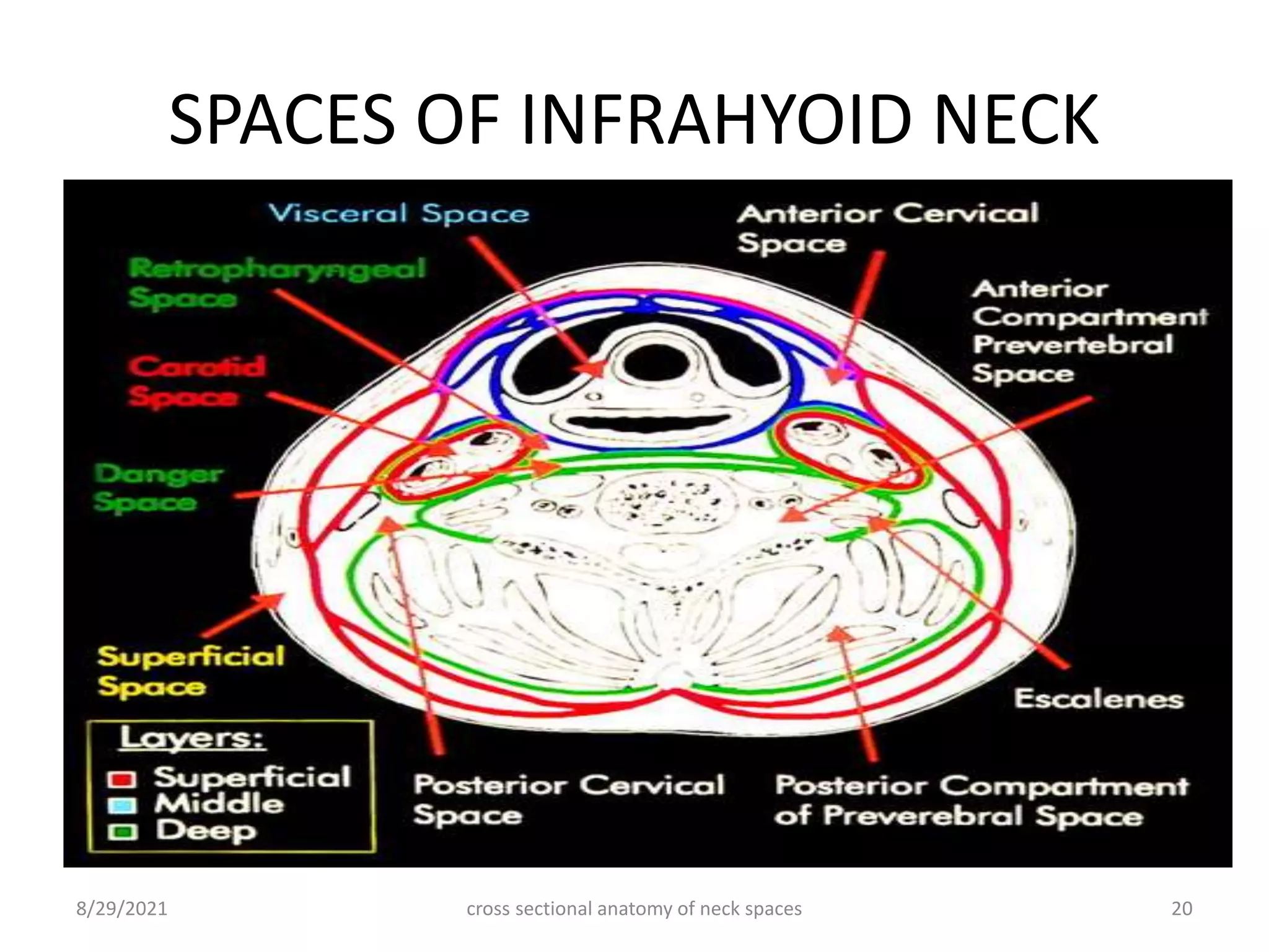
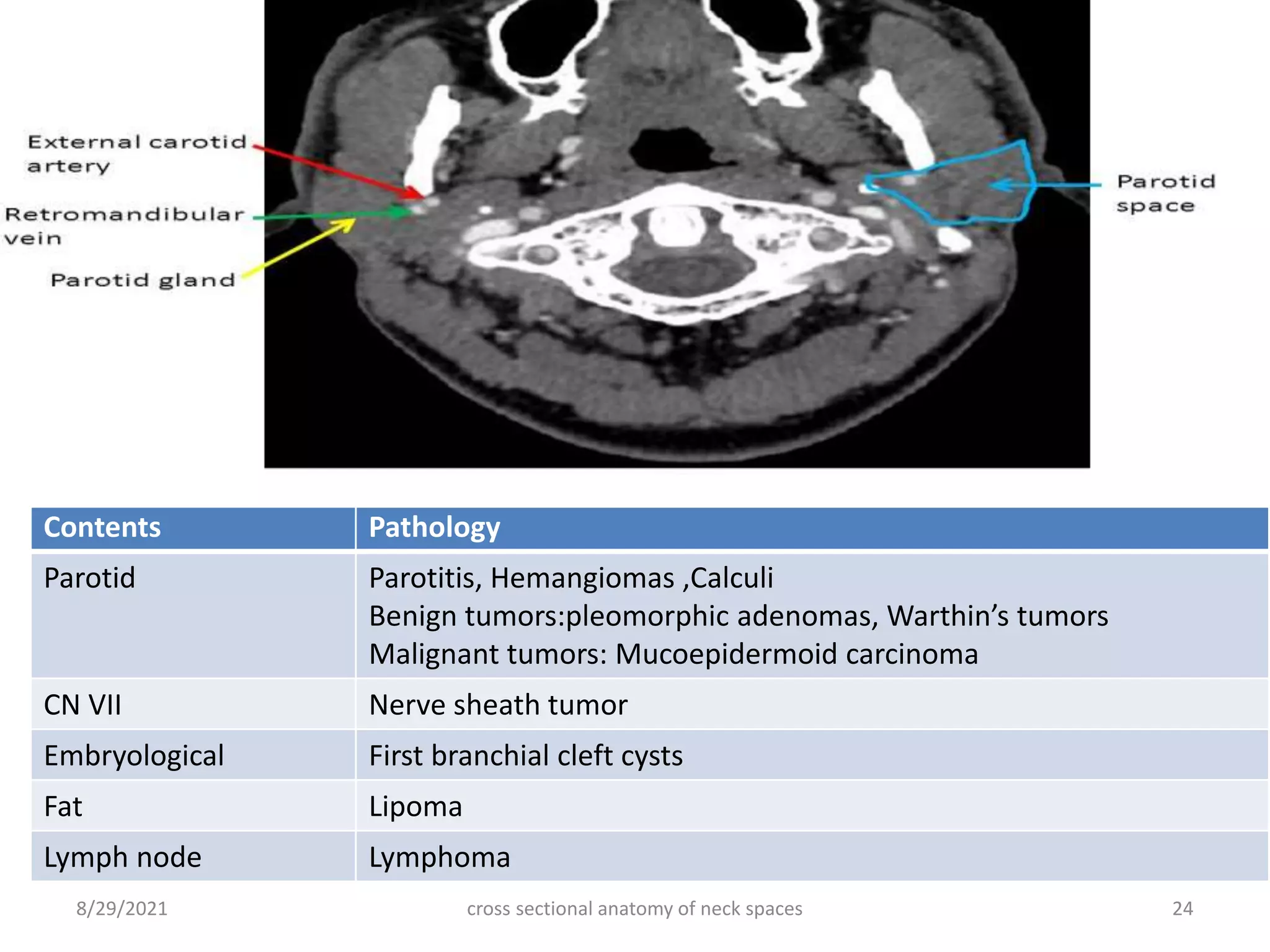
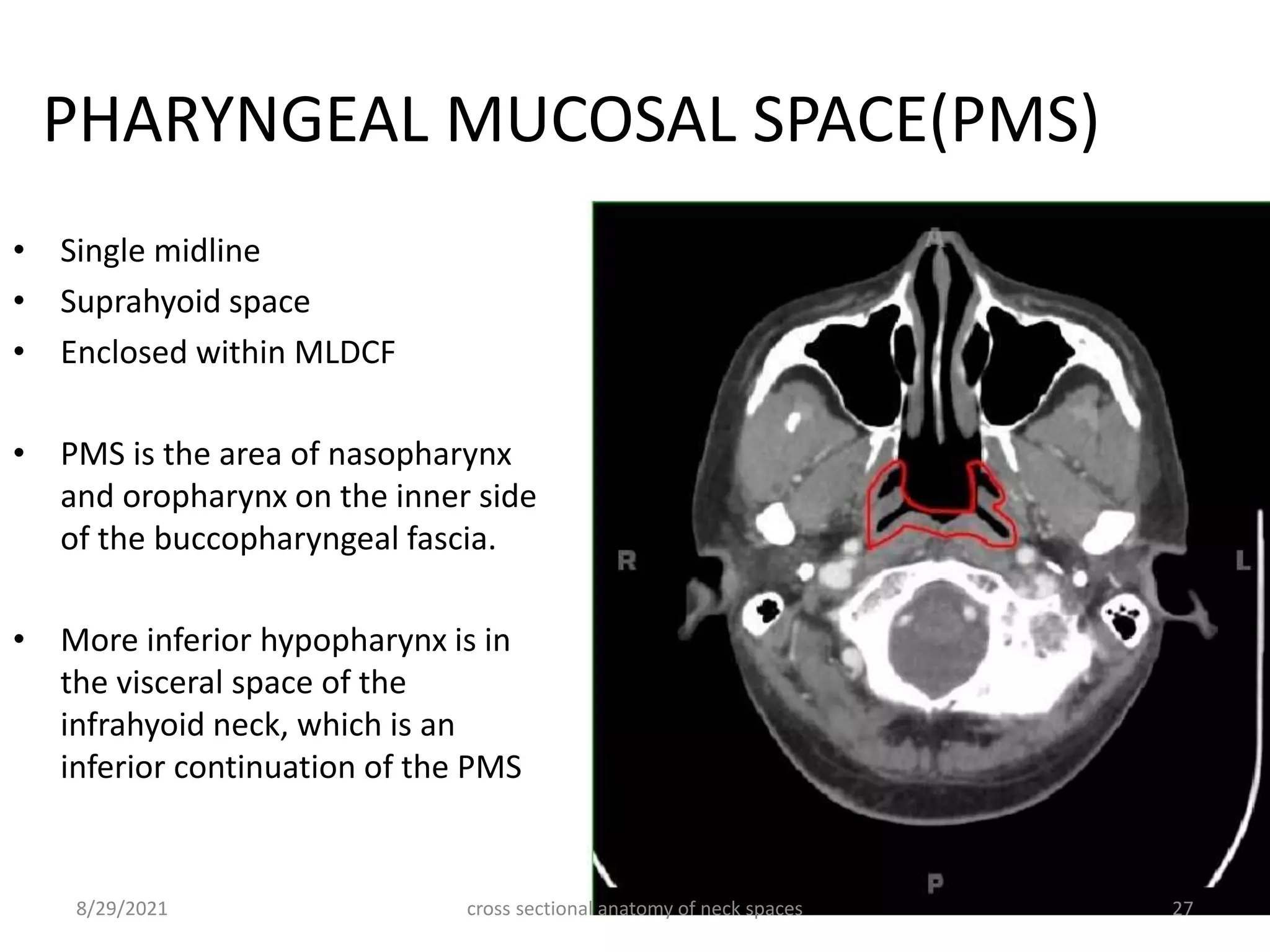
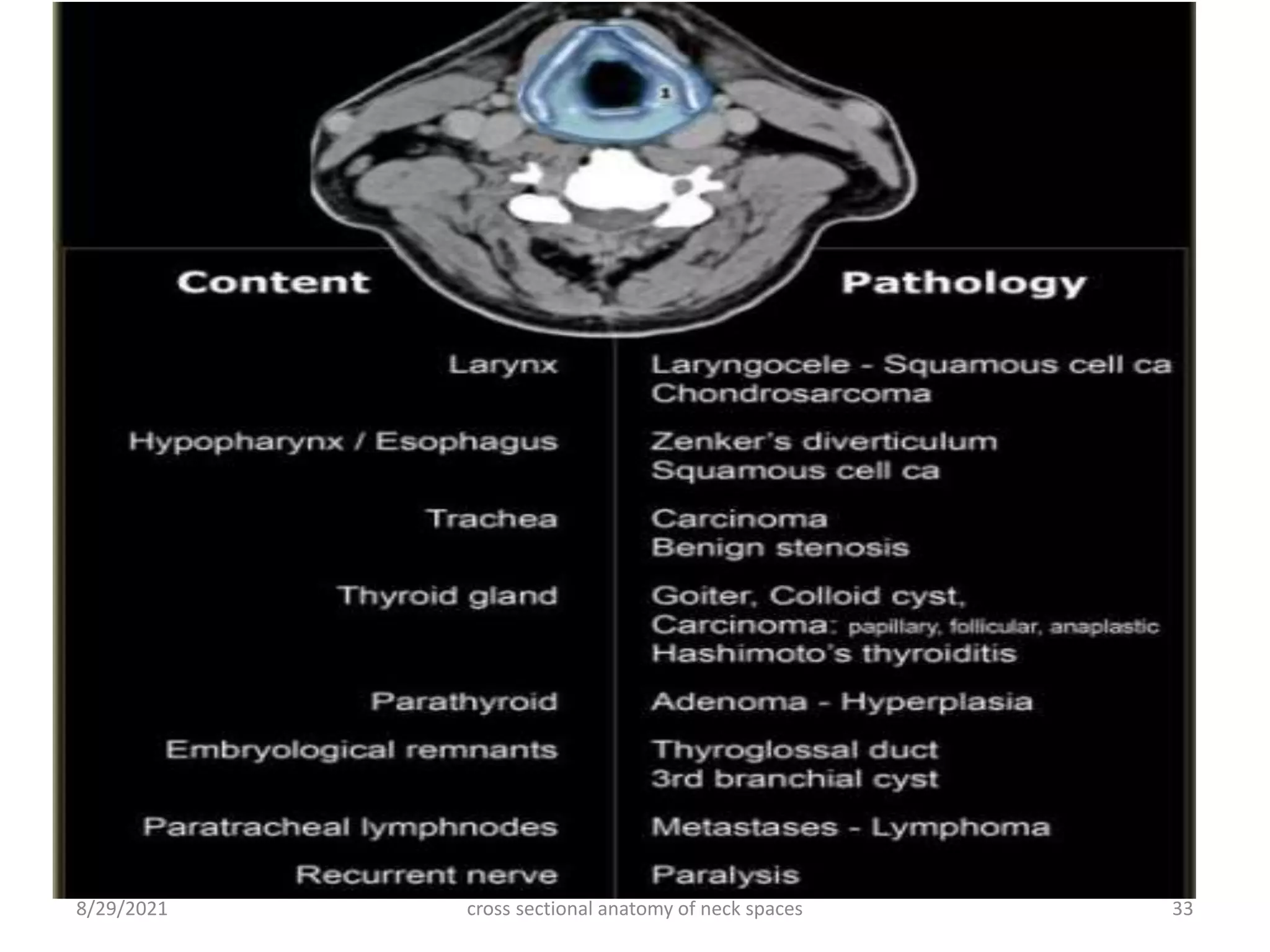
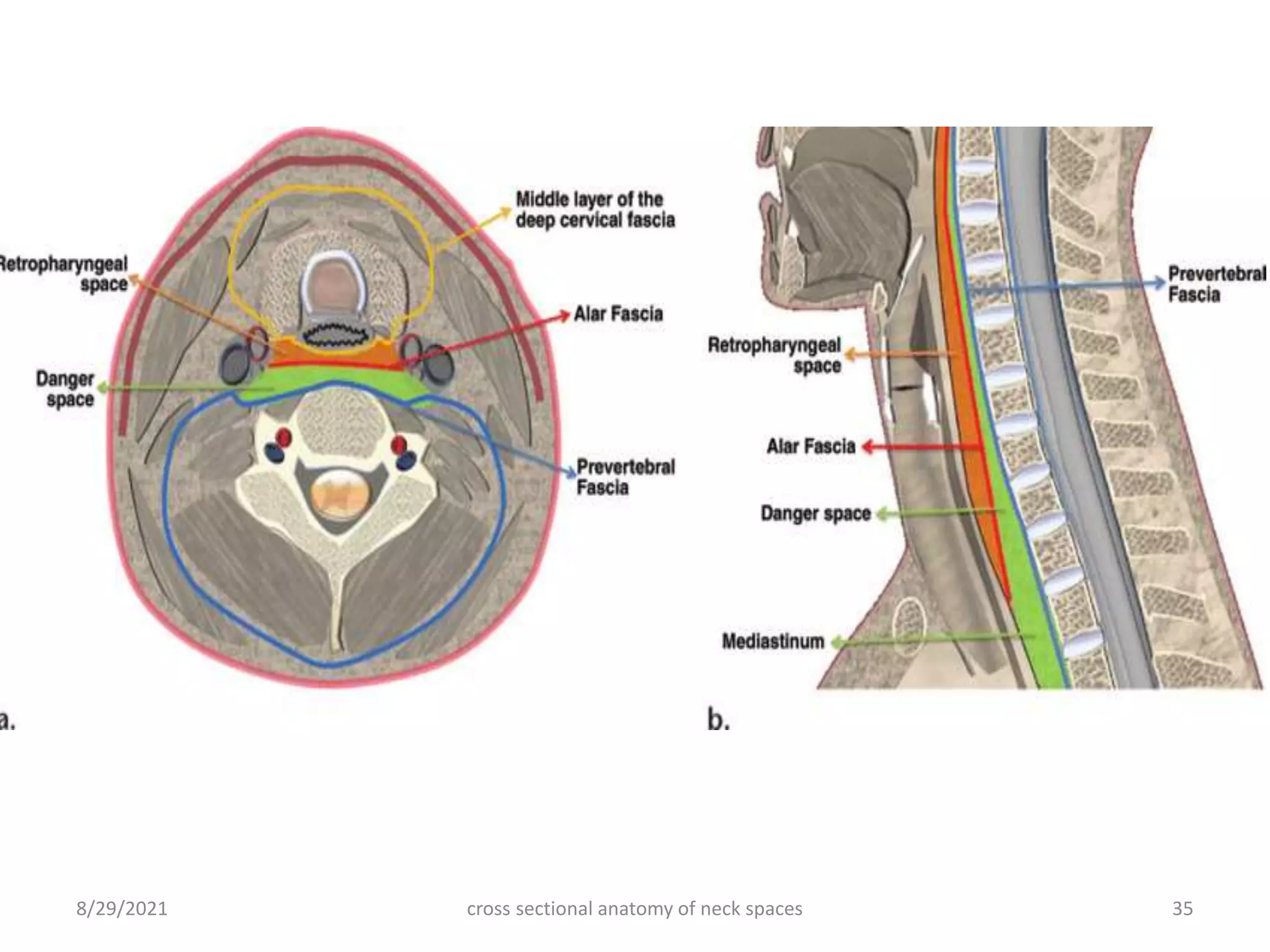
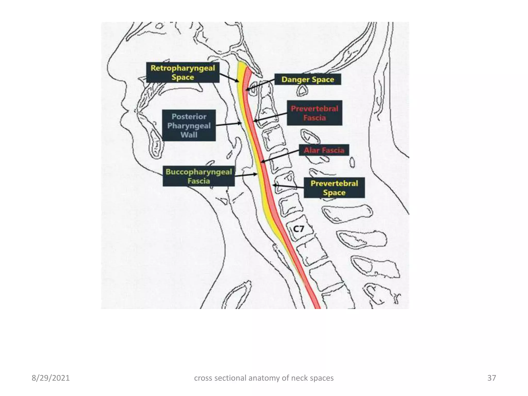
CROSS SECTIONAL ANATOMY OF
NECK SPACES
Dr Raman Ghimire
Resident Radiology
NAMS
INTRODUCTION
• The neck is composed of :
 Cervix(anteriorly)-means “neck”: cylinder of soft tissue
 Nucha(posteriorly) :vertebral column and its associated musculature
• Extent:
 Superior: line connecting the occiput and the tip of the chin
 Inferior :course of the first rib at the thoracic inlet.
• The neck has a complex anatomy where many critical anatomic structures are
compartmentalized into a small space.
• The traditional classification of the head and neck into nasopharynx, oropharynx
and hypopharynx is useful in the evaluation of superficial mucosal lesions but is
much less helpful in localizing deep-seated head and neck lesions.
• Clinicians divide the superficial structures of the neck into anterior and posterior
triangles using superficial musculature as landmarks.
cross sectional anatomy of neck spaces 2
8/29/2021
Sagittal T2-weighted image of the extracranial head and neck showing the division of the
anatomical and functional units of the upper aerodigestive tract
cross sectional anatomy of neck spaces 3
8/29/2021
cross sectional anatomy of neck spaces 4
8/29/2021
cross sectional anatomy of neck spaces 5
8/29/2021
FASCIA AND SPACES
• Anatomists pioneered the work on fascial anatomy and spaces.
• With the advent of cross-sectional imaging clinicians, surgeons and
radiologists adopted the spatial organization of the head and neck into fascial
compartments.
• The basis for dividing the neck into spaces and compartments is the arrangement
of the superficial and deep layers of the cervical fascia.
• The importance of these spaces is that they limit to some degree the spread of
most infections and some tumors.
• This concept can be used to formulate differential diagnosis, determine the extent
of disease and facilitate surgical and therapy planning.
cross sectional anatomy of neck spaces 6
8/29/2021
FASCIAE OF NECK
Fascia
Superficial
Deep
Superficial
( Investing)
Middle
(Visceral)
Deep
(Prevertebral)
cross sectional anatomy of neck spaces 7
8/29/2021
Superficial cervical fascia (SCF)
• Layer of fatty loose
connective tissue that
encircles the neck like a
collar.
• Encircles the deep fascia.
• Contains:
 Platysma muscles
 Muscles of facial expression
 Subcutaneous nerves and
lymphatics
 Portions of the anterior and
external jugular veins
cross sectional anatomy of neck spaces 8
8/29/2021
Deep cervical fascia (DCF)
• Made up of thinner but
denser, better-defined layers.
 Superficial layer (SLDCF)
 Middle layer (MLDCF)
 Deep layer (DLDCF)
All three layers contribute to
the carotid sheath.
cross sectional anatomy of neck spaces 9
8/29/2021
Superficial layer of deep cervical fascia (SLDCF)/
Investing layer
• Forms a complete collar around the
neck and envelops the
sternocleidomastoid, trapezius and
attaches to the skull base, mandible,
hyoid, clavicle, sternum and scapula.
• Capsule: parotid and submandibular
gland
• Spaces:
 Masticator space (including Buccal
space)
 Submandibular space (sub
mental,sub lingual ,submaxillary
sapce)
 Parotid space
cross sectional anatomy of neck spaces 10
8/29/2021
cross sectional anatomy of neck spaces 11
8/29/2021
• Caudally in the midline the SLDCF splits into two layers enclosing the
manubrium. This creates the variably sized suprasternal space of Burns
(or Gruber).
• Contains fat and a communicating vein between the left and right
anterior jugular veins.
cross sectional anatomy of neck spaces 12
8/29/2021
Middle layer of deep cervical fascia (MLDCF)
/Visceral/Pre-tracheal layer
• The MLDCF encloses the
anterior neck.
• Extent:
 IHN: from hyoid bone to
superior mediastinum.
 SHN: continues as
buccopharyngeal fascia and
encloses naso/oropharynx
• Muscular part: encases the
infrahyoid strap muscles
• Visceral part : envelops the
trachea, larynx, pharynx,
esophagus, and the thyroid
gland
cross sectional anatomy of neck spaces 13
8/29/2021
• Spaces:
 Pharyngeal mucosal space.
 Parapharyngeal space.
 Visceral space
 Retropharyngeal space
(along with DLDCF)
cross sectional anatomy of neck spaces 14
8/29/2021
Deep layer of deep cervical fascia (DLDCF)
/Prevertebral layer
• DLCF extends from the skull base to the T4 vertebra where it fuses with the anterior
longitudinal ligament in the posterior mediastinum.
• The deep layer encircles the vertebrae, paravertebral and paraspinal muscles,
vertebral artery and vein, phrenic nerve and trunks of the brachial plexus.
• The vertebrae are completely enclosed by this layer which attaches firmly to the
transverse processes dividing the space into an anterior prevertebral and posterior
paraspinal portion.
• Anteromedial to the scalene muscles, the deep layer splits into two leaves; the
ventral leaf being the alar fascia, and the dorsal leaf being the prevertebral fascia
• Alar layer blends with the visceral layer along the posterior margin of the
esophagus at the level between the C6 and T4 vertebrae; the prevertebral layer
extends from the skull base to the coccyx.
cross sectional anatomy of neck spaces 15
8/29/2021
• This fascia separates the
lower neck from the
thorax and is called
Sibson’s fascia.
Spaces:
 Perivertebral space
 Danger space
 Retropharyngeal space
(MLDCF+DLDCF)
 Carotid space.
(all 3 layers)
cross sectional anatomy of neck spaces 16
8/29/2021
SPACES OF NECK
Suprahyoid Neck (SHN)
• Parotid space
• Masticator space
(+ buccal space)
• Submandibular space
(+ sublingual space
and submental space)
• Pharyngeal mucosal
Space
• Parapharyngeal
space(or prestyloid
parapharyngeal space )
Infrahyoid Neck (IHN)
• Visceral space
• Anterior cervical space
Entire length of neck
(SHN+IHN)
• Carotid space (SHN:
poststyloid
parapharyngeal space,
IHN: carotid sheath)
• Retropharyngeal space
• Danger space
• Perivertebral space
• posterior cervical space
cross sectional anatomy of neck spaces 17
8/29/2021
SPACES OF SUPRAHYOID NECK
cross sectional anatomy of neck spaces 18
8/29/2021
8/29/2021 cross sectional anatomy of neck spaces 19
SPACES OF INFRAHYOID NECK
cross sectional anatomy of neck spaces 20
8/29/2021
MASTICATOR SPACE
• Paired
• Suprahyoid space
• Enclosed within SLDCF
• Contains :
 Ramus and body of mandible
 Trigeminal nerve(mandibular
division)
 Muscles of mastication.
MS
PPS
PS
cross sectional anatomy of neck spaces 21
8/29/2021
Contents Pathology
Muscle Rhabdomyosarcoma
CN V3 Schwannoma,neurofibroma
Mandible Dental tumors ,cysts ,abscesses
and osteomyelitis.
Vessels Hemangiomas, lymphangiomas
Fat Lipoma
Extension of SCC
8/29/2021 cross sectional anatomy of neck spaces 22
• Masticator space malignancy or infection can spread perineurally via the
foramen ovale and along the course of the mandibular division of
the trigeminal nerve into the middle cranial fossa.
PAROTID SPACE
• Paired
• Suprahyoid space
• Enclosed within SLDCF
cross sectional anatomy of neck spaces 23
8/29/2021
Contents Pathology
Parotid Parotitis, Hemangiomas ,Calculi
Benign tumors:pleomorphic adenomas, Warthin’s tumors
Malignant tumors: Mucoepidermoid carcinoma
CN VII Nerve sheath tumor
Embryological First branchial cleft cysts
Fat Lipoma
Lymph node Lymphoma
8/29/2021 cross sectional anatomy of neck spaces 24
8/29/2021 cross sectional anatomy of neck spaces 25
SUBMANDIBULAR SPACE
• Paired
• Suprahyoid space
• Enclosed within SLDCF
• Submental space and sublingual
space also considered within SMS as
these spaces communicate freely
Anatomy of the submandibular and sublingual spaces in the coronal plane: picture illustration
and T2-weighted MR image
Contents Pathology
Submandibular gland Sialolithisis and sialadenitis
Benign:pleomorphic adenomas,
malignant masses :mucoepidermoid and adenocystic carcinomas
Sub mental
Ia/submandibular Ib
Lymph nodes
Lymphoma ,Metastasis,TB
Facial artery /vein Vascular malformations
Embryological Branchial cleft cysts, cystic hygromas, dermoids, epidermoids and
thyroglossal duct cysts
Fat Lipoma,liposarcoma
Ranula
Abscess, ludwig angina
Squamous cell carcinoma extension
cross sectional anatomy of neck spaces 26
8/29/2021
PHARYNGEAL MUCOSAL SPACE(PMS)
• Single midline
• Suprahyoid space
• Enclosed within MLDCF
• PMS is the area of nasopharynx
and oropharynx on the inner side
of the buccopharyngeal fascia.
• More inferior hypopharynx is in
the visceral space of the
infrahyoid neck, which is an
inferior continuation of the PMS
cross sectional anatomy of neck spaces 27
8/29/2021
PMS
PPS
RPS
cross sectional anatomy of neck spaces 28
8/29/2021
Contents Pathology
Squamous mucosa SCC
Tornwaldt cyst
lymphoid tissue of
Waldeyer's ring
Tonsillitis , Peritonsillar abscess
non-Hodgkin’s lymphoma (NHL)
Minor salivary glands Benign and malignant tumor
Pharyngeal constrictor
muscle
Cartilaginous portion
of the Eustachian
tube
Juvenile Angiofibroma
cross sectional anatomy of neck spaces 29
8/29/2021
8/29/2021 cross sectional anatomy of neck spaces 30
PARAPHARYNGEAL SPACE(PPS)
• Paired
• Suprahyoid space
• Enclosed within MLDCF
• Its a inverted pyramid space
from inferior surface of
petrous bone to the hyoid
bone
CONTENTS Pathology
Fat Lipoma
Vessels: pharyngeal venous plexus,
internal mammary artery and ascending
pharyngeal artery
Branch of mandibular nerve Trigeminal schwannoma
Minor or ectopic salivary gland/rests Salivary gland tumor pleomorphic
adenoma
Embryological Cystic hygroma/lymphangioma
second branchial cleft cyst
Parapharyngeal abscess
cross sectional anatomy of neck spaces 31
8/29/2021
cross sectional anatomy of neck spaces 32
VISCERAL SPACE
• The visceral space includes all
structures within the confines of
the MLDCF .
• The visceral space extends from
the hyoid bone superiorly to
the superior mediastinum (level
of aortic arch / T4
• Infrahyoid continuation of PMS
8/29/2021
cross sectional anatomy of neck spaces 33
8/29/2021
RETROPHARYNGEAL SPACE
• Boundaries:
Anterior: MLDCF
Posterior:DLDCF(alar)
• Extends from the skull base upto
T4 level.
• Contents:
 Suprahyoid RPS :Nodes and fat
 Infrahyoid RPS : fat
cross sectional anatomy of neck spaces 34
8/29/2021
cross sectional anatomy of neck spaces 35
8/29/2021
DANGER SPACE (DS)
• The DS is formed by the splitting of
the deep layer into an anterolateral
alar slip and a posterior prevertebral
portion.
• Boundaries:
 Anterior: Alar fascia
 Posterior: Prevertebral fascia
 Superior: clivus
 Inferior: posterior mediastinum at
the level of the diaphragm
• Contents :Small amount of loose
fatty connecctive tissue
• In healthy patients, it is
indistinguishable from
the retropharyngeal space
cross sectional anatomy of neck spaces 36
8/29/2021
cross sectional anatomy of neck spaces 37
8/29/2021
PERIVERTEBRAL SPACE(PVS)
• Posterior midline space
• Enclosed by DLDCF
• Extends from the base of the skull
to the level of coccyx (SHN+IHN)
• Prevertebral space: prevertebral
and scalene muscle, roots of
brachial plexus ,vertebral bodies
and pedicles, phrenic nerve,the
vertebral arteries and veins.
• Paraspinal Space: paraspinal
musculature, vertebral laminae
and spinous processes.
cross sectional anatomy of neck spaces 38
8/29/2021
cross sectional anatomy of neck spaces 39
8/29/2021
A.Contrast-enhanced axial CT shows fluid in RPS and posterior displacement of the longus
capitis muscles (B) Axial contrast-enhanced axial CT shows anterior displacemnet of longus
capitis muscles with fluid collection within the prevertebral space
• Prevertebral soft tissue thickness(PVST):
 RPS and PVS should not exceed >7 mm at C2 for adult and children
 C6 soft-tissue space should no be >4 mm (children) and >22 mm for
(adults)
A 24-year-old patient after a motor vehicle crash with closed head injury; no fracture
was identified in the cervical spine or craniocervical junction. A, Midsagittal MDCT
image of the cervical spine demonstrates abnormal PVST thickening at C1 and C2
(asterisks). B, Correlation with midsagittal short τ inversion recovery MR image
obtained the next day confirms the presence of extensive PVST edema and/or
cross sectional anatomy of neck spaces 40
8/29/2021
cross sectional anatomy of neck spaces 41
8/29/2021
CAROTID SPACE
• Carotid space is a paired space
defined by the carotid sheath.
• Carotid sheath is formed by all
three layers of DCF.
• This space extends from the skull
base to the aortic arch at the
thoracic inlet/T4.
• The suprahyoid CS is also known as
the poststyloid PPS.
• Contents:
SHN: CN IX-XII, ICA,IJV
IHN: CN X, CCA,IJV
cross sectional anatomy of neck spaces 42
8/29/2021
cross sectional anatomy of neck spaces 43
8/29/2021
POSTERIOR CERVICAL SPACE
• Fat filled deep spaces of the
neck located posterolaterally.
• Posterolateral part of the neck
extending from the mastoid tip
and base of skull to the clavicles
• Most of the volume is in the
infrahyoid neck, with some
extension into the suprahyoid neck .
• Between SLDCF and DLDCF
• Lesion in PCS typically cause
anteromedial displacement of the
carotid space and posteromedial
displacement of the prevertebral
space
cross sectional anatomy of neck spaces 44
8/29/2021
cross sectional anatomy of neck spaces 45
8/29/2021
ANTERIOR CERVICAL SPACE
• The anterior cervical space is a
small infrahyoid compartment of
the head and neck.
• It is a fat containing space and is
not enclosed by fascia.
• Content : Fat
• Pathology : lipoma , second
branchial cleft cyst
cross sectional anatomy of neck spaces 46
8/29/2021
REFERENCES
• CT AND MR IMAGING OF THE WHOLE BODY;6TH EDITION
• GRAINGER & ALLISON’S DIAGNOSTIC RADIOLOGY; 6TH EDITION
• https://radiologyassistant.nl/head-neck/infrahyoid-neck
• https://radiopaedia.org/articles/deep-cervical-fascia
cross sectional anatomy of neck spaces 47
8/29/2021
THANK YOU
cross sectional anatomy of neck spaces 48
8/29/2021

Cross sectional anatomy of neck spaces

  • 1.
    CROSS SECTIONAL ANATOMYOF NECK SPACES Dr Raman Ghimire Resident Radiology NAMS
  • 2.
    INTRODUCTION • The neckis composed of :  Cervix(anteriorly)-means “neck”: cylinder of soft tissue  Nucha(posteriorly) :vertebral column and its associated musculature • Extent:  Superior: line connecting the occiput and the tip of the chin  Inferior :course of the first rib at the thoracic inlet. • The neck has a complex anatomy where many critical anatomic structures are compartmentalized into a small space. • The traditional classification of the head and neck into nasopharynx, oropharynx and hypopharynx is useful in the evaluation of superficial mucosal lesions but is much less helpful in localizing deep-seated head and neck lesions. • Clinicians divide the superficial structures of the neck into anterior and posterior triangles using superficial musculature as landmarks. cross sectional anatomy of neck spaces 2 8/29/2021
  • 3.
    Sagittal T2-weighted imageof the extracranial head and neck showing the division of the anatomical and functional units of the upper aerodigestive tract cross sectional anatomy of neck spaces 3 8/29/2021
  • 4.
    cross sectional anatomyof neck spaces 4 8/29/2021
  • 5.
    cross sectional anatomyof neck spaces 5 8/29/2021
  • 6.
    FASCIA AND SPACES •Anatomists pioneered the work on fascial anatomy and spaces. • With the advent of cross-sectional imaging clinicians, surgeons and radiologists adopted the spatial organization of the head and neck into fascial compartments. • The basis for dividing the neck into spaces and compartments is the arrangement of the superficial and deep layers of the cervical fascia. • The importance of these spaces is that they limit to some degree the spread of most infections and some tumors. • This concept can be used to formulate differential diagnosis, determine the extent of disease and facilitate surgical and therapy planning. cross sectional anatomy of neck spaces 6 8/29/2021
  • 7.
    FASCIAE OF NECK Fascia Superficial Deep Superficial (Investing) Middle (Visceral) Deep (Prevertebral) cross sectional anatomy of neck spaces 7 8/29/2021
  • 8.
    Superficial cervical fascia(SCF) • Layer of fatty loose connective tissue that encircles the neck like a collar. • Encircles the deep fascia. • Contains:  Platysma muscles  Muscles of facial expression  Subcutaneous nerves and lymphatics  Portions of the anterior and external jugular veins cross sectional anatomy of neck spaces 8 8/29/2021
  • 9.
    Deep cervical fascia(DCF) • Made up of thinner but denser, better-defined layers.  Superficial layer (SLDCF)  Middle layer (MLDCF)  Deep layer (DLDCF) All three layers contribute to the carotid sheath. cross sectional anatomy of neck spaces 9 8/29/2021
  • 10.
    Superficial layer ofdeep cervical fascia (SLDCF)/ Investing layer • Forms a complete collar around the neck and envelops the sternocleidomastoid, trapezius and attaches to the skull base, mandible, hyoid, clavicle, sternum and scapula. • Capsule: parotid and submandibular gland • Spaces:  Masticator space (including Buccal space)  Submandibular space (sub mental,sub lingual ,submaxillary sapce)  Parotid space cross sectional anatomy of neck spaces 10 8/29/2021
  • 11.
    cross sectional anatomyof neck spaces 11 8/29/2021
  • 12.
    • Caudally inthe midline the SLDCF splits into two layers enclosing the manubrium. This creates the variably sized suprasternal space of Burns (or Gruber). • Contains fat and a communicating vein between the left and right anterior jugular veins. cross sectional anatomy of neck spaces 12 8/29/2021
  • 13.
    Middle layer ofdeep cervical fascia (MLDCF) /Visceral/Pre-tracheal layer • The MLDCF encloses the anterior neck. • Extent:  IHN: from hyoid bone to superior mediastinum.  SHN: continues as buccopharyngeal fascia and encloses naso/oropharynx • Muscular part: encases the infrahyoid strap muscles • Visceral part : envelops the trachea, larynx, pharynx, esophagus, and the thyroid gland cross sectional anatomy of neck spaces 13 8/29/2021
  • 14.
    • Spaces:  Pharyngealmucosal space.  Parapharyngeal space.  Visceral space  Retropharyngeal space (along with DLDCF) cross sectional anatomy of neck spaces 14 8/29/2021
  • 15.
    Deep layer ofdeep cervical fascia (DLDCF) /Prevertebral layer • DLCF extends from the skull base to the T4 vertebra where it fuses with the anterior longitudinal ligament in the posterior mediastinum. • The deep layer encircles the vertebrae, paravertebral and paraspinal muscles, vertebral artery and vein, phrenic nerve and trunks of the brachial plexus. • The vertebrae are completely enclosed by this layer which attaches firmly to the transverse processes dividing the space into an anterior prevertebral and posterior paraspinal portion. • Anteromedial to the scalene muscles, the deep layer splits into two leaves; the ventral leaf being the alar fascia, and the dorsal leaf being the prevertebral fascia • Alar layer blends with the visceral layer along the posterior margin of the esophagus at the level between the C6 and T4 vertebrae; the prevertebral layer extends from the skull base to the coccyx. cross sectional anatomy of neck spaces 15 8/29/2021
  • 16.
    • This fasciaseparates the lower neck from the thorax and is called Sibson’s fascia. Spaces:  Perivertebral space  Danger space  Retropharyngeal space (MLDCF+DLDCF)  Carotid space. (all 3 layers) cross sectional anatomy of neck spaces 16 8/29/2021
  • 17.
    SPACES OF NECK SuprahyoidNeck (SHN) • Parotid space • Masticator space (+ buccal space) • Submandibular space (+ sublingual space and submental space) • Pharyngeal mucosal Space • Parapharyngeal space(or prestyloid parapharyngeal space ) Infrahyoid Neck (IHN) • Visceral space • Anterior cervical space Entire length of neck (SHN+IHN) • Carotid space (SHN: poststyloid parapharyngeal space, IHN: carotid sheath) • Retropharyngeal space • Danger space • Perivertebral space • posterior cervical space cross sectional anatomy of neck spaces 17 8/29/2021
  • 18.
    SPACES OF SUPRAHYOIDNECK cross sectional anatomy of neck spaces 18 8/29/2021
  • 19.
    8/29/2021 cross sectionalanatomy of neck spaces 19
  • 20.
    SPACES OF INFRAHYOIDNECK cross sectional anatomy of neck spaces 20 8/29/2021
  • 21.
    MASTICATOR SPACE • Paired •Suprahyoid space • Enclosed within SLDCF • Contains :  Ramus and body of mandible  Trigeminal nerve(mandibular division)  Muscles of mastication. MS PPS PS cross sectional anatomy of neck spaces 21 8/29/2021
  • 22.
    Contents Pathology Muscle Rhabdomyosarcoma CNV3 Schwannoma,neurofibroma Mandible Dental tumors ,cysts ,abscesses and osteomyelitis. Vessels Hemangiomas, lymphangiomas Fat Lipoma Extension of SCC 8/29/2021 cross sectional anatomy of neck spaces 22 • Masticator space malignancy or infection can spread perineurally via the foramen ovale and along the course of the mandibular division of the trigeminal nerve into the middle cranial fossa.
  • 23.
    PAROTID SPACE • Paired •Suprahyoid space • Enclosed within SLDCF cross sectional anatomy of neck spaces 23 8/29/2021
  • 24.
    Contents Pathology Parotid Parotitis,Hemangiomas ,Calculi Benign tumors:pleomorphic adenomas, Warthin’s tumors Malignant tumors: Mucoepidermoid carcinoma CN VII Nerve sheath tumor Embryological First branchial cleft cysts Fat Lipoma Lymph node Lymphoma 8/29/2021 cross sectional anatomy of neck spaces 24
  • 25.
    8/29/2021 cross sectionalanatomy of neck spaces 25 SUBMANDIBULAR SPACE • Paired • Suprahyoid space • Enclosed within SLDCF • Submental space and sublingual space also considered within SMS as these spaces communicate freely Anatomy of the submandibular and sublingual spaces in the coronal plane: picture illustration and T2-weighted MR image
  • 26.
    Contents Pathology Submandibular glandSialolithisis and sialadenitis Benign:pleomorphic adenomas, malignant masses :mucoepidermoid and adenocystic carcinomas Sub mental Ia/submandibular Ib Lymph nodes Lymphoma ,Metastasis,TB Facial artery /vein Vascular malformations Embryological Branchial cleft cysts, cystic hygromas, dermoids, epidermoids and thyroglossal duct cysts Fat Lipoma,liposarcoma Ranula Abscess, ludwig angina Squamous cell carcinoma extension cross sectional anatomy of neck spaces 26 8/29/2021
  • 27.
    PHARYNGEAL MUCOSAL SPACE(PMS) •Single midline • Suprahyoid space • Enclosed within MLDCF • PMS is the area of nasopharynx and oropharynx on the inner side of the buccopharyngeal fascia. • More inferior hypopharynx is in the visceral space of the infrahyoid neck, which is an inferior continuation of the PMS cross sectional anatomy of neck spaces 27 8/29/2021
  • 28.
    PMS PPS RPS cross sectional anatomyof neck spaces 28 8/29/2021
  • 29.
    Contents Pathology Squamous mucosaSCC Tornwaldt cyst lymphoid tissue of Waldeyer's ring Tonsillitis , Peritonsillar abscess non-Hodgkin’s lymphoma (NHL) Minor salivary glands Benign and malignant tumor Pharyngeal constrictor muscle Cartilaginous portion of the Eustachian tube Juvenile Angiofibroma cross sectional anatomy of neck spaces 29 8/29/2021
  • 30.
    8/29/2021 cross sectionalanatomy of neck spaces 30 PARAPHARYNGEAL SPACE(PPS) • Paired • Suprahyoid space • Enclosed within MLDCF • Its a inverted pyramid space from inferior surface of petrous bone to the hyoid bone
  • 31.
    CONTENTS Pathology Fat Lipoma Vessels:pharyngeal venous plexus, internal mammary artery and ascending pharyngeal artery Branch of mandibular nerve Trigeminal schwannoma Minor or ectopic salivary gland/rests Salivary gland tumor pleomorphic adenoma Embryological Cystic hygroma/lymphangioma second branchial cleft cyst Parapharyngeal abscess cross sectional anatomy of neck spaces 31 8/29/2021
  • 32.
    cross sectional anatomyof neck spaces 32 VISCERAL SPACE • The visceral space includes all structures within the confines of the MLDCF . • The visceral space extends from the hyoid bone superiorly to the superior mediastinum (level of aortic arch / T4 • Infrahyoid continuation of PMS 8/29/2021
  • 33.
    cross sectional anatomyof neck spaces 33 8/29/2021
  • 34.
    RETROPHARYNGEAL SPACE • Boundaries: Anterior:MLDCF Posterior:DLDCF(alar) • Extends from the skull base upto T4 level. • Contents:  Suprahyoid RPS :Nodes and fat  Infrahyoid RPS : fat cross sectional anatomy of neck spaces 34 8/29/2021
  • 35.
    cross sectional anatomyof neck spaces 35 8/29/2021
  • 36.
    DANGER SPACE (DS) •The DS is formed by the splitting of the deep layer into an anterolateral alar slip and a posterior prevertebral portion. • Boundaries:  Anterior: Alar fascia  Posterior: Prevertebral fascia  Superior: clivus  Inferior: posterior mediastinum at the level of the diaphragm • Contents :Small amount of loose fatty connecctive tissue • In healthy patients, it is indistinguishable from the retropharyngeal space cross sectional anatomy of neck spaces 36 8/29/2021
  • 37.
    cross sectional anatomyof neck spaces 37 8/29/2021
  • 38.
    PERIVERTEBRAL SPACE(PVS) • Posteriormidline space • Enclosed by DLDCF • Extends from the base of the skull to the level of coccyx (SHN+IHN) • Prevertebral space: prevertebral and scalene muscle, roots of brachial plexus ,vertebral bodies and pedicles, phrenic nerve,the vertebral arteries and veins. • Paraspinal Space: paraspinal musculature, vertebral laminae and spinous processes. cross sectional anatomy of neck spaces 38 8/29/2021
  • 39.
    cross sectional anatomyof neck spaces 39 8/29/2021 A.Contrast-enhanced axial CT shows fluid in RPS and posterior displacement of the longus capitis muscles (B) Axial contrast-enhanced axial CT shows anterior displacemnet of longus capitis muscles with fluid collection within the prevertebral space
  • 40.
    • Prevertebral softtissue thickness(PVST):  RPS and PVS should not exceed >7 mm at C2 for adult and children  C6 soft-tissue space should no be >4 mm (children) and >22 mm for (adults) A 24-year-old patient after a motor vehicle crash with closed head injury; no fracture was identified in the cervical spine or craniocervical junction. A, Midsagittal MDCT image of the cervical spine demonstrates abnormal PVST thickening at C1 and C2 (asterisks). B, Correlation with midsagittal short τ inversion recovery MR image obtained the next day confirms the presence of extensive PVST edema and/or cross sectional anatomy of neck spaces 40 8/29/2021
  • 41.
    cross sectional anatomyof neck spaces 41 8/29/2021
  • 42.
    CAROTID SPACE • Carotidspace is a paired space defined by the carotid sheath. • Carotid sheath is formed by all three layers of DCF. • This space extends from the skull base to the aortic arch at the thoracic inlet/T4. • The suprahyoid CS is also known as the poststyloid PPS. • Contents: SHN: CN IX-XII, ICA,IJV IHN: CN X, CCA,IJV cross sectional anatomy of neck spaces 42 8/29/2021
  • 43.
    cross sectional anatomyof neck spaces 43 8/29/2021
  • 44.
    POSTERIOR CERVICAL SPACE •Fat filled deep spaces of the neck located posterolaterally. • Posterolateral part of the neck extending from the mastoid tip and base of skull to the clavicles • Most of the volume is in the infrahyoid neck, with some extension into the suprahyoid neck . • Between SLDCF and DLDCF • Lesion in PCS typically cause anteromedial displacement of the carotid space and posteromedial displacement of the prevertebral space cross sectional anatomy of neck spaces 44 8/29/2021
  • 45.
    cross sectional anatomyof neck spaces 45 8/29/2021
  • 46.
    ANTERIOR CERVICAL SPACE •The anterior cervical space is a small infrahyoid compartment of the head and neck. • It is a fat containing space and is not enclosed by fascia. • Content : Fat • Pathology : lipoma , second branchial cleft cyst cross sectional anatomy of neck spaces 46 8/29/2021
  • 47.
    REFERENCES • CT ANDMR IMAGING OF THE WHOLE BODY;6TH EDITION • GRAINGER & ALLISON’S DIAGNOSTIC RADIOLOGY; 6TH EDITION • https://radiologyassistant.nl/head-neck/infrahyoid-neck • https://radiopaedia.org/articles/deep-cervical-fascia cross sectional anatomy of neck spaces 47 8/29/2021
  • 48.
    THANK YOU cross sectionalanatomy of neck spaces 48 8/29/2021

Editor's Notes

  • #13 Space of burns: if this space is entered during a tracheostomy, inadvertent transection of the communicating vein may result in considerable blood loss
  • #14 Strap muscle: TOSS
  • #18 Differs in different sources
  • #19 SMS..missing
  • #22 Muscles of mastication :masseter ,temporalis ,pterigoid Anterior buccal space: space betn buccinator & platysma
  • #24 Retromandibular vein (temporomaxillary vein, posterior facial vein), formed by the union of the superficial temporal and maxillary veins Drains into internal /external jugular.
  • #26 Medially submental . Post :PPS
  • #35 Retropharyngeal abscess…… extension of SCC,lymphoma
  • #36 There are two other spaces in close proximity to the retropharyngeal space: the danger space and the prevertebral space. They are often confused with the retropharyngeal space.
  • #37 It is only visible when distended by fluid or pus, below the level of T1-T6, since the retropharyngeal space variably ends at this level.
  • #43 IHN: cca ,ijv,vagus
  • #46 MPNST: malignant peripheral nerve sheath tumor