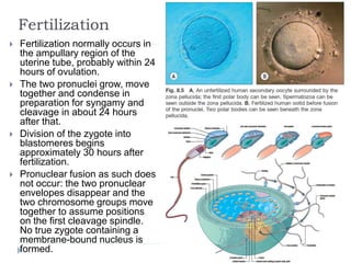
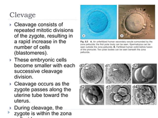
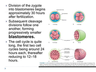
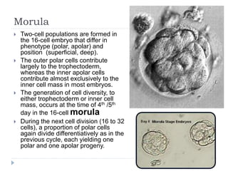
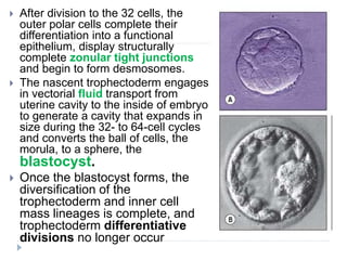
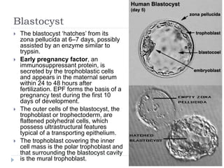
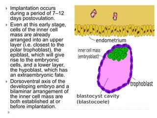
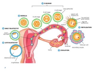
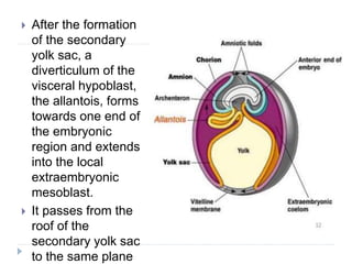
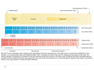
SlideShare a Scribd company logo
Blastulation
Dr. Komal Parmar
Fertilization
 Fertilization normally occurs in
the ampullary region of the
uterine tube, probably within 24
hours of ovulation.
 The two pronuclei grow, move
together and condense in
preparation for syngamy and
cleavage in about 24 hours
after that.
 Division of the zygote into
blastomeres begins
approximately 30 hours after
fertilization.
 Pronuclear fusion as such does
not occur: the two pronuclear
envelopes disappear and the
two chromosome groups move
together to assume positions
on the first cleavage spindle.
No true zygote containing a
membrane-bound nucleus is
formed.
Clevage
 Cleavage consists of
repeated mitotic divisions
of the zygote, resulting in
a rapid increase in the
number of cells
(blastomeres).
 These embryonic cells
become smaller with each
successive cleavage
division.
 Cleavage occurs as the
zygote passes along the
uterine tube toward the
uterus.
 During cleavage, the
zygote is within the zona
 Division of the zygote
into blastomeres begins
approximately 30 hours
after fertilization.
 Subsequent cleavage
divisions follow one
another, forming
progressively smaller
blastomeres.
 The cell cycle is quite
long, the first two cell
cycles being around 24
hours each, thereafter
reducing to 12–18
hours.
 Cell division is asynchronous
 Daughter cells may retain a cytoplasmic link through
much of the immediately subsequent cell cycle via a
midbody, as a result of the delayed completion of
cytokinesis.
 No centrioles are present until 16 to 32 cells are seen,
but amorphous pericentriolar material is present and
serves to organize the mitotic spindles.
 All cleavage divisions after fertilization are dependent
upon continuing protein synthesis which up to the 8 cell
stage is derived from the available maternal mRNA and
thereafter from the embryonic mRNA. Hence the initial
cell divisions are independent of the mRNA synthesis.
 Early cleavage, up to the formation of eight cells, requires
pyruvate or lactate as metabolic substrates, but
thereafter more glucose is metabolized and may be
required.
 Up to the formation of eight cells, cells are essentially
spherical, touch each other loosely, and have no
specialized intercellular junctions or significant
 After the nine-cell stage, the blastomeres change their
shape and tightly align themselves against each other to
form a compact ball of cells: Compaction.
 Cells flatten on each other to maximize intercellular
contact, initiate the formation of gap and focal tight
junctions.
 As a result of the process of compaction, the embryo
forms a primitive protoepithelial cyst, which consists of
eight polarized cells, in which the apices face outward
and basolateral surfaces face internally.
 Cell surface and the calcium dependent cell–cell
adhesion glycoprotein, E-cadherin (also called LCAM or
uvomorulin).
Morula
 Two-cell populations are formed in
the 16-cell embryo that differ in
phenotype (polar, apolar) and
position (superficial, deep).
 The outer polar cells contribute
largely to the trophectoderm,
whereas the inner apolar cells
contribute almost exclusively to the
inner cell mass in most embryos.
 The generation of cell diversity, to
either trophectoderm or inner cell
mass, occurs at the time of 4th /5th
day in the 16-cell morula
 During the next cell division (16 to 32
cells), a proportion of polar cells
again divide differentiatively as in the
previous cycle, each yielding one
polar and one apolar progeny.
 After division to the 32 cells, the
outer polar cells complete their
differentiation into a functional
epithelium, display structurally
complete zonular tight junctions
and begin to form desmosomes.
 The nascent trophectoderm engages
in vectorial fluid transport from
uterine cavity to the inside of embryo
to generate a cavity that expands in
size during the 32- to 64-cell cycles
and converts the ball of cells, the
morula, to a sphere, the
blastocyst.
 Once the blastocyst forms, the
diversification of the
trophectoderm and inner cell
mass lineages is complete, and
trophectoderm differentiative
divisions no longer occur
Blastocyst
 The blastocyst ‘hatches’ from its
zona pellucida at 6–7 days, possibly
assisted by an enzyme similar to
trypsin.
 Early pregnancy factor, an
immunosuppressant protein, is
secreted by the trophoblastic cells
and appears in the maternal serum
within 24 to 48 hours after
fertilization. EPF forms the basis of a
pregnancy test during the first 10
days of development.
 The outer cells of the blastocyst, the
trophoblast or trophectoderm, are
flattened polyhedral cells, which
possess ultrastructural features
typical of a transporting epithelium.
 The trophoblast covering the inner
cell mass is the polar trophoblast and
that surrounding the blastocyst cavity
is the mural trophoblast.
 Implantation occurs
during a period of 7–12
days postovulation.
 Even at this early stage,
cells of the inner cell
mass are already
arranged into an upper
layer (i.e. closest to the
polar trophoblast), the
epiblast, which will give
rise to the embryonic
cells, and a lower layer,
the hypoblast, which has
an extraembryonic fate.
 Dorsoventral axis of the
developing embryo and a
bilaminar arrangement of
the inner cell mass are
both established at or
before implantation.
Attachment to the uterine wall
 Implantation includes the
following stages:
 Dissolution of the zona
pellucida;
 orientation and adhesion of
the blastocyst onto the
endometrium;
 trophoblastic penetration into
the endometrium;
 migration of the blastocyst into
the endometrium;
 spread and proliferation of the
trophoblast, which envelops and
specifically disrupts and invades
the maternal tissues.
 The site of implantation is
normally in the endometrium of
the posterior wall of the uterus,
nearer to the fundus than to the
cervix and may be in the
median plane or to one or other
Formation Of Extraembryonic Tissues
 Epiblast and amniotic cavity
 closest to the implanting face of the
trophoblast,
 A basal lamina surrounds a
spherical cluster of epiblast cells,
and isolates them from all other
cells.
 Epiblast cells adjacent to the
hypoblast become taller and more
columnar than those adjacent to
the trophoblast, and this causes
the epiblast sphere to become
flattened and the centre of the
sphere to be shifted towards the
polar trophoblast.
 Amniotic fluid accumulates
and the amniotic cavity is
formed.
 Hypoblast and yolk sac
 Hypoblast is the term used to
delineate the lower layer of
cells of the early bilaminar
disc.
 During early implantation,
the hypoblast extends
beyond the edges of the
epiblast and can now be
subdivided into those cells in
contact with the epiblast
basal lamina, the visceral
hypoblast, and those cells in
contact with the mural
trophoblast, the parietal
hypoblast.
 The parietal hypoblast cells
are proliferating and
spreading along the mural
trophoblast.
 The cavity that the layer
initially surrounds is termed
the primary yolk sac, or
alternatively the primary
 At the same time, a space
appears between the parietal
hypoblast (Heuser’s
membrane) and the mural
trophoblast that limits the
circumference of the
hypoblastic cavity.
 The resultant smaller cavity
inside lined by hypoblast is
termed the secondary yolk sac.
 The visceral hypoblast cells
are believed to be important in
many aspects of the early
specification of cell lines.
 The cells induce the formation
of the primitive streak, thus
establishing the first axis of the
embryonic disc.
 They are also believed to be
necessary for successful
induction of the head region
and for the successful
specification of the primordial
 After the formation
of the secondary
yolk sac, a
diverticulum of the
visceral hypoblast,
the allantois, forms
towards one end of
the embryonic
region and extends
into the local
extraembryonic
mesoblast.
 It passes from the
roof of the
secondary yolk sac
to the same plane
Extraembryonic mesoblast
 Extraembryonic tissues
encompass all tissues that do not
contribute directly to the future
body of the definitive embryo.
 Extraembryonic mesoblast:
 It will come to cover the amnion,
secondary yolk sac and the
internal wall of the mural
trophoblast, and will form the
connecting stalk of the embryo
with its contained allanto-enteric
diverticulum.
 The first mesoblastic
extraembryonic layer gives rise to
the layer known as
extraembryonic mesoblast,
arranged as a mesothelium with
underlying extraembryonic
mesenchymal cells.
 Later extraembryonic mesoblast populations mushroom
beneath the cytotrophoblastic cells at the embryonic pole,
forming the cores of the developing villus stems, and villi and
the angioblastic cells that will give rise to the capillaries within
them and the earliest blood cells.
 .
• Initially, the
extraembryonic mesoblast
connects the amnion to
the chorion over a wide
area.
• Continued development
and expansion of the
extraembryonic coelom
means that this
attachment becomes
increasingly circumvented
to a connecting stalk,
which is a permanent
connection between the
future caudal end of the
embryonic disc and the
chorion.
• The connecting stalk forms
a pathway along which
vascular anastomoses
around the allantois
establish communication
with those of the chorion
Clinical Correlates
MOSAICISM
 If nondisjunction (failure of a chromosome pair to separate)
occurs during an early cleavage division of a zygote, an
embryo with two or more cell lines with
differentchromosome complements is produced.
 Example: Down’s Syndrome
PREIMPLANTATION GENETIC
DIAGNOSIS
 Can be carried out 3 to 5 days after IVF of the
oocyte.
 One or two cells (blastomeres) are removed from
the embryo known to be at risk for a single gene
defect or chromosomal anomaly.
ABNORMAL EMBRYOS AND
SPONTANEOUS ABORTIONS
 Many zygotes, morulae, and blastocysts abort
spontaneously.
 Early implantation of the blastocyst is a critical
period of development that may fail to occur owing
to inadequate production of progesterone and
estrogen by the corpus luteum.
 Early spontaneous abortions occur for a variety of
reasons, one being the presence of chromosomal
abnormalities. More than half of all known
spontaneous abortions occur because of these
abnormalities.
Ectopic implantation
 The conceptus may be arrested at any point during its migration through
the uterine tube and implant in its wall.
 Previous pelvic inflammation damages the tubal epithelium and may
predispose to such delay in tubal transport.
 The presence of an intrauterine contraceptive device or the use of
progesterone-based oral contraceptives may also predispose to ectopic
pregnancy, probably because of alteration in the normal tubal transport
mechanisms.
Abnormal Implantation within Uterus
 Implantation near the internal os results in the
condition of placenta praevia, with its attendant risk
of severe antepartum haemorrhage.
Twinning: Identical Twins
Staging and Study of Embryo
Embryology
 The study of spatial and temporal
developmental processes that take
place within an embryo as it
develops from a single cell into a
recognizable organism is known as
embryology.
 Much of our knowledge of the early
developmental processes is
derived from experimental studies
on amniote embryos, particularly
the chick, mouse and rat.
 A classification of human embryos
into 23 stages occurring during the
first 8 weeks after ovulation was
developed most successfully by
Streeter (1942) and the task was
continued by O’Rahilly & Müller
(1987).
 An embryo was initially staged by
comparing its development with
that of other embryos.
Embryonic Staging Fetal Staging
 The embryonic period has
been defined by Streeter
as 8 weeks
postfertilization, or 56
days.
 This timescale is divided
into 23 Carnegie stages, a
term introduced by
O’Rahilly & Müller (1987)
to replace developmental
‘horizons’.
 The designation of stage
is based on external and
internal morphological
criteria and not on length
or age.
 Staging of fetal development and
growth is based on an estimate of the
duration of a pregnancy.
 Whereas development of a human from
fertilization to full term averages 266
days, or 9.5 lunar months (28 day
units), the start of pregnancy is
traditionally determined clinically by
counting days from the last menstrual
period; estimated in this manner,
pregnancy averages 280 days, or 10
lunar months (40 weeks).
 In obstetric practice the duration of the
period of gestation is regarded as nine
calendar months, which is
approximately 270 days.
 The period of pregnancy is divided into
thirds, termed trimesters. The first and
second trimesters each cover a period
of 12 weeks, and the third trimester
covers the period from 24 weeks to
Blastulation
Blastulation
Blastulation

More Related Content

What's hot

Gastrulation in Sea Urchin
Gastrulation in Sea UrchinGastrulation in Sea Urchin
Gastrulation in Sea Urchin
Bushra Jajja
 
Cleavage: Definition, types, and mechanism
Cleavage: Definition, types, and mechanismCleavage: Definition, types, and mechanism
Cleavage: Definition, types, and mechanism
Dr. Mafatlal Kher
 
2. gastrulation
2. gastrulation2. gastrulation
2. gastrulation
Dr. Mohammad Mahmoud
 
cleavage and gastrulation in amphibians
cleavage and gastrulation in amphibianscleavage and gastrulation in amphibians
cleavage and gastrulation in amphibians
Amina Ayub
 
Post fertilization events
Post fertilization eventsPost fertilization events
Post fertilization events
Anushi Jain
 
15 Molecular event in fertilization
15 Molecular event  in fertilization15 Molecular event  in fertilization
15 Molecular event in fertilization
Shailendra shera
 
Gastrulation chick
Gastrulation chickGastrulation chick
Gastrulation chick
jyoti malik
 
Molecular event during fertilization
Molecular event during fertilizationMolecular event during fertilization
Molecular event during fertilization
KAUSHAL SAHU
 
Role of CYTOPLASM AND NUCLEUS IN DEVELOPMENT
Role of CYTOPLASM AND NUCLEUS IN DEVELOPMENTRole of CYTOPLASM AND NUCLEUS IN DEVELOPMENT
Role of CYTOPLASM AND NUCLEUS IN DEVELOPMENT
Iram Qaiser
 
Cleavage, Types of cleavage
Cleavage, Types of cleavage Cleavage, Types of cleavage
Cleavage, Types of cleavage
SoniaBajaj10
 
Development of chick embryo- structure of egg,cleavages,fate map and primitiv...
Development of chick embryo- structure of egg,cleavages,fate map and primitiv...Development of chick embryo- structure of egg,cleavages,fate map and primitiv...
Development of chick embryo- structure of egg,cleavages,fate map and primitiv...
SoniaBajaj10
 
Gastrulation
GastrulationGastrulation
Gastrulation
Amani Riyadh
 
Extra embryonic membranes
Extra embryonic membranesExtra embryonic membranes
Extra embryonic membranes
jyoti malik
 
Neurulation in Chick Development
Neurulation in Chick DevelopmentNeurulation in Chick Development
Neurulation in Chick Development
Iram Qaiser
 
Development of chick
Development of chickDevelopment of chick
Development of chick
VaishaliSharma256
 
Maturation of sperm
Maturation of spermMaturation of sperm
Maturation of sperm
drammarmehdi
 
Early Vertebrate development
Early Vertebrate developmentEarly Vertebrate development
Early Vertebrate development
AzkaSidHu1
 
Neurulation developmental biology
Neurulation developmental biologyNeurulation developmental biology
Neurulation developmental biology
Kashif Manzoor
 
Cell fate (fate maps)
Cell fate (fate maps)Cell fate (fate maps)
Cell fate (fate maps)
Shashank Patel
 

What's hot (20)

PATTERN FORMATION
PATTERN FORMATIONPATTERN FORMATION
PATTERN FORMATION
 
Gastrulation in Sea Urchin
Gastrulation in Sea UrchinGastrulation in Sea Urchin
Gastrulation in Sea Urchin
 
Cleavage: Definition, types, and mechanism
Cleavage: Definition, types, and mechanismCleavage: Definition, types, and mechanism
Cleavage: Definition, types, and mechanism
 
2. gastrulation
2. gastrulation2. gastrulation
2. gastrulation
 
cleavage and gastrulation in amphibians
cleavage and gastrulation in amphibianscleavage and gastrulation in amphibians
cleavage and gastrulation in amphibians
 
Post fertilization events
Post fertilization eventsPost fertilization events
Post fertilization events
 
15 Molecular event in fertilization
15 Molecular event  in fertilization15 Molecular event  in fertilization
15 Molecular event in fertilization
 
Gastrulation chick
Gastrulation chickGastrulation chick
Gastrulation chick
 
Molecular event during fertilization
Molecular event during fertilizationMolecular event during fertilization
Molecular event during fertilization
 
Role of CYTOPLASM AND NUCLEUS IN DEVELOPMENT
Role of CYTOPLASM AND NUCLEUS IN DEVELOPMENTRole of CYTOPLASM AND NUCLEUS IN DEVELOPMENT
Role of CYTOPLASM AND NUCLEUS IN DEVELOPMENT
 
Cleavage, Types of cleavage
Cleavage, Types of cleavage Cleavage, Types of cleavage
Cleavage, Types of cleavage
 
Development of chick embryo- structure of egg,cleavages,fate map and primitiv...
Development of chick embryo- structure of egg,cleavages,fate map and primitiv...Development of chick embryo- structure of egg,cleavages,fate map and primitiv...
Development of chick embryo- structure of egg,cleavages,fate map and primitiv...
 
Gastrulation
GastrulationGastrulation
Gastrulation
 
Extra embryonic membranes
Extra embryonic membranesExtra embryonic membranes
Extra embryonic membranes
 
Neurulation in Chick Development
Neurulation in Chick DevelopmentNeurulation in Chick Development
Neurulation in Chick Development
 
Development of chick
Development of chickDevelopment of chick
Development of chick
 
Maturation of sperm
Maturation of spermMaturation of sperm
Maturation of sperm
 
Early Vertebrate development
Early Vertebrate developmentEarly Vertebrate development
Early Vertebrate development
 
Neurulation developmental biology
Neurulation developmental biologyNeurulation developmental biology
Neurulation developmental biology
 
Cell fate (fate maps)
Cell fate (fate maps)Cell fate (fate maps)
Cell fate (fate maps)
 

Similar to Blastulation

Obg ppt
Obg pptObg ppt
Obg ppt
naz khan
 
2nd week-implantation.ppt
2nd week-implantation.ppt2nd week-implantation.ppt
2nd week-implantation.ppt
ShrutiYadav335673
 
Embriología. Taller #3 Desarrollo embrionario temprano
Embriología. Taller #3 Desarrollo embrionario tempranoEmbriología. Taller #3 Desarrollo embrionario temprano
Embriología. Taller #3 Desarrollo embrionario temprano
Profe Ariel
 
Embryology part 2
Embryology part 2Embryology part 2
Embryology part 2
Amirrasa Kateb
 
1st and 2nd week of human development
1st and 2nd week of human development1st and 2nd week of human development
1st and 2nd week of human development
Dr. Muhammad ishaque abbasi
 
Second week of development
Second week of developmentSecond week of development
Second week of development
Dr. Vibhash Kumar Vaidya
 
PLACENTA FORMATION,STRUCTURE, APPLIED IMPORTANCE.ppt
PLACENTA FORMATION,STRUCTURE, APPLIED IMPORTANCE.pptPLACENTA FORMATION,STRUCTURE, APPLIED IMPORTANCE.ppt
PLACENTA FORMATION,STRUCTURE, APPLIED IMPORTANCE.ppt
vishwanathhbn
 
FOETAL AND PLACENTA DEVLOPMENT
FOETAL AND PLACENTA DEVLOPMENTFOETAL AND PLACENTA DEVLOPMENT
FOETAL AND PLACENTA DEVLOPMENT
Snehlata Parashar
 
Embryology Course II - 2nd and 3rd Weeks of Development
Embryology Course II - 2nd and 3rd Weeks of DevelopmentEmbryology Course II - 2nd and 3rd Weeks of Development
Embryology Course II - 2nd and 3rd Weeks of Development
Rawa Muhsin
 
Amnion & early development
Amnion & early developmentAmnion & early development
Amnion & early development
JoonuCharles
 
2nd week of development.pdf
2nd week of development.pdf2nd week of development.pdf
2nd week of development.pdf
Dr. Faiza Munir Ch
 
Cleavage, Blastocyte formation and Implantation.pptx
Cleavage, Blastocyte formation and Implantation.pptxCleavage, Blastocyte formation and Implantation.pptx
Cleavage, Blastocyte formation and Implantation.pptx
FavourUgochukwu
 
The Placenta and Fetal Membranes.pptx
The Placenta and Fetal Membranes.pptxThe Placenta and Fetal Membranes.pptx
The Placenta and Fetal Membranes.pptx
IndrajithIrissappan
 
EBRYOGENESIS BY HASSAN SHAH ARYANI GROUP 18.pptx
EBRYOGENESIS BY HASSAN SHAH ARYANI GROUP 18.pptxEBRYOGENESIS BY HASSAN SHAH ARYANI GROUP 18.pptx
EBRYOGENESIS BY HASSAN SHAH ARYANI GROUP 18.pptx
hassanaryani
 
fertilization.pptx
fertilization.pptxfertilization.pptx
fertilization.pptx
Sujatha A
 
First week of development after fertilization.pptx
First week of development after fertilization.pptxFirst week of development after fertilization.pptx
First week of development after fertilization.pptx
iqra osman
 
Placenta and amniotic fluid
Placenta and amniotic fluidPlacenta and amniotic fluid
Placenta and amniotic fluidShail Pandher
 

Similar to Blastulation (20)

Obg ppt
Obg pptObg ppt
Obg ppt
 
2nd week-implantation.ppt
2nd week-implantation.ppt2nd week-implantation.ppt
2nd week-implantation.ppt
 
Embriología. Taller #3 Desarrollo embrionario temprano
Embriología. Taller #3 Desarrollo embrionario tempranoEmbriología. Taller #3 Desarrollo embrionario temprano
Embriología. Taller #3 Desarrollo embrionario temprano
 
Lecture4
Lecture4Lecture4
Lecture4
 
Germ Layers Ppdf
Germ Layers PpdfGerm Layers Ppdf
Germ Layers Ppdf
 
Embryology part 2
Embryology part 2Embryology part 2
Embryology part 2
 
1st and 2nd week of human development
1st and 2nd week of human development1st and 2nd week of human development
1st and 2nd week of human development
 
Second week of development
Second week of developmentSecond week of development
Second week of development
 
PLACENTA FORMATION,STRUCTURE, APPLIED IMPORTANCE.ppt
PLACENTA FORMATION,STRUCTURE, APPLIED IMPORTANCE.pptPLACENTA FORMATION,STRUCTURE, APPLIED IMPORTANCE.ppt
PLACENTA FORMATION,STRUCTURE, APPLIED IMPORTANCE.ppt
 
Alaqah 1
Alaqah 1Alaqah 1
Alaqah 1
 
FOETAL AND PLACENTA DEVLOPMENT
FOETAL AND PLACENTA DEVLOPMENTFOETAL AND PLACENTA DEVLOPMENT
FOETAL AND PLACENTA DEVLOPMENT
 
Embryology Course II - 2nd and 3rd Weeks of Development
Embryology Course II - 2nd and 3rd Weeks of DevelopmentEmbryology Course II - 2nd and 3rd Weeks of Development
Embryology Course II - 2nd and 3rd Weeks of Development
 
Amnion & early development
Amnion & early developmentAmnion & early development
Amnion & early development
 
2nd week of development.pdf
2nd week of development.pdf2nd week of development.pdf
2nd week of development.pdf
 
Cleavage, Blastocyte formation and Implantation.pptx
Cleavage, Blastocyte formation and Implantation.pptxCleavage, Blastocyte formation and Implantation.pptx
Cleavage, Blastocyte formation and Implantation.pptx
 
The Placenta and Fetal Membranes.pptx
The Placenta and Fetal Membranes.pptxThe Placenta and Fetal Membranes.pptx
The Placenta and Fetal Membranes.pptx
 
EBRYOGENESIS BY HASSAN SHAH ARYANI GROUP 18.pptx
EBRYOGENESIS BY HASSAN SHAH ARYANI GROUP 18.pptxEBRYOGENESIS BY HASSAN SHAH ARYANI GROUP 18.pptx
EBRYOGENESIS BY HASSAN SHAH ARYANI GROUP 18.pptx
 
fertilization.pptx
fertilization.pptxfertilization.pptx
fertilization.pptx
 
First week of development after fertilization.pptx
First week of development after fertilization.pptxFirst week of development after fertilization.pptx
First week of development after fertilization.pptx
 
Placenta and amniotic fluid
Placenta and amniotic fluidPlacenta and amniotic fluid
Placenta and amniotic fluid
 

More from Komal Parmar

Perineum- 2
Perineum- 2Perineum- 2
Perineum- 2
Komal Parmar
 
Sole of foot soft tissue and muscles
Sole of foot  soft tissue and musclesSole of foot  soft tissue and muscles
Sole of foot soft tissue and muscles
Komal Parmar
 
Ankle joint and joints of foot
Ankle joint and joints of footAnkle joint and joints of foot
Ankle joint and joints of foot
Komal Parmar
 
Abdomen surface marking and radiology
Abdomen  surface marking and radiologyAbdomen  surface marking and radiology
Abdomen surface marking and radiology
Komal Parmar
 
Radiological anatomy of lower limb
Radiological anatomy of lower limbRadiological anatomy of lower limb
Radiological anatomy of lower limb
Komal Parmar
 
Mid brain 2
Mid brain 2Mid brain 2
Mid brain 2
Komal Parmar
 
Cerebrum
CerebrumCerebrum
Cerebrum
Komal Parmar
 
Pelvic Skeleton
Pelvic SkeletonPelvic Skeleton
Pelvic Skeleton
Komal Parmar
 
Fetal circulation
Fetal circulation Fetal circulation
Fetal circulation
Komal Parmar
 
Development of Arteries
Development of ArteriesDevelopment of Arteries
Development of Arteries
Komal Parmar
 
Muscle Microanatomy
Muscle MicroanatomyMuscle Microanatomy
Muscle Microanatomy
Komal Parmar
 
Cell Menbrane
Cell MenbraneCell Menbrane
Cell Menbrane
Komal Parmar
 
Perineum
PerineumPerineum
Perineum
Komal Parmar
 
Pelvic Diaphragm
Pelvic DiaphragmPelvic Diaphragm
Pelvic Diaphragm
Komal Parmar
 
Microtomy
MicrotomyMicrotomy
Microtomy
Komal Parmar
 
Tissue Processing for Histopathological Analysis
Tissue Processing for Histopathological AnalysisTissue Processing for Histopathological Analysis
Tissue Processing for Histopathological Analysis
Komal Parmar
 
GIT blood supply
GIT blood supplyGIT blood supply
GIT blood supply
Komal Parmar
 
Skin
SkinSkin
Lymphatic system- Thymus and MALT
Lymphatic system- Thymus and MALTLymphatic system- Thymus and MALT
Lymphatic system- Thymus and MALT
Komal Parmar
 
Lymphatic system- Lymph nodes and Spleen
Lymphatic system- Lymph nodes and SpleenLymphatic system- Lymph nodes and Spleen
Lymphatic system- Lymph nodes and Spleen
Komal Parmar
 

More from Komal Parmar (20)

Perineum- 2
Perineum- 2Perineum- 2
Perineum- 2
 
Sole of foot soft tissue and muscles
Sole of foot  soft tissue and musclesSole of foot  soft tissue and muscles
Sole of foot soft tissue and muscles
 
Ankle joint and joints of foot
Ankle joint and joints of footAnkle joint and joints of foot
Ankle joint and joints of foot
 
Abdomen surface marking and radiology
Abdomen  surface marking and radiologyAbdomen  surface marking and radiology
Abdomen surface marking and radiology
 
Radiological anatomy of lower limb
Radiological anatomy of lower limbRadiological anatomy of lower limb
Radiological anatomy of lower limb
 
Mid brain 2
Mid brain 2Mid brain 2
Mid brain 2
 
Cerebrum
CerebrumCerebrum
Cerebrum
 
Pelvic Skeleton
Pelvic SkeletonPelvic Skeleton
Pelvic Skeleton
 
Fetal circulation
Fetal circulation Fetal circulation
Fetal circulation
 
Development of Arteries
Development of ArteriesDevelopment of Arteries
Development of Arteries
 
Muscle Microanatomy
Muscle MicroanatomyMuscle Microanatomy
Muscle Microanatomy
 
Cell Menbrane
Cell MenbraneCell Menbrane
Cell Menbrane
 
Perineum
PerineumPerineum
Perineum
 
Pelvic Diaphragm
Pelvic DiaphragmPelvic Diaphragm
Pelvic Diaphragm
 
Microtomy
MicrotomyMicrotomy
Microtomy
 
Tissue Processing for Histopathological Analysis
Tissue Processing for Histopathological AnalysisTissue Processing for Histopathological Analysis
Tissue Processing for Histopathological Analysis
 
GIT blood supply
GIT blood supplyGIT blood supply
GIT blood supply
 
Skin
SkinSkin
Skin
 
Lymphatic system- Thymus and MALT
Lymphatic system- Thymus and MALTLymphatic system- Thymus and MALT
Lymphatic system- Thymus and MALT
 
Lymphatic system- Lymph nodes and Spleen
Lymphatic system- Lymph nodes and SpleenLymphatic system- Lymph nodes and Spleen
Lymphatic system- Lymph nodes and Spleen
 

Recently uploaded

Physiology of Chemical Sensation of smell.pdf
Physiology of Chemical Sensation of smell.pdfPhysiology of Chemical Sensation of smell.pdf
Physiology of Chemical Sensation of smell.pdf
MedicoseAcademics
 
Cervical & Brachial Plexus By Dr. RIG.pptx
Cervical & Brachial Plexus By Dr. RIG.pptxCervical & Brachial Plexus By Dr. RIG.pptx
Cervical & Brachial Plexus By Dr. RIG.pptx
Dr. Rabia Inam Gandapore
 
Tom Selleck Health: A Comprehensive Look at the Iconic Actor’s Wellness Journey
Tom Selleck Health: A Comprehensive Look at the Iconic Actor’s Wellness JourneyTom Selleck Health: A Comprehensive Look at the Iconic Actor’s Wellness Journey
Tom Selleck Health: A Comprehensive Look at the Iconic Actor’s Wellness Journey
greendigital
 
Flu Vaccine Alert in Bangalore Karnataka
Flu Vaccine Alert in Bangalore KarnatakaFlu Vaccine Alert in Bangalore Karnataka
Flu Vaccine Alert in Bangalore Karnataka
addon Scans
 
Surgical Site Infections, pathophysiology, and prevention.pptx
Surgical Site Infections, pathophysiology, and prevention.pptxSurgical Site Infections, pathophysiology, and prevention.pptx
Surgical Site Infections, pathophysiology, and prevention.pptx
jval Landero
 
POST OPERATIVE OLIGURIA and its management
POST OPERATIVE OLIGURIA and its managementPOST OPERATIVE OLIGURIA and its management
POST OPERATIVE OLIGURIA and its management
touseefaziz1
 
The POPPY STUDY (Preconception to post-partum cardiovascular function in prim...
The POPPY STUDY (Preconception to post-partum cardiovascular function in prim...The POPPY STUDY (Preconception to post-partum cardiovascular function in prim...
The POPPY STUDY (Preconception to post-partum cardiovascular function in prim...
Catherine Liao
 
Pulmonary Thromboembolism - etilogy, types, medical- Surgical and nursing man...
Pulmonary Thromboembolism - etilogy, types, medical- Surgical and nursing man...Pulmonary Thromboembolism - etilogy, types, medical- Surgical and nursing man...
Pulmonary Thromboembolism - etilogy, types, medical- Surgical and nursing man...
VarunMahajani
 
micro teaching on communication m.sc nursing.pdf
micro teaching on communication m.sc nursing.pdfmicro teaching on communication m.sc nursing.pdf
micro teaching on communication m.sc nursing.pdf
Anurag Sharma
 
Lung Cancer: Artificial Intelligence, Synergetics, Complex System Analysis, S...
Lung Cancer: Artificial Intelligence, Synergetics, Complex System Analysis, S...Lung Cancer: Artificial Intelligence, Synergetics, Complex System Analysis, S...
Lung Cancer: Artificial Intelligence, Synergetics, Complex System Analysis, S...
Oleg Kshivets
 
Phone Us ❤85270-49040❤ #ℂall #gIRLS In Surat By Surat @ℂall @Girls Hotel With...
Phone Us ❤85270-49040❤ #ℂall #gIRLS In Surat By Surat @ℂall @Girls Hotel With...Phone Us ❤85270-49040❤ #ℂall #gIRLS In Surat By Surat @ℂall @Girls Hotel With...
Phone Us ❤85270-49040❤ #ℂall #gIRLS In Surat By Surat @ℂall @Girls Hotel With...
Savita Shen $i11
 
basicmodesofventilation2022-220313203758.pdf
basicmodesofventilation2022-220313203758.pdfbasicmodesofventilation2022-220313203758.pdf
basicmodesofventilation2022-220313203758.pdf
aljamhori teaching hospital
 
How to Give Better Lectures: Some Tips for Doctors
How to Give Better Lectures: Some Tips for DoctorsHow to Give Better Lectures: Some Tips for Doctors
How to Give Better Lectures: Some Tips for Doctors
LanceCatedral
 
Couples presenting to the infertility clinic- Do they really have infertility...
Couples presenting to the infertility clinic- Do they really have infertility...Couples presenting to the infertility clinic- Do they really have infertility...
Couples presenting to the infertility clinic- Do they really have infertility...
Sujoy Dasgupta
 
Ophthalmology Clinical Tests for OSCE exam
Ophthalmology Clinical Tests for OSCE examOphthalmology Clinical Tests for OSCE exam
Ophthalmology Clinical Tests for OSCE exam
KafrELShiekh University
 
New Directions in Targeted Therapeutic Approaches for Older Adults With Mantl...
New Directions in Targeted Therapeutic Approaches for Older Adults With Mantl...New Directions in Targeted Therapeutic Approaches for Older Adults With Mantl...
New Directions in Targeted Therapeutic Approaches for Older Adults With Mantl...
i3 Health
 
Prix Galien International 2024 Forum Program
Prix Galien International 2024 Forum ProgramPrix Galien International 2024 Forum Program
Prix Galien International 2024 Forum Program
Levi Shapiro
 
Alcohol_Dr. Jeenal Mistry MD Pharmacology.pdf
Alcohol_Dr. Jeenal Mistry MD Pharmacology.pdfAlcohol_Dr. Jeenal Mistry MD Pharmacology.pdf
Alcohol_Dr. Jeenal Mistry MD Pharmacology.pdf
Dr Jeenal Mistry
 
Ocular injury ppt Upendra pal optometrist upums saifai etawah
Ocular injury  ppt  Upendra pal  optometrist upums saifai etawahOcular injury  ppt  Upendra pal  optometrist upums saifai etawah
Ocular injury ppt Upendra pal optometrist upums saifai etawah
pal078100
 
TEST BANK for Operations Management, 14th Edition by William J. Stevenson, Ve...
TEST BANK for Operations Management, 14th Edition by William J. Stevenson, Ve...TEST BANK for Operations Management, 14th Edition by William J. Stevenson, Ve...
TEST BANK for Operations Management, 14th Edition by William J. Stevenson, Ve...
kevinkariuki227
 

Recently uploaded (20)

Physiology of Chemical Sensation of smell.pdf
Physiology of Chemical Sensation of smell.pdfPhysiology of Chemical Sensation of smell.pdf
Physiology of Chemical Sensation of smell.pdf
 
Cervical & Brachial Plexus By Dr. RIG.pptx
Cervical & Brachial Plexus By Dr. RIG.pptxCervical & Brachial Plexus By Dr. RIG.pptx
Cervical & Brachial Plexus By Dr. RIG.pptx
 
Tom Selleck Health: A Comprehensive Look at the Iconic Actor’s Wellness Journey
Tom Selleck Health: A Comprehensive Look at the Iconic Actor’s Wellness JourneyTom Selleck Health: A Comprehensive Look at the Iconic Actor’s Wellness Journey
Tom Selleck Health: A Comprehensive Look at the Iconic Actor’s Wellness Journey
 
Flu Vaccine Alert in Bangalore Karnataka
Flu Vaccine Alert in Bangalore KarnatakaFlu Vaccine Alert in Bangalore Karnataka
Flu Vaccine Alert in Bangalore Karnataka
 
Surgical Site Infections, pathophysiology, and prevention.pptx
Surgical Site Infections, pathophysiology, and prevention.pptxSurgical Site Infections, pathophysiology, and prevention.pptx
Surgical Site Infections, pathophysiology, and prevention.pptx
 
POST OPERATIVE OLIGURIA and its management
POST OPERATIVE OLIGURIA and its managementPOST OPERATIVE OLIGURIA and its management
POST OPERATIVE OLIGURIA and its management
 
The POPPY STUDY (Preconception to post-partum cardiovascular function in prim...
The POPPY STUDY (Preconception to post-partum cardiovascular function in prim...The POPPY STUDY (Preconception to post-partum cardiovascular function in prim...
The POPPY STUDY (Preconception to post-partum cardiovascular function in prim...
 
Pulmonary Thromboembolism - etilogy, types, medical- Surgical and nursing man...
Pulmonary Thromboembolism - etilogy, types, medical- Surgical and nursing man...Pulmonary Thromboembolism - etilogy, types, medical- Surgical and nursing man...
Pulmonary Thromboembolism - etilogy, types, medical- Surgical and nursing man...
 
micro teaching on communication m.sc nursing.pdf
micro teaching on communication m.sc nursing.pdfmicro teaching on communication m.sc nursing.pdf
micro teaching on communication m.sc nursing.pdf
 
Lung Cancer: Artificial Intelligence, Synergetics, Complex System Analysis, S...
Lung Cancer: Artificial Intelligence, Synergetics, Complex System Analysis, S...Lung Cancer: Artificial Intelligence, Synergetics, Complex System Analysis, S...
Lung Cancer: Artificial Intelligence, Synergetics, Complex System Analysis, S...
 
Phone Us ❤85270-49040❤ #ℂall #gIRLS In Surat By Surat @ℂall @Girls Hotel With...
Phone Us ❤85270-49040❤ #ℂall #gIRLS In Surat By Surat @ℂall @Girls Hotel With...Phone Us ❤85270-49040❤ #ℂall #gIRLS In Surat By Surat @ℂall @Girls Hotel With...
Phone Us ❤85270-49040❤ #ℂall #gIRLS In Surat By Surat @ℂall @Girls Hotel With...
 
basicmodesofventilation2022-220313203758.pdf
basicmodesofventilation2022-220313203758.pdfbasicmodesofventilation2022-220313203758.pdf
basicmodesofventilation2022-220313203758.pdf
 
How to Give Better Lectures: Some Tips for Doctors
How to Give Better Lectures: Some Tips for DoctorsHow to Give Better Lectures: Some Tips for Doctors
How to Give Better Lectures: Some Tips for Doctors
 
Couples presenting to the infertility clinic- Do they really have infertility...
Couples presenting to the infertility clinic- Do they really have infertility...Couples presenting to the infertility clinic- Do they really have infertility...
Couples presenting to the infertility clinic- Do they really have infertility...
 
Ophthalmology Clinical Tests for OSCE exam
Ophthalmology Clinical Tests for OSCE examOphthalmology Clinical Tests for OSCE exam
Ophthalmology Clinical Tests for OSCE exam
 
New Directions in Targeted Therapeutic Approaches for Older Adults With Mantl...
New Directions in Targeted Therapeutic Approaches for Older Adults With Mantl...New Directions in Targeted Therapeutic Approaches for Older Adults With Mantl...
New Directions in Targeted Therapeutic Approaches for Older Adults With Mantl...
 
Prix Galien International 2024 Forum Program
Prix Galien International 2024 Forum ProgramPrix Galien International 2024 Forum Program
Prix Galien International 2024 Forum Program
 
Alcohol_Dr. Jeenal Mistry MD Pharmacology.pdf
Alcohol_Dr. Jeenal Mistry MD Pharmacology.pdfAlcohol_Dr. Jeenal Mistry MD Pharmacology.pdf
Alcohol_Dr. Jeenal Mistry MD Pharmacology.pdf
 
Ocular injury ppt Upendra pal optometrist upums saifai etawah
Ocular injury  ppt  Upendra pal  optometrist upums saifai etawahOcular injury  ppt  Upendra pal  optometrist upums saifai etawah
Ocular injury ppt Upendra pal optometrist upums saifai etawah
 
TEST BANK for Operations Management, 14th Edition by William J. Stevenson, Ve...
TEST BANK for Operations Management, 14th Edition by William J. Stevenson, Ve...TEST BANK for Operations Management, 14th Edition by William J. Stevenson, Ve...
TEST BANK for Operations Management, 14th Edition by William J. Stevenson, Ve...
 

Blastulation

  • 2. Fertilization  Fertilization normally occurs in the ampullary region of the uterine tube, probably within 24 hours of ovulation.  The two pronuclei grow, move together and condense in preparation for syngamy and cleavage in about 24 hours after that.  Division of the zygote into blastomeres begins approximately 30 hours after fertilization.  Pronuclear fusion as such does not occur: the two pronuclear envelopes disappear and the two chromosome groups move together to assume positions on the first cleavage spindle. No true zygote containing a membrane-bound nucleus is formed.
  • 3. Clevage  Cleavage consists of repeated mitotic divisions of the zygote, resulting in a rapid increase in the number of cells (blastomeres).  These embryonic cells become smaller with each successive cleavage division.  Cleavage occurs as the zygote passes along the uterine tube toward the uterus.  During cleavage, the zygote is within the zona
  • 4.  Division of the zygote into blastomeres begins approximately 30 hours after fertilization.  Subsequent cleavage divisions follow one another, forming progressively smaller blastomeres.  The cell cycle is quite long, the first two cell cycles being around 24 hours each, thereafter reducing to 12–18 hours.
  • 5.  Cell division is asynchronous  Daughter cells may retain a cytoplasmic link through much of the immediately subsequent cell cycle via a midbody, as a result of the delayed completion of cytokinesis.  No centrioles are present until 16 to 32 cells are seen, but amorphous pericentriolar material is present and serves to organize the mitotic spindles.  All cleavage divisions after fertilization are dependent upon continuing protein synthesis which up to the 8 cell stage is derived from the available maternal mRNA and thereafter from the embryonic mRNA. Hence the initial cell divisions are independent of the mRNA synthesis.  Early cleavage, up to the formation of eight cells, requires pyruvate or lactate as metabolic substrates, but thereafter more glucose is metabolized and may be required.  Up to the formation of eight cells, cells are essentially spherical, touch each other loosely, and have no specialized intercellular junctions or significant
  • 6.  After the nine-cell stage, the blastomeres change their shape and tightly align themselves against each other to form a compact ball of cells: Compaction.  Cells flatten on each other to maximize intercellular contact, initiate the formation of gap and focal tight junctions.  As a result of the process of compaction, the embryo forms a primitive protoepithelial cyst, which consists of eight polarized cells, in which the apices face outward and basolateral surfaces face internally.  Cell surface and the calcium dependent cell–cell adhesion glycoprotein, E-cadherin (also called LCAM or uvomorulin).
  • 7. Morula  Two-cell populations are formed in the 16-cell embryo that differ in phenotype (polar, apolar) and position (superficial, deep).  The outer polar cells contribute largely to the trophectoderm, whereas the inner apolar cells contribute almost exclusively to the inner cell mass in most embryos.  The generation of cell diversity, to either trophectoderm or inner cell mass, occurs at the time of 4th /5th day in the 16-cell morula  During the next cell division (16 to 32 cells), a proportion of polar cells again divide differentiatively as in the previous cycle, each yielding one polar and one apolar progeny.
  • 8.  After division to the 32 cells, the outer polar cells complete their differentiation into a functional epithelium, display structurally complete zonular tight junctions and begin to form desmosomes.  The nascent trophectoderm engages in vectorial fluid transport from uterine cavity to the inside of embryo to generate a cavity that expands in size during the 32- to 64-cell cycles and converts the ball of cells, the morula, to a sphere, the blastocyst.  Once the blastocyst forms, the diversification of the trophectoderm and inner cell mass lineages is complete, and trophectoderm differentiative divisions no longer occur
  • 9. Blastocyst  The blastocyst ‘hatches’ from its zona pellucida at 6–7 days, possibly assisted by an enzyme similar to trypsin.  Early pregnancy factor, an immunosuppressant protein, is secreted by the trophoblastic cells and appears in the maternal serum within 24 to 48 hours after fertilization. EPF forms the basis of a pregnancy test during the first 10 days of development.  The outer cells of the blastocyst, the trophoblast or trophectoderm, are flattened polyhedral cells, which possess ultrastructural features typical of a transporting epithelium.  The trophoblast covering the inner cell mass is the polar trophoblast and that surrounding the blastocyst cavity is the mural trophoblast.
  • 10.  Implantation occurs during a period of 7–12 days postovulation.  Even at this early stage, cells of the inner cell mass are already arranged into an upper layer (i.e. closest to the polar trophoblast), the epiblast, which will give rise to the embryonic cells, and a lower layer, the hypoblast, which has an extraembryonic fate.  Dorsoventral axis of the developing embryo and a bilaminar arrangement of the inner cell mass are both established at or before implantation.
  • 11. Attachment to the uterine wall  Implantation includes the following stages:  Dissolution of the zona pellucida;  orientation and adhesion of the blastocyst onto the endometrium;  trophoblastic penetration into the endometrium;  migration of the blastocyst into the endometrium;  spread and proliferation of the trophoblast, which envelops and specifically disrupts and invades the maternal tissues.  The site of implantation is normally in the endometrium of the posterior wall of the uterus, nearer to the fundus than to the cervix and may be in the median plane or to one or other
  • 12.
  • 13. Formation Of Extraembryonic Tissues  Epiblast and amniotic cavity  closest to the implanting face of the trophoblast,  A basal lamina surrounds a spherical cluster of epiblast cells, and isolates them from all other cells.  Epiblast cells adjacent to the hypoblast become taller and more columnar than those adjacent to the trophoblast, and this causes the epiblast sphere to become flattened and the centre of the sphere to be shifted towards the polar trophoblast.  Amniotic fluid accumulates and the amniotic cavity is formed.
  • 14.  Hypoblast and yolk sac  Hypoblast is the term used to delineate the lower layer of cells of the early bilaminar disc.  During early implantation, the hypoblast extends beyond the edges of the epiblast and can now be subdivided into those cells in contact with the epiblast basal lamina, the visceral hypoblast, and those cells in contact with the mural trophoblast, the parietal hypoblast.  The parietal hypoblast cells are proliferating and spreading along the mural trophoblast.  The cavity that the layer initially surrounds is termed the primary yolk sac, or alternatively the primary
  • 15.  At the same time, a space appears between the parietal hypoblast (Heuser’s membrane) and the mural trophoblast that limits the circumference of the hypoblastic cavity.  The resultant smaller cavity inside lined by hypoblast is termed the secondary yolk sac.  The visceral hypoblast cells are believed to be important in many aspects of the early specification of cell lines.  The cells induce the formation of the primitive streak, thus establishing the first axis of the embryonic disc.  They are also believed to be necessary for successful induction of the head region and for the successful specification of the primordial
  • 16.
  • 17.  After the formation of the secondary yolk sac, a diverticulum of the visceral hypoblast, the allantois, forms towards one end of the embryonic region and extends into the local extraembryonic mesoblast.  It passes from the roof of the secondary yolk sac to the same plane
  • 18. Extraembryonic mesoblast  Extraembryonic tissues encompass all tissues that do not contribute directly to the future body of the definitive embryo.  Extraembryonic mesoblast:  It will come to cover the amnion, secondary yolk sac and the internal wall of the mural trophoblast, and will form the connecting stalk of the embryo with its contained allanto-enteric diverticulum.  The first mesoblastic extraembryonic layer gives rise to the layer known as extraembryonic mesoblast, arranged as a mesothelium with underlying extraembryonic mesenchymal cells.
  • 19.  Later extraembryonic mesoblast populations mushroom beneath the cytotrophoblastic cells at the embryonic pole, forming the cores of the developing villus stems, and villi and the angioblastic cells that will give rise to the capillaries within them and the earliest blood cells.  .
  • 20. • Initially, the extraembryonic mesoblast connects the amnion to the chorion over a wide area. • Continued development and expansion of the extraembryonic coelom means that this attachment becomes increasingly circumvented to a connecting stalk, which is a permanent connection between the future caudal end of the embryonic disc and the chorion. • The connecting stalk forms a pathway along which vascular anastomoses around the allantois establish communication with those of the chorion
  • 22. MOSAICISM  If nondisjunction (failure of a chromosome pair to separate) occurs during an early cleavage division of a zygote, an embryo with two or more cell lines with differentchromosome complements is produced.  Example: Down’s Syndrome
  • 23. PREIMPLANTATION GENETIC DIAGNOSIS  Can be carried out 3 to 5 days after IVF of the oocyte.  One or two cells (blastomeres) are removed from the embryo known to be at risk for a single gene defect or chromosomal anomaly.
  • 24. ABNORMAL EMBRYOS AND SPONTANEOUS ABORTIONS  Many zygotes, morulae, and blastocysts abort spontaneously.  Early implantation of the blastocyst is a critical period of development that may fail to occur owing to inadequate production of progesterone and estrogen by the corpus luteum.  Early spontaneous abortions occur for a variety of reasons, one being the presence of chromosomal abnormalities. More than half of all known spontaneous abortions occur because of these abnormalities.
  • 25. Ectopic implantation  The conceptus may be arrested at any point during its migration through the uterine tube and implant in its wall.  Previous pelvic inflammation damages the tubal epithelium and may predispose to such delay in tubal transport.  The presence of an intrauterine contraceptive device or the use of progesterone-based oral contraceptives may also predispose to ectopic pregnancy, probably because of alteration in the normal tubal transport mechanisms.
  • 26. Abnormal Implantation within Uterus  Implantation near the internal os results in the condition of placenta praevia, with its attendant risk of severe antepartum haemorrhage.
  • 28.
  • 29. Staging and Study of Embryo
  • 30. Embryology  The study of spatial and temporal developmental processes that take place within an embryo as it develops from a single cell into a recognizable organism is known as embryology.  Much of our knowledge of the early developmental processes is derived from experimental studies on amniote embryos, particularly the chick, mouse and rat.  A classification of human embryos into 23 stages occurring during the first 8 weeks after ovulation was developed most successfully by Streeter (1942) and the task was continued by O’Rahilly & Müller (1987).  An embryo was initially staged by comparing its development with that of other embryos.
  • 31.
  • 32. Embryonic Staging Fetal Staging  The embryonic period has been defined by Streeter as 8 weeks postfertilization, or 56 days.  This timescale is divided into 23 Carnegie stages, a term introduced by O’Rahilly & Müller (1987) to replace developmental ‘horizons’.  The designation of stage is based on external and internal morphological criteria and not on length or age.  Staging of fetal development and growth is based on an estimate of the duration of a pregnancy.  Whereas development of a human from fertilization to full term averages 266 days, or 9.5 lunar months (28 day units), the start of pregnancy is traditionally determined clinically by counting days from the last menstrual period; estimated in this manner, pregnancy averages 280 days, or 10 lunar months (40 weeks).  In obstetric practice the duration of the period of gestation is regarded as nine calendar months, which is approximately 270 days.  The period of pregnancy is divided into thirds, termed trimesters. The first and second trimesters each cover a period of 12 weeks, and the third trimester covers the period from 24 weeks to