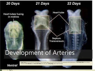
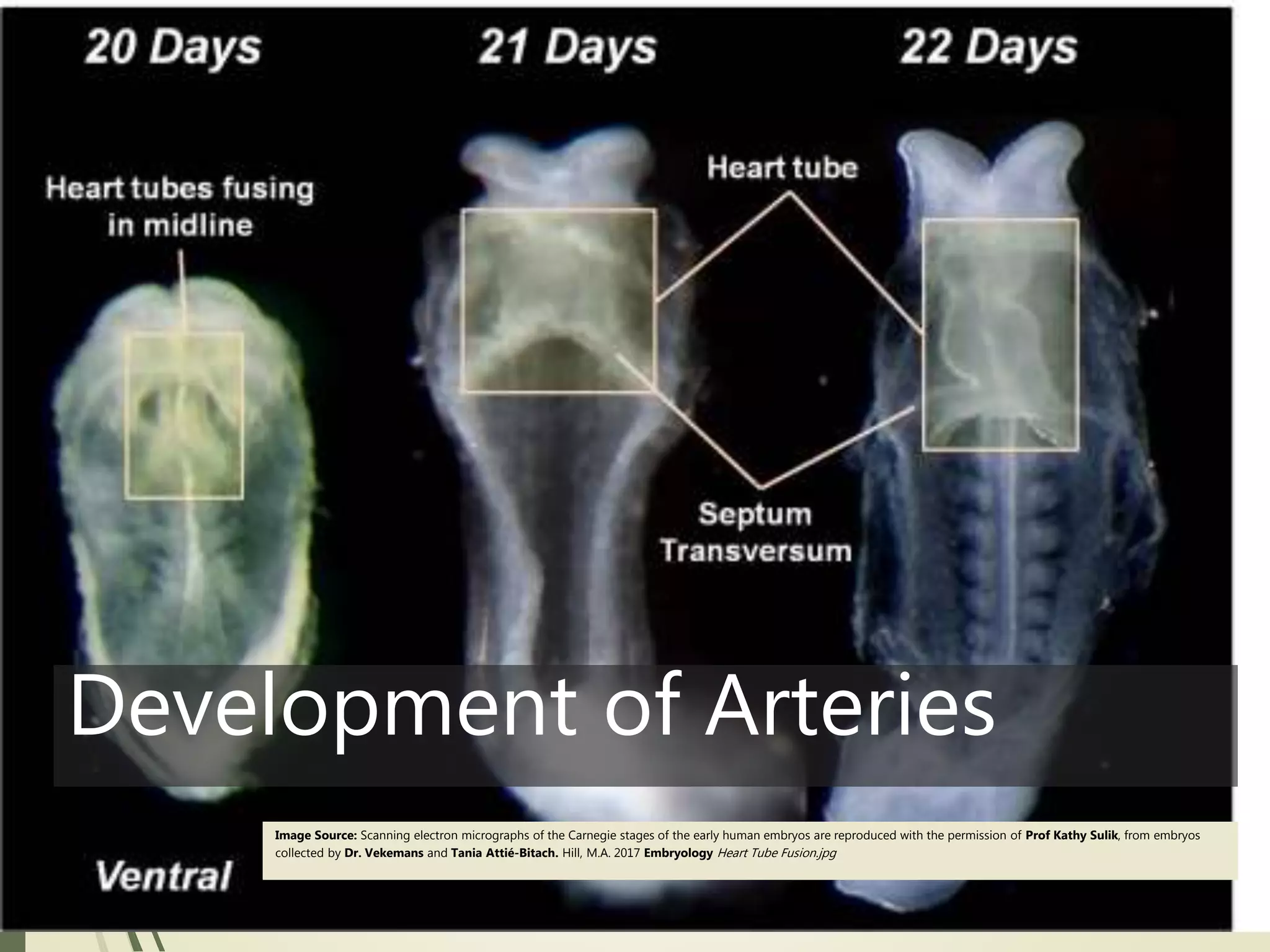
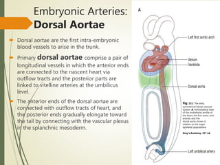
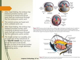
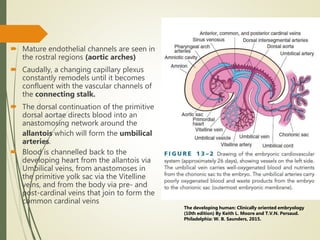
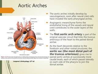
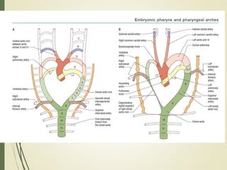
The three main arteries that develop in early human embryos are:
1) The dorsal aortae, which are the first major blood vessels to form and connect the heart to the developing vascular system.
2) The aortic arch arteries, which develop from the aortic sac to supply the pharyngeal arches as the embryo grows.
3) The umbilical arteries, which develop from the dorsal aortae and connect to the placenta to allow nutrient exchange with the mother.
As the embryo develops, these major arteries and the accompanying vascular networks are refined through vasculogenesis and angiogenesis guided by growth factors to establish the adult circulatory system.
Lateral ventricle of Brain. By Dr.N.Mugunthan.M.Smgmcri1234
Lateral ventricle of brain. Lecture by Dr.N.Mugunthan.
Associate Professor,
Mahatma Gandhi Medical College & Research Institute,
Sri Balaji Vidyapeeth, Pondicherry.
Anatomy of Blood vessels of abdomen pelvic cavities. Portacaval & Cavacaval A...Eneutron
1. The abdominal aorta
a. the parietal branches
b. the visceral branches
2. The common iliac arteries and veins
3. The external iliac artery and veins
4. The internal iliac artery and veins
5. The inferior vena cava
6. The portal vein
7. The cavacacal Anastomoses
8. The portacaval Anastomoses
9. The Fetal Circulation
4 th ventricle- Anatomical and surgical perspectivesuresh Bishokarma
4th ventricle connects the entire ventricular system of brain. Its connection with cisterns magna and cerebella pontine cistern via foramen of magenta and Luschka. CSF absorbs into the arachnoid granulation.
4 th ventricle- Anatomical and surgical perspectivesuresh Bishokarma
4th ventricle connects the entire ventricular system of brain. Its connection with cisterns magna and cerebella pontine cistern via foramen of magenta and Luschka. CSF absorbs into the arachnoid granulation.
Development of heart in embryology.
● Formation and position of the heart tube.
● Formation and position of the heart loop
● Mechanism of cardiac looping
● Formation of the embryonic ventricle
● Development of the sinus venosus
● Formation of the cardiac septa
● Atrial septation
● The atrio-ventricular canal
● The muscular interventricular septum
● The septum in truncus arteriosus and the cordis conus
This presentation is made for the purpose of understanding the congenital malformation of the heart as it relates to the embryology of the heart during development.
Histological review of the cardiac muscle-maha hammady.pptxMaha Hammady
Histological review of the cardiac muscle-maha hammady.pptx
for references and more details, check my article :
https://www.researchgate.net/publication/378439219_Enhancing_Our_Understanding_A_Comprehensive_Exploration_of_Heart_Histology_and_Cardiomyocyte_Molecular_Structure
Slide 1: Title Slide
Extrachromosomal Inheritance
Slide 2: Introduction to Extrachromosomal Inheritance
Definition: Extrachromosomal inheritance refers to the transmission of genetic material that is not found within the nucleus.
Key Components: Involves genes located in mitochondria, chloroplasts, and plasmids.
Slide 3: Mitochondrial Inheritance
Mitochondria: Organelles responsible for energy production.
Mitochondrial DNA (mtDNA): Circular DNA molecule found in mitochondria.
Inheritance Pattern: Maternally inherited, meaning it is passed from mothers to all their offspring.
Diseases: Examples include Leber’s hereditary optic neuropathy (LHON) and mitochondrial myopathy.
Slide 4: Chloroplast Inheritance
Chloroplasts: Organelles responsible for photosynthesis in plants.
Chloroplast DNA (cpDNA): Circular DNA molecule found in chloroplasts.
Inheritance Pattern: Often maternally inherited in most plants, but can vary in some species.
Examples: Variegation in plants, where leaf color patterns are determined by chloroplast DNA.
Slide 5: Plasmid Inheritance
Plasmids: Small, circular DNA molecules found in bacteria and some eukaryotes.
Features: Can carry antibiotic resistance genes and can be transferred between cells through processes like conjugation.
Significance: Important in biotechnology for gene cloning and genetic engineering.
Slide 6: Mechanisms of Extrachromosomal Inheritance
Non-Mendelian Patterns: Do not follow Mendel’s laws of inheritance.
Cytoplasmic Segregation: During cell division, organelles like mitochondria and chloroplasts are randomly distributed to daughter cells.
Heteroplasmy: Presence of more than one type of organellar genome within a cell, leading to variation in expression.
Slide 7: Examples of Extrachromosomal Inheritance
Four O’clock Plant (Mirabilis jalapa): Shows variegated leaves due to different cpDNA in leaf cells.
Petite Mutants in Yeast: Result from mutations in mitochondrial DNA affecting respiration.
Slide 8: Importance of Extrachromosomal Inheritance
Evolution: Provides insight into the evolution of eukaryotic cells.
Medicine: Understanding mitochondrial inheritance helps in diagnosing and treating mitochondrial diseases.
Agriculture: Chloroplast inheritance can be used in plant breeding and genetic modification.
Slide 9: Recent Research and Advances
Gene Editing: Techniques like CRISPR-Cas9 are being used to edit mitochondrial and chloroplast DNA.
Therapies: Development of mitochondrial replacement therapy (MRT) for preventing mitochondrial diseases.
Slide 10: Conclusion
Summary: Extrachromosomal inheritance involves the transmission of genetic material outside the nucleus and plays a crucial role in genetics, medicine, and biotechnology.
Future Directions: Continued research and technological advancements hold promise for new treatments and applications.
Slide 11: Questions and Discussion
Invite Audience: Open the floor for any questions or further discussion on the topic.
Richard's aventures in two entangled wonderlandsRichard Gill
Since the loophole-free Bell experiments of 2020 and the Nobel prizes in physics of 2022, critics of Bell's work have retreated to the fortress of super-determinism. Now, super-determinism is a derogatory word - it just means "determinism". Palmer, Hance and Hossenfelder argue that quantum mechanics and determinism are not incompatible, using a sophisticated mathematical construction based on a subtle thinning of allowed states and measurements in quantum mechanics, such that what is left appears to make Bell's argument fail, without altering the empirical predictions of quantum mechanics. I think however that it is a smoke screen, and the slogan "lost in math" comes to my mind. I will discuss some other recent disproofs of Bell's theorem using the language of causality based on causal graphs. Causal thinking is also central to law and justice. I will mention surprising connections to my work on serial killer nurse cases, in particular the Dutch case of Lucia de Berk and the current UK case of Lucy Letby.
A brief information about the SCOP protein database used in bioinformatics.
The Structural Classification of Proteins (SCOP) database is a comprehensive and authoritative resource for the structural and evolutionary relationships of proteins. It provides a detailed and curated classification of protein structures, grouping them into families, superfamilies, and folds based on their structural and sequence similarities.
Deep Behavioral Phenotyping in Systems Neuroscience for Functional Atlasing a...Ana Luísa Pinho
Functional Magnetic Resonance Imaging (fMRI) provides means to characterize brain activations in response to behavior. However, cognitive neuroscience has been limited to group-level effects referring to the performance of specific tasks. To obtain the functional profile of elementary cognitive mechanisms, the combination of brain responses to many tasks is required. Yet, to date, both structural atlases and parcellation-based activations do not fully account for cognitive function and still present several limitations. Further, they do not adapt overall to individual characteristics. In this talk, I will give an account of deep-behavioral phenotyping strategies, namely data-driven methods in large task-fMRI datasets, to optimize functional brain-data collection and improve inference of effects-of-interest related to mental processes. Key to this approach is the employment of fast multi-functional paradigms rich on features that can be well parametrized and, consequently, facilitate the creation of psycho-physiological constructs to be modelled with imaging data. Particular emphasis will be given to music stimuli when studying high-order cognitive mechanisms, due to their ecological nature and quality to enable complex behavior compounded by discrete entities. I will also discuss how deep-behavioral phenotyping and individualized models applied to neuroimaging data can better account for the subject-specific organization of domain-general cognitive systems in the human brain. Finally, the accumulation of functional brain signatures brings the possibility to clarify relationships among tasks and create a univocal link between brain systems and mental functions through: (1) the development of ontologies proposing an organization of cognitive processes; and (2) brain-network taxonomies describing functional specialization. To this end, tools to improve commensurability in cognitive science are necessary, such as public repositories, ontology-based platforms and automated meta-analysis tools. I will thus discuss some brain-atlasing resources currently under development, and their applicability in cognitive as well as clinical neuroscience.
(May 29th, 2024) Advancements in Intravital Microscopy- Insights for Preclini...Scintica Instrumentation
Intravital microscopy (IVM) is a powerful tool utilized to study cellular behavior over time and space in vivo. Much of our understanding of cell biology has been accomplished using various in vitro and ex vivo methods; however, these studies do not necessarily reflect the natural dynamics of biological processes. Unlike traditional cell culture or fixed tissue imaging, IVM allows for the ultra-fast high-resolution imaging of cellular processes over time and space and were studied in its natural environment. Real-time visualization of biological processes in the context of an intact organism helps maintain physiological relevance and provide insights into the progression of disease, response to treatments or developmental processes.
In this webinar we give an overview of advanced applications of the IVM system in preclinical research. IVIM technology is a provider of all-in-one intravital microscopy systems and solutions optimized for in vivo imaging of live animal models at sub-micron resolution. The system’s unique features and user-friendly software enables researchers to probe fast dynamic biological processes such as immune cell tracking, cell-cell interaction as well as vascularization and tumor metastasis with exceptional detail. This webinar will also give an overview of IVM being utilized in drug development, offering a view into the intricate interaction between drugs/nanoparticles and tissues in vivo and allows for the evaluation of therapeutic intervention in a variety of tissues and organs. This interdisciplinary collaboration continues to drive the advancements of novel therapeutic strategies.
Richard's entangled aventures in wonderlandRichard Gill
Since the loophole-free Bell experiments of 2020 and the Nobel prizes in physics of 2022, critics of Bell's work have retreated to the fortress of super-determinism. Now, super-determinism is a derogatory word - it just means "determinism". Palmer, Hance and Hossenfelder argue that quantum mechanics and determinism are not incompatible, using a sophisticated mathematical construction based on a subtle thinning of allowed states and measurements in quantum mechanics, such that what is left appears to make Bell's argument fail, without altering the empirical predictions of quantum mechanics. I think however that it is a smoke screen, and the slogan "lost in math" comes to my mind. I will discuss some other recent disproofs of Bell's theorem using the language of causality based on causal graphs. Causal thinking is also central to law and justice. I will mention surprising connections to my work on serial killer nurse cases, in particular the Dutch case of Lucia de Berk and the current UK case of Lucy Letby.
Nutraceutical market, scope and growth: Herbal drug technologyLokesh Patil
As consumer awareness of health and wellness rises, the nutraceutical market—which includes goods like functional meals, drinks, and dietary supplements that provide health advantages beyond basic nutrition—is growing significantly. As healthcare expenses rise, the population ages, and people want natural and preventative health solutions more and more, this industry is increasing quickly. Further driving market expansion are product formulation innovations and the use of cutting-edge technology for customized nutrition. With its worldwide reach, the nutraceutical industry is expected to keep growing and provide significant chances for research and investment in a number of categories, including vitamins, minerals, probiotics, and herbal supplements.
This presentation explores a brief idea about the structural and functional attributes of nucleotides, the structure and function of genetic materials along with the impact of UV rays and pH upon them.
Nucleic Acid-its structural and functional complexity.
Development of Arteries
1. Development of Arteries
Image Source: Scanning electron micrographs of the Carnegie stages of the early human embryos are reproduced with the permission of Prof Kathy Sulik, from embryos
collected by Dr. Vekemans and Tania Attié-Bitach. Hill, M.A. 2017 Embryology Heart Tube Fusion.jpg
2. Overview
Outline of time course
Vasculogenesis and Angiogenesis
Growth factors involved
Brief mention of early vascular systems- Vitelline, Systemic
and Placental.
Aortic Arch and Pharyngeal arteries
Umbilical Arteries
Segmental Arteries
Vitelline Arteries
Anomalies of arterial tree.
3. Introduction
Development of the heart and vascular system begins very early in
mesoderm both within (embryonic) and outside (extra embryonic,
yolk sac and placental) the embryo.
•forms initially in splanchnic mesoderm of prechordal plate
region - cardiogenic region
• growth and folding of the embryo moves heart
ventrally and downward into anatomical position
•heart tube connects to blood vessels forming in splanchnic
and extraembryonic mesoderm
•Week 2-3 pair of thin-walled tubes
•Week 3 paired heart tubes fuse, truncus arteriosus outflow,
heart contracting
•Week 4 heart tube continues to elongate, curving to form S
shape
•Week 5 septation starts, atrial and ventricular
• Septation continues, atrial septa remains open until
after birth, foramen ovale.
•Week 37-38 at birth, pressure difference closes foramen
ovale leaving a fossa ovals
Time course
4. Angioblastic mesenchyme
Extraembryonic
mesenchyme in the
splanchnopleure of the
yolk sac
in the body
stalk(containing the
allantois)
the somatopleure of the
chorion
The peripheral cells flatten
as a vascular endothelium,
whereas the central cells
transform into primitive
red blood corpuscles.
Later, contiguous islands
merge, forming a
continuous network of fine
vessels.
Figure 6.15 Extraembryonic blood vessel formation in the villi,
chorion, connecting stalk, and wall of the yolk sac in a
presomite embryo of approximately 19 days. Langman’s
Embryology, 12th ed.
5. Growth factors involved
Vascular Endothelial Growth Factor (VEGF) and Placental
Growth Factor (PGF) and Platelet derived Growth Factor
(PGDF)
Growing blood vessels follow a gradient generated by target
tissues/regions of Vascular Endothelial Growth Factor (VEGF) to
establish a vascular bed.
Notch signaling acts as an inhibitor for this system, preventing
sprouting of blood vessels. Notch is a transmembrane receptor
protein involved in regulating cell differentiation in many
developing systems.
6. Figure 6.14 Blood vessels form in two
ways: vasculogenesis (top), in
which vessels arise from blood
and angiogenesis (bottom), in which
new vessels sprout from existing
During vasculogenesis, fibroblast
growth factor 2 (FGF-2) binds to its
receptor on subpopulations of
mesoderm cells and induces them to
form hemangioblasts. Then, under
influence of vascular endothelial
growth factor (VEGF) acting through
two different receptors, these cells
become endothelial and coalesce to
form vessels.
Angiogenesis is also regulated
by VEGF, which stimulates
proliferation of endothelial cells at
points where new vessels will sprout
from existing ones. Final modeling
stabilization of the vasculature are
accomplished by platelet-derived
growth factor (PDGF) and
transforming growth factor β (TGF-β).
Langman’s Embryology, 12th ed
7. Specification into arteries, veins and lymphatic begins soon
after the angioblast induction.
For arterial development
SHH VEGF Notch pathway ephrinB2
EPHB4- vein specific gene
Prox-1- lymphatics
8. Till second week:
Diffusion through
the extraembryonic
coelom and
umbilical vesicle.
Beginning of 3rd
week: Blood vessel
formation begins in
the extraembryonic
mesoderm of the
umbilical vesicle,
connecting stalk,
and chorion.
EARLY DEVELOPMENT OF
CARDIOVASCULAR SYSTEM: Vasculogenesis
Makoto Kamei, W Brian Saunders, Kayla J Bayless, Louis Dye, George E Davis, Brant M Weinstein Endothelial tubes
assemble from intracellular vacuoles in vivo.Nature: 2006, 442(7101);453-6 PubMed 16799567
Hill, M.A. 2017 Embryology Embryonic
Circulations.jpg. Retrieved November 24,
2017,
from https://embryology.med.unsw.edu.a
u/embryology/index.php/File:Embryonic_
Circulations.jpg
9. Blood formation: End of the 3rd
Week and thereafter
Blood cells develop from specialized endothelial cells (hemangiogenic
epithelium) of vessels as they grow on the umbilical vesicle (yolk sac) and allantois
at the end of the third week
Later (week 5) throughout embryonic mesenchyme
The definitive hematopoietic stem cells are derived from mesoderm surrounding
the aorta in a site near the developing mesonephric kidney called the aortagonad-
mesonephros region (AGM).
These cells colonize the liver, which becomes the major hematopoietic organ of the
embryo for a period of 2nd to 7th month of development.
Figure 1. The first definitive multipotent hematopoietic stem
cells (HSCs) are generated within the embryonic aorta-gonad-
mesonephros (AGM) region. The AGM extends from the
umbilicus to the anterior limb bud of the human embryo and
contains the dorsal aorta. Within the dorsal aorta, a cluster of
CD34+ hematopoietic cells is associated with the ventral floor
of the aorta.
Printed in the R&D Systems 2003 Catalog
10. Angiogenesis
None of the main vessels of the adult arises as a single trunk in the embryo.
A capillary network is first laid down along the course of each vessel
The larger arteries and veins are defined by selection and enlargement of definite paths in this network.)
Angioblasts, arising in splanchnic and somitic tissues, add endothelial sprouts and branches to earlier vessels.
Fig: 51- Dorsal view of
chick embryo with ten
pairs of mesodermal
somites: Bailey FR. and
Miller AM. Text-Book of
Embryology (1921) New
York: William Wood and
Co.
12. Dorsal aortae are the first intra-embryonic
blood vessels to arise in the trunk.
Primary dorsal aortae comprise a pair of
longitudinal vessels in which the anterior ends
are connected to the nascent heart via
outflow tracts and the posterior parts are
linked to vitelline arteries at the umbilicus
level.
The anterior ends of the dorsal aortae are
connected with outflow tracts of heart, and
the posterior ends gradually elongate toward
the tail by connecting with the vascular plexus
in the splanchnic mesoderm.
Embryonic Arteries:
Dorsal Aortae
Fig. 13.1 The early,
symmetrical blood vascular
system. A, Ventrolateral view
of the endothelial profile of
the heart, the first aortic arch
arteries and the
dorsal aorta shown in
relation to the major
epithelial populations
Gray’s Anatomy. 41st ed
13. After head folding, the embryo has
bilateral primitive aortae, each
consisting of ventral and dorsal
parts that are continuous through
the first embryonic aortic arch.
The ventral aortae are fused and
form a dilated aortic sac.
After head folding, the embryo has
bilateral primitive aortae, each
consisting of ventral and dorsal
parts that are continuous through
the first embryonic aortic arches.
The dorsal aortae run caudally, one
on each side of the notochord. In
the fourth week they fuse from
about the level of the fourth
thoracic to that of the fourth lumbar
segment to form a single definitive
descending aorta.
Langman’s Embryology, 12th ed
14. Mature endothelial channels are seen in
the rostral regions (aortic arches)
Caudally, a changing capillary plexus
constantly remodels until it becomes
confluent with the vascular channels of
the connecting stalk.
The dorsal continuation of the primitive
dorsal aortae directs blood into an
anastomosing network around the
allantois which will form the umbilical
arteries.
Blood is channelled back to the
developing heart from the allantois via
Umbilical veins, from anastomoses in
the primitive yolk sac via the Vitelline
veins, and from the body via pre- and
post-cardinal veins that join to form the
common cardinal veins
The developing human: Clinically oriented embryology
(10th edition) By Keith L. Moore and T.V.N. Persaud.
Philadelphia: W. B. Saunders, 2015.
16. Aortic Arches
The aortic arches initially develop by
vasculogenesis, soon after neural crest cells
have invaded the early pharyngeal arches.
Angiogenic mesenchyme forms the
endothelial lining of the vessels and neural
crest contributes to the outer layers of the
walls.
The first aortic arch artery is part of the
original vascular circuit that links the truncus
arteriosus of the heart to the paired dorsal
aortae.
As the heart descends relative to the
forebrain and other rostral structures, the
aortic sac (the most distal part of
the truncus arteriosus) gives rise to
paired aortic arches at successively more
caudal levels, each of which passes laterally
on each side of the pharynx to join the
dorsal aortae.
Langman’s Embryology, 12th ed
17. Aortic Sac
The pharyngeal arches and their
vessels appear in a cranial-to-
caudal sequence.
Five arches are numbered I, II, III,
IV, and VI
Division of the truncus arteriosus
by the aorticopulmonary septum
divides the outflow channel of the
heart into the ventral aorta and
the pulmonary trunk.
The aortic sac then forms right
and left horns, which
subsequently give rise to the
brachiocephalic artery and the
proximal segment of the aortic
arch, respectively.
Langman’s Embryology, 12th ed
18. Arch Arteries
1st and 2nd
By day 27, most
of the first aortic
arch has
disappeared,
although a small
portion persists
to form the
maxillary artery.
Second aortic
arch soon
disappears. The
remaining
portions of this
arch are- the
hyoid and
stapedial arteries.
Tanoue, Shuichi & Kiyosue, Hiro &
Mori, Hiromu & Hori, Yuzo &
Okahara, Mika & Sagara, Yoshiko.
(2013). Maxillary Artery: Functional
and Imaging Anatomy for Safe and
Effective Transcatheter Treatment.
Radiographics : a review publication
of the Radiological Society of North
America, Inc. 33. e209-24.
10.1148/rg.337125173.
19. The stapedial artery passes through
the ring of the stapes and divides
into supraorbital, infraorbital, and
mandibular branches which follow
the three divisions of the trigeminal
nerve. The infraorbital and
mandibular branches arise from a
common stem, the terminal part of
which anastomoses with
the external carotid artery.
On the obliteration of the stapedial
artery, this anastomosis enlarges
and forms the internal maxillary
artery
The common stem of the
infraorbital and mandibular
branches passes between the two
roots of the auriculotemporal
nerve and becomes the middle
meningeal artery
the original supraorbital branch of
the stapedial is represented by the
orbital twigs of the middle
meningeal.
The Persistent Stapedial Artery
Richard Silbergleit, Douglas J. Quint, Bharat A. Mehta, Suresh C. Patel, Joseph J. Metes and Samir E. Noujaim
American Journal of Neuroradiology March 2000, 21 (3) 572-577
20. Third & Fourth Arch Artery
In the 29-day embryo, the first and second aortic arches have disappeared
The third, fourth, and sixth arches are large.
The conotruncal region has divided so that the sixth arches are now continuous
with the pulmonary trunk.
Langman’s Embryology, 12th ed
21.
22. The third aortic arch forms the common
carotid artery and
The first part of the The external carotid
artery is a sprout of the third aortic arch.
Fourth Arch Artery: On the left, it forms part
of the arch of the aorta, between the left
common carotid and the left subclavian
arteries.
On the right, it forms the most proximal
segment of the right subclavian artery,
23. 6th Aortic Arch
also known as the pulmonary arch
On the right side, the proximal part becomes the proximal segment of the right pulmonary artery.
On the left, the distal part persists during intrauterine life as the ductus arteriosus
Fig. 4.2 (a) Cardiac neural crest cells (CNCCs) in the outflow
cushions. Cross-section through the distal and middle outflow
tract shows position of the condensed CNCC-derived
mesenchyme within the cushions to form the future aorta (a)
and pulmonary trunk (p). (b) Stages of outflow septation in the
chick. (a) U-shaped septation complex straddles the aortic sac
between the 4th and 6th aortic arch arteries. (b) The septation
complex grows at the expense of the prongs. (c) Septation of
the conus (Adapted from Hutson and Kirby
24.
25. Somatic arteries (Intersegmental)
Each primitive dorsal aorta gives off somatic arteries
(intersegmental branches to the body wall)
posterior intercostal, subcostal and lumbar arteries
Gray’s Anatomy. 41st ed
26. Early umbilical arteries
Direct caudal continuation of the primitive dorsal
aortae.
They are present in the body stalk before any
vitelline or visceral branches emerge.
After the dorsal aortae fuse, the umbilical
arteries arise from their ventrolateral aspects
Later, the proximal part of each umbilical artery
is joined by a new vessel, that leaves the aorta
at its termination.
This, possibly the fifth lumbar intersegmental
artery, constitutes the dorsal root of the
umbilical artery (the original stem, the ventral
root).
The dorsal root gives off the axial artery of the
lower limb, branches to the pelvic viscera and,
more proximally, the external iliac artery.
The ventral root disappears entirely, and the
umbilical artery now arises from that part of its
dorsal root distal to the external iliac artery, i.e.
the internal iliac artery
Henry Gray (1918) Anatomy of the Human Body
27. Adult derivatives of umbilical
arteries
medial umbilical ligament
a branch of the anterior division of
the internal iliac artery.
gives rise to the superior vesical
arteries.
28. Lateral splanchnic arteries
the mesonephros, metanephros,
testis or ovary
the suprarenal gland.
Diagrams showing arterial mesonephric blood supply
and its relationship to eventual metanephros in a 4–6-
week embryo and 8–10-week embryo. SMA: Superior
mesenteric artery, IMA: inferior mesenteric artery, 1:
cranial group mesonephric arteries, 2: middle group
mesonephric arteries, RG: reproductive gland, MSN:
mesonephros, MTN: metanephros, RAU: rete arteriosum
urogenitale, SMA: superior mesenteric artery, IMA:
inferior mesenteric artery, GA: gonadal artery, AG:
adrenal gland, AA: adrenal artery, MTN: metanephros,
RA: renal artery, PAA: polar accessory artery. The
adrenal artery usually develops from the cranial group
of mesonephric arteries whereas the gonadal artery
forms from arteries of the caudal group (RAU). The
definite renal artery comes either from the last branch of
the middle group or the first of the caudal group.
Persisting mesonephric arteries form accessory vessels
(PAA) as shown in the example
29. Ventral splanchnic arteries
Distributed to the capillary plexus in the
wall of the yolk sac.
After fusion of the dorsal aortae, they
merge as unpaired trunks.
dorsal and ventral splanchnic anastomoses
reduced to three, the coeliac trunk and the
superior and inferior mesenteric arteries
above the diaphragm, a variable number
of ventral splanchnic arteries persist,
usually four or five, supplying the thoracic
oesophagus.
The dorsal splanchnic anastomosis persists
in the gastroepiploic, pancreaticoduodenal
and primary branches of the colic arteries
the ventral splanchnic anastomosis forms
the right and left gastric and the hepatic
arteries
Gray’s Anatomy. 41st ed
30. derived from two sources:
(1) angioblasts formed
from sprouts off the sinus
venosus that are
distributed over the heart
surface by cell migration
(2) the epicardium itself.
Neural crest cells also
contribute smooth muscle
cells along the proximal
segments of these arteries.
Connection of the coronary
arteries to the aorta occurs
by ingrowth of arterial
endothelial cells from the
arteries into the aorta.
Coronary Arteries
Fig. 5.2 Embryologic development of coronary vasculature (From Lluri
and Aboulhosn)