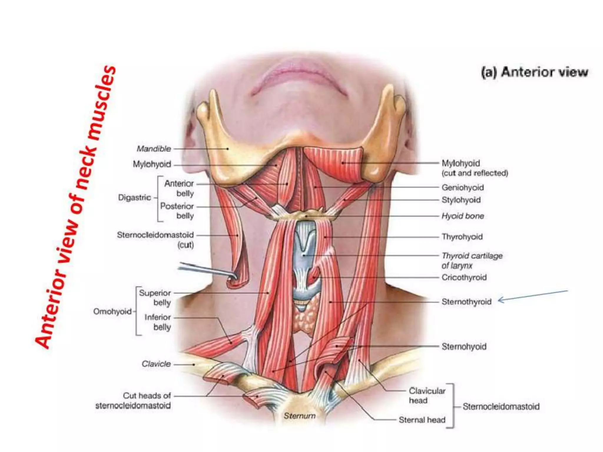
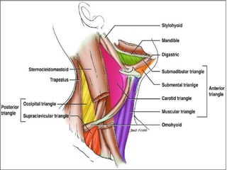
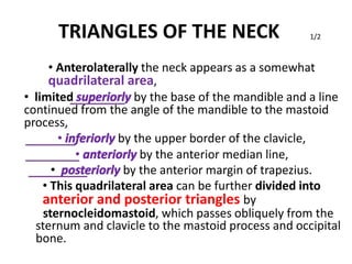
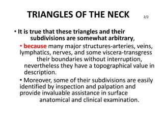
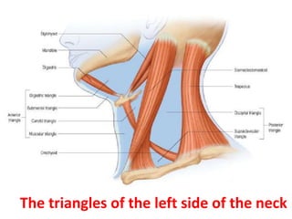
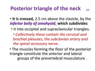
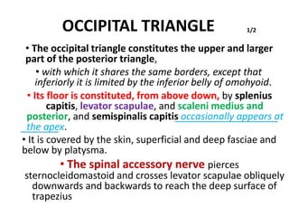
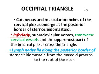
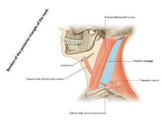
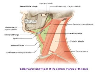
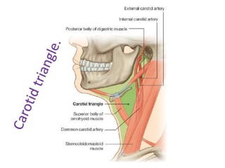
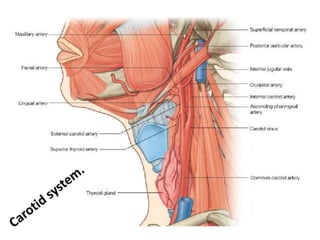
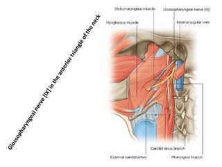
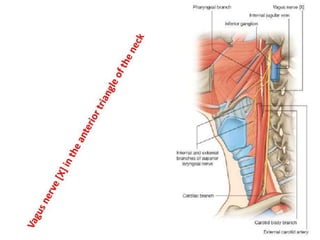
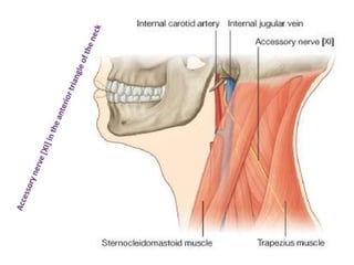
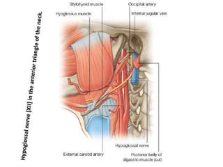
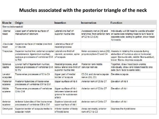
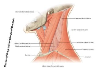
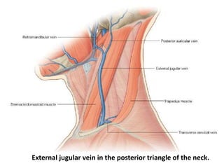
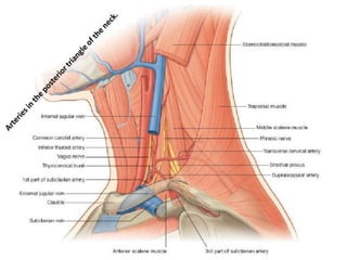
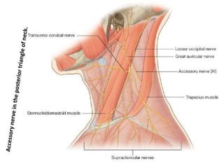
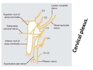
TRIANGLES OF THE NECK 1/2
• Anterolaterally the neck appears as a somewhat
quadrilateral area,
• limited superiorly by the base of the mandible and a line
continued from the angle of the mandible to the mastoid
process,
• inferiorly by the upper border of the clavicle,
• anteriorly by the anterior median line,
• posteriorly by the anterior margin of trapezius.
• This quadrilateral area can be further divided into
anterior and posterior triangles by
sternocleidomastoid, which passes obliquely from the
sternum and clavicle to the mastoid process and occipital
bone.
TRIANGLES OF THE NECK
• It is true that these triangles and their
subdivisions are somewhat arbitrary,
2/2
• because many major structures-arteries, veins,
lymphatics, nerves, and some viscera-transgress
their boundaries without interruption,
nevertheless they have a topographical value in
description.
• Moreover, some of their subdivisions are easily
identified by inspection and palpation and
provide invaluable assistance in surface
anatomical and clinical examination.
The triangles of the left side of the neck
Anterior triangle of the neck
• is bounded anteriorly by the median line of the neck,
• posteriorly by the anterior margin of sterno-cleidomastoid.
• Its base is the inferior border of the mandible
• and its projection to the mastoid process,
• and its apex is at the manubrium sterni.
• It can be subdivided into;
• suprahyoid and infrahyoid areas above and below the
hyoid bone,
• and into
• digastric, submental, muscular and carotid triangles
by the passage of digastric and omohyoid across the
anterior triangle
Subdivisions of the anterior triangle of the neck-a regional approach
The triangles of the left side of the neck
Dissection of the left anterior
triangle.
Platysma has been divided
transversely:
its upper part has been turned
upwards on to the face,
and its lower part turned
backwards, exposing the lower
part of sternocleidomastoid.
DIGASTRIC TRIANGLE
• The digastric triangle is bordered
1/2
• above by the base of the mandible and its projection to the
mastoid process,
• posteroinferiorly by the posterior belly of digastric and by
stylohyoid,
• anteroinferiorly by the anterior belly of digastric.
• It is covered by the skin, superficial fascia, platysma and
deep fascia, which contain branches of the facial and
transverse cutaneous cervical nerves.
• Its floor is formed by mylohyoid and hyoglossus.
• The anterior region of the digastric triangle contains the
submandibular gland,which has the facial vein superficial
to it and the facial artery deep to it.
• The submental and mylohyoid arteries and nerves lie on
mylohyoid.
DIGASTRIC TRIANGLE 2/2
• The submandibular lymph nodes are variably related to
the submandibular gland.
• The posterior region of the digastric triangle contains
the lower part of the parotid gland.
• The external carotid artery,
- passing deep to stylohyoid,
- curves above the muscle,
- and overlaps its superficial surface as it ascends deep to the
parotid gland before entering it.
The internal carotid artery, internal jugular vein and vagus
nerve lie deeper and are separated from the external
carotid artery by
styloglossus, stylopharyngeus and the glossopharyngeal
nerve.
SUBMENTAL TRIANGLE
• The single submental triangle is demarcated
by the anterior bellies of both digastric
muscles.
• Its apex is at the chin,
• its base is the body of the hyoid bone
• and its floor is formed by both mylohyoid
muscles.
• It contains lymph nodes and small veins that
unite to form the anterior jugular vein.
The triangles of the left side of the neck
MUSCULAR TRIANGLE
• The muscular triangle is bounded,
• anteriorly by the median line of the neck from
the hyoid bone to the sternum,
• inferoposteriorly by the anterior margin of
sternocleidomastoid,
• posterosuperiorly by the superior belly of
omohyoid.
• The triangle contains omohyoid, sternohyoid,
sternothyroid and thyrohyoid
The triangles of the left side of the neck
CAROTID TRIANGLE
• The carotid triangle is limited,
• posteriorly by sternocleidomastoid,
1/4
• anteroinferiorly by the superior belly of omohyoid,
• and superiorly by stylohyoid and the posterior belly of
digastric.
• In the living (except the obese) the triangle is usually a
small visible triangular depression, sometimes best seen
with the head and cervical vertebral column slightly
extended and the head contralaterally rotated.
• The carotid triangle is covered by the skin, superficial
fascia, platysma and deep fascia containing branches
of the facial and cutaneous cervical nerves.
CAROTID TRIANGLE 2/4
• The hyoid bone forms its anterior angle and adjacent
floor and can be located on simple inspection,
verified by palpation.
• Parts of thyrohyoid, hyoglossus and inferior and
middle pharyngeal constrictor muscles form its floor.
• The carotid triangle contains the upper part of the
common carotid artery and its division into external
and internal carotid arteries.
• Overlapped by the anterior margin of
sternocleidomastoid, the external carotid artery is
first anteromedial, then anterior to the internal carotid
artery.
• Branches of the external carotid artery are
encountered in the carotid triangle.
CAROTID TRIANGLE
• . Thus the superior thyroid artery runs
anteroinferiorly,
3/4
• the lingual artery anteriorly with a characteristic
upward loop,
• the facial artery anterosuperiorly,
• the occipital artery posterosuperiorly
• and the ascending pharyngeal artery medial
to the internal carotid artery. Arterial pulsation
greets the examining finger.
• The superior thyroid, lingual, facial, ascending
pharyngeal and sometimes the occipital, veins,
correspond to the branches of the external carotid
artery, and all drain into the internal jugular vein.
CAROTID TRIANGLE
• The hypoglossal nerve
4/4
• crosses the external and internal carotid arteries.
• It curves round the origin of the lower sternocleidomastoid
branch of the occipital artery,
• and at this point the superior root of the ansa cervicalis
leaves it to descend anteriorly in the carotid sheath.
• The internal laryngeal nerve and, below it, the external
laryngeal nerve, lie medial to the external carotid artery
below the hyoid bone.
• Many structures in this region, such as all or part of the
internal jugular vein, associated deep cervical lymph nodes,
and the vagus nerve, may be variably obscured by
sternocleidomastoid, and, pedantically, are thus 'outside
the triangle‘.
The triangles of the left side of the neck
Posterior triangle of the neck
• The posterior triangle is delimited;
• anteriorly by sternocleidomastoid,
• posteriorly by the anterior edge of trapezius,
• and inferiorly by the middle third of the clavicle.
• Its apex is between the attachments of
1/2
sternocleidomastoid and trapezius to the occiput and is
often blunted, so that the 'triangle' becomes quadrilateral.
• The roof of the posterior triangle is formed by the
investing layer of the deep cervical fascia.
• The floor of the triangle is formed by the prevertebral
fascia overlying splenius capitis, levator scapulae and the
scalene muscles.
Posterior triangle of the neck 2/2
• It is crossed, 2.5 cm above the clavicle, by the
inferior belly of omohyoid, which subdivides
• it into occipital and supraclavicular triangles.
• Collectively these contain the cervical and
brachial plexuses, the subclavian artery and
the spinal accessory nerve.
• The muscles forming the floor of the posterior
triangle constitute the anterior and lateral
groups of the prevertebral musculature .
The triangles of the left side of the neck
OCCIPITAL TRIANGLE 1/2
• The occipital triangle constitutes the upper and larger
part of the posterior triangle,
• with which it shares the same borders, except that
inferiorly it is limited by the inferior belly of omohyoid.
• Its floor is constituted, from above down, by splenius
capitis, levator scapulae, and scaleni medius and
posterior, and semispinalis capitis occasionally appears at
the apex.
• It is covered by the skin, superficial and deep fasciae and
below by platysma.
• The spinal accessory nerve pierces
sternocleidomastoid and crosses levator scapulae obliquely
downwards and backwards to reach the deep surface of
trapezius
OCCIPITAL TRIANGLE 2/2
• Cutaneous and muscular branches of the
cervical plexus emerge at the posterior
border of sternocleidomastoid.
• Inferiorly, supraclavicular nerves, transverse
cervical vessels and the uppermost part of
the brachial plexus cross the triangle.
• Lymph nodes lie along the posterior border of
sternocleidomastoid from the mastoid process
to the root of the neck
SUPRACLAVICULAR TRIANGLE
• The supraclavicular triangle
1/3
• is the lower and smaller division of the posterior triangle,
with which it shares the same boundaries, except that
superiorly it is limited by omohyoid.
• It corresponds in the living neck with the lower part of a deep,
prominent hollow, namely, the greater supraclavicular foss
• Its floor contains the first rib, scalenus medius and the first
slip of serratus anterior.
• Its size varies with the extent of the clavicular attachments
of sternocleidomastoid and trapezius and also the level of
the inferior belly of omohyoid.
• The triangle is covered by the skin, superficial and deep
fasciae and platysma and crossed by the supraclavicular
nerves.
SUPRACLAVICULAR TRIANGLE
• Just above the clavicle,
2/3
• the third part of the subclavian artery curves inferolaterally
from the lateral margin of scalenus anterior across the firs
rib to the axilla.
• The subclavian vein is behind the clavicle and is not usually
in the triangle; but it may rise as high as the artery an
even accompany it behind scalenus anterior.
• The brachial plexus is partly superior, and partly posterior to
the artery and is always closely related to it.
• The trunks of the brachial plexus may easily be palpated
here if the neck is contralaterally flexed and the examining
finger is drawn across the trunks at right angles to their
length.
SUPRACLAVICULAR TRIANGLE 3/3
• With the musculature relaxed, pulsation of the subclavian
artery may be felt and the arterial flow can be controlled b
retroclavicular compression against the first rib.
• The suprascapular vessels pass transversely behind the
clavicle, below the transverse cervical artery and vein.
• The external jugular vein
- descends behind the posterior border of sternocleidomastoid to end
in the subclavian vein.
- It receives the transverse cervical and suprascapular veins, which
form a plexus in front of the third part of the subclavian artery
- occasionally it is joined by a small vein crossing the clavicle anteriorly
from the cephalic vein.
• The nerve to subclavius crosses the triangle.
• The triangle contains some lymph nodes.
Muscles in the anterior triangle of the neck
Borders and subdivisions of the anterior triangle of the neck
Branches of the external carotid artery
Muscles associated with the posterior triangle of the neck
External jugular vein in the posterior triangle of the neck.
Prevertebral and lateral muscles
MUSCLES
PLATYSMA
•
1/3
• Platysma is a broad sheet of muscle of varying prominence.
• arises from the fascia covering the upper parts of pectoralis
major and deltoid.
• Its fibres cross the clavicle and ascend medially in the side
of the neck.
• Anterior fibres interlace across the midline with the fibres
of the contralateral muscle, below and behind the
symphysis menti.
• Other fibres attach to the lower border of the mandible or
to the lower lip or cross the mandible to attach to skin and
subcutaneous tissue of the lower face.
PLATYSMA
• Vascular supply
2/3
• Platysma receives its blood supply from
- the submental branch of the facial artery
- and the suprascapular artery from the thyrocervical
trunk of the subclavian artery.
• Innervation
• Platysma is innervated by the cervical branch of
the facial nerve which descends on the deep
surface of the muscle close to the angle of the
mandible
PLATYSMA3/3 Action
• Contraction diminishes the concavity
between the jaw and the side of the neck and
produces tense oblique ridges in the skin of
the neck.
• Platysma may assist in depressing the
mandible, and via its labial and modiolar
attachments it can draw down the lower lip
and corners of the mouth in expressions of
horror or surprise.
Muscles that form the floor of the oral cavity, superior view
Origins and insertions on the mandible and hyoid
Fascial spaces
• Between the fascial layers in the neck are spaces that may provide a conduit fo
the spread of infections from the neck to the mediastinum.
• Three spaces could be involved in this process:
1. pretracheal space the first is the pretracheal space between the investing layer
of cervical fascia (covering the posterior surface of the infrahyoid muscles) and
the pretracheal fascia (covering the anterior surface of the trachea and the
thyroid gland), which passes between the neck and the anterior part of the
superior mediastinum;
2. the second is the retropharyngeal space between the buccopharyngeal fascia
(on the posterior surface of the pharynx and esophagus) and the prevertebral
fascia (on the anterior surface of the transverse processes and bodies of the
cervical vertebrae), which extends from the base of the skull to the upper part of
the posterior mediastinum
3. the third space is within the prevertebral layer covering the anterior surface of
the transverse processes and bodies of the cervical vertebrae. This layer splits
into two laminae to create a fascial space that begins at the base of the skull and
extends through the posterior mediastinum to the diaphragm
 triangles of the neck-dr.meher

triangles of the neck-dr.meher

  • 3.
    TRIANGLES OF THENECK 1/2 • Anterolaterally the neck appears as a somewhat quadrilateral area, • limited superiorly by the base of the mandible and a line continued from the angle of the mandible to the mastoid process, • inferiorly by the upper border of the clavicle, • anteriorly by the anterior median line, • posteriorly by the anterior margin of trapezius. • This quadrilateral area can be further divided into anterior and posterior triangles by sternocleidomastoid, which passes obliquely from the sternum and clavicle to the mastoid process and occipital bone.
  • 4.
    TRIANGLES OF THENECK • It is true that these triangles and their subdivisions are somewhat arbitrary, 2/2 • because many major structures-arteries, veins, lymphatics, nerves, and some viscera-transgress their boundaries without interruption, nevertheless they have a topographical value in description. • Moreover, some of their subdivisions are easily identified by inspection and palpation and provide invaluable assistance in surface anatomical and clinical examination.
  • 5.
    The triangles ofthe left side of the neck
  • 6.
    Anterior triangle ofthe neck • is bounded anteriorly by the median line of the neck, • posteriorly by the anterior margin of sterno-cleidomastoid. • Its base is the inferior border of the mandible • and its projection to the mastoid process, • and its apex is at the manubrium sterni. • It can be subdivided into; • suprahyoid and infrahyoid areas above and below the hyoid bone, • and into • digastric, submental, muscular and carotid triangles by the passage of digastric and omohyoid across the anterior triangle
  • 7.
    Subdivisions of theanterior triangle of the neck-a regional approach
  • 8.
    The triangles ofthe left side of the neck
  • 9.
    Dissection of theleft anterior triangle. Platysma has been divided transversely: its upper part has been turned upwards on to the face, and its lower part turned backwards, exposing the lower part of sternocleidomastoid.
  • 10.
    DIGASTRIC TRIANGLE • Thedigastric triangle is bordered 1/2 • above by the base of the mandible and its projection to the mastoid process, • posteroinferiorly by the posterior belly of digastric and by stylohyoid, • anteroinferiorly by the anterior belly of digastric. • It is covered by the skin, superficial fascia, platysma and deep fascia, which contain branches of the facial and transverse cutaneous cervical nerves. • Its floor is formed by mylohyoid and hyoglossus. • The anterior region of the digastric triangle contains the submandibular gland,which has the facial vein superficial to it and the facial artery deep to it. • The submental and mylohyoid arteries and nerves lie on mylohyoid.
  • 11.
    DIGASTRIC TRIANGLE 2/2 •The submandibular lymph nodes are variably related to the submandibular gland. • The posterior region of the digastric triangle contains the lower part of the parotid gland. • The external carotid artery, - passing deep to stylohyoid, - curves above the muscle, - and overlaps its superficial surface as it ascends deep to the parotid gland before entering it. The internal carotid artery, internal jugular vein and vagus nerve lie deeper and are separated from the external carotid artery by styloglossus, stylopharyngeus and the glossopharyngeal nerve.
  • 13.
    SUBMENTAL TRIANGLE • Thesingle submental triangle is demarcated by the anterior bellies of both digastric muscles. • Its apex is at the chin, • its base is the body of the hyoid bone • and its floor is formed by both mylohyoid muscles. • It contains lymph nodes and small veins that unite to form the anterior jugular vein.
  • 14.
    The triangles ofthe left side of the neck
  • 16.
    MUSCULAR TRIANGLE • Themuscular triangle is bounded, • anteriorly by the median line of the neck from the hyoid bone to the sternum, • inferoposteriorly by the anterior margin of sternocleidomastoid, • posterosuperiorly by the superior belly of omohyoid. • The triangle contains omohyoid, sternohyoid, sternothyroid and thyrohyoid
  • 17.
    The triangles ofthe left side of the neck
  • 19.
    CAROTID TRIANGLE • Thecarotid triangle is limited, • posteriorly by sternocleidomastoid, 1/4 • anteroinferiorly by the superior belly of omohyoid, • and superiorly by stylohyoid and the posterior belly of digastric. • In the living (except the obese) the triangle is usually a small visible triangular depression, sometimes best seen with the head and cervical vertebral column slightly extended and the head contralaterally rotated. • The carotid triangle is covered by the skin, superficial fascia, platysma and deep fascia containing branches of the facial and cutaneous cervical nerves.
  • 20.
    CAROTID TRIANGLE 2/4 •The hyoid bone forms its anterior angle and adjacent floor and can be located on simple inspection, verified by palpation. • Parts of thyrohyoid, hyoglossus and inferior and middle pharyngeal constrictor muscles form its floor. • The carotid triangle contains the upper part of the common carotid artery and its division into external and internal carotid arteries. • Overlapped by the anterior margin of sternocleidomastoid, the external carotid artery is first anteromedial, then anterior to the internal carotid artery. • Branches of the external carotid artery are encountered in the carotid triangle.
  • 21.
    CAROTID TRIANGLE • .Thus the superior thyroid artery runs anteroinferiorly, 3/4 • the lingual artery anteriorly with a characteristic upward loop, • the facial artery anterosuperiorly, • the occipital artery posterosuperiorly • and the ascending pharyngeal artery medial to the internal carotid artery. Arterial pulsation greets the examining finger. • The superior thyroid, lingual, facial, ascending pharyngeal and sometimes the occipital, veins, correspond to the branches of the external carotid artery, and all drain into the internal jugular vein.
  • 22.
    CAROTID TRIANGLE • Thehypoglossal nerve 4/4 • crosses the external and internal carotid arteries. • It curves round the origin of the lower sternocleidomastoid branch of the occipital artery, • and at this point the superior root of the ansa cervicalis leaves it to descend anteriorly in the carotid sheath. • The internal laryngeal nerve and, below it, the external laryngeal nerve, lie medial to the external carotid artery below the hyoid bone. • Many structures in this region, such as all or part of the internal jugular vein, associated deep cervical lymph nodes, and the vagus nerve, may be variably obscured by sternocleidomastoid, and, pedantically, are thus 'outside the triangle‘.
  • 23.
    The triangles ofthe left side of the neck
  • 24.
    Posterior triangle ofthe neck • The posterior triangle is delimited; • anteriorly by sternocleidomastoid, • posteriorly by the anterior edge of trapezius, • and inferiorly by the middle third of the clavicle. • Its apex is between the attachments of 1/2 sternocleidomastoid and trapezius to the occiput and is often blunted, so that the 'triangle' becomes quadrilateral. • The roof of the posterior triangle is formed by the investing layer of the deep cervical fascia. • The floor of the triangle is formed by the prevertebral fascia overlying splenius capitis, levator scapulae and the scalene muscles.
  • 27.
    Posterior triangle ofthe neck 2/2 • It is crossed, 2.5 cm above the clavicle, by the inferior belly of omohyoid, which subdivides • it into occipital and supraclavicular triangles. • Collectively these contain the cervical and brachial plexuses, the subclavian artery and the spinal accessory nerve. • The muscles forming the floor of the posterior triangle constitute the anterior and lateral groups of the prevertebral musculature .
  • 28.
    The triangles ofthe left side of the neck
  • 29.
    OCCIPITAL TRIANGLE 1/2 •The occipital triangle constitutes the upper and larger part of the posterior triangle, • with which it shares the same borders, except that inferiorly it is limited by the inferior belly of omohyoid. • Its floor is constituted, from above down, by splenius capitis, levator scapulae, and scaleni medius and posterior, and semispinalis capitis occasionally appears at the apex. • It is covered by the skin, superficial and deep fasciae and below by platysma. • The spinal accessory nerve pierces sternocleidomastoid and crosses levator scapulae obliquely downwards and backwards to reach the deep surface of trapezius
  • 30.
    OCCIPITAL TRIANGLE 2/2 •Cutaneous and muscular branches of the cervical plexus emerge at the posterior border of sternocleidomastoid. • Inferiorly, supraclavicular nerves, transverse cervical vessels and the uppermost part of the brachial plexus cross the triangle. • Lymph nodes lie along the posterior border of sternocleidomastoid from the mastoid process to the root of the neck
  • 32.
    SUPRACLAVICULAR TRIANGLE • Thesupraclavicular triangle 1/3 • is the lower and smaller division of the posterior triangle, with which it shares the same boundaries, except that superiorly it is limited by omohyoid. • It corresponds in the living neck with the lower part of a deep, prominent hollow, namely, the greater supraclavicular foss • Its floor contains the first rib, scalenus medius and the first slip of serratus anterior. • Its size varies with the extent of the clavicular attachments of sternocleidomastoid and trapezius and also the level of the inferior belly of omohyoid. • The triangle is covered by the skin, superficial and deep fasciae and platysma and crossed by the supraclavicular nerves.
  • 33.
    SUPRACLAVICULAR TRIANGLE • Justabove the clavicle, 2/3 • the third part of the subclavian artery curves inferolaterally from the lateral margin of scalenus anterior across the firs rib to the axilla. • The subclavian vein is behind the clavicle and is not usually in the triangle; but it may rise as high as the artery an even accompany it behind scalenus anterior. • The brachial plexus is partly superior, and partly posterior to the artery and is always closely related to it. • The trunks of the brachial plexus may easily be palpated here if the neck is contralaterally flexed and the examining finger is drawn across the trunks at right angles to their length.
  • 34.
    SUPRACLAVICULAR TRIANGLE 3/3 •With the musculature relaxed, pulsation of the subclavian artery may be felt and the arterial flow can be controlled b retroclavicular compression against the first rib. • The suprascapular vessels pass transversely behind the clavicle, below the transverse cervical artery and vein. • The external jugular vein - descends behind the posterior border of sternocleidomastoid to end in the subclavian vein. - It receives the transverse cervical and suprascapular veins, which form a plexus in front of the third part of the subclavian artery - occasionally it is joined by a small vein crossing the clavicle anteriorly from the cephalic vein. • The nerve to subclavius crosses the triangle. • The triangle contains some lymph nodes.
  • 36.
    Muscles in theanterior triangle of the neck
  • 37.
    Borders and subdivisionsof the anterior triangle of the neck
  • 43.
    Branches of theexternal carotid artery
  • 54.
    Muscles associated withthe posterior triangle of the neck
  • 56.
    External jugular veinin the posterior triangle of the neck.
  • 60.
  • 62.
  • 63.
    PLATYSMA • 1/3 • Platysma isa broad sheet of muscle of varying prominence. • arises from the fascia covering the upper parts of pectoralis major and deltoid. • Its fibres cross the clavicle and ascend medially in the side of the neck. • Anterior fibres interlace across the midline with the fibres of the contralateral muscle, below and behind the symphysis menti. • Other fibres attach to the lower border of the mandible or to the lower lip or cross the mandible to attach to skin and subcutaneous tissue of the lower face.
  • 64.
    PLATYSMA • Vascular supply 2/3 •Platysma receives its blood supply from - the submental branch of the facial artery - and the suprascapular artery from the thyrocervical trunk of the subclavian artery. • Innervation • Platysma is innervated by the cervical branch of the facial nerve which descends on the deep surface of the muscle close to the angle of the mandible
  • 65.
    PLATYSMA3/3 Action • Contractiondiminishes the concavity between the jaw and the side of the neck and produces tense oblique ridges in the skin of the neck. • Platysma may assist in depressing the mandible, and via its labial and modiolar attachments it can draw down the lower lip and corners of the mouth in expressions of horror or surprise.
  • 66.
    Muscles that formthe floor of the oral cavity, superior view
  • 68.
    Origins and insertionson the mandible and hyoid
  • 69.
    Fascial spaces • Betweenthe fascial layers in the neck are spaces that may provide a conduit fo the spread of infections from the neck to the mediastinum. • Three spaces could be involved in this process: 1. pretracheal space the first is the pretracheal space between the investing layer of cervical fascia (covering the posterior surface of the infrahyoid muscles) and the pretracheal fascia (covering the anterior surface of the trachea and the thyroid gland), which passes between the neck and the anterior part of the superior mediastinum; 2. the second is the retropharyngeal space between the buccopharyngeal fascia (on the posterior surface of the pharynx and esophagus) and the prevertebral fascia (on the anterior surface of the transverse processes and bodies of the cervical vertebrae), which extends from the base of the skull to the upper part of the posterior mediastinum 3. the third space is within the prevertebral layer covering the anterior surface of the transverse processes and bodies of the cervical vertebrae. This layer splits into two laminae to create a fascial space that begins at the base of the skull and extends through the posterior mediastinum to the diaphragm