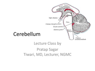
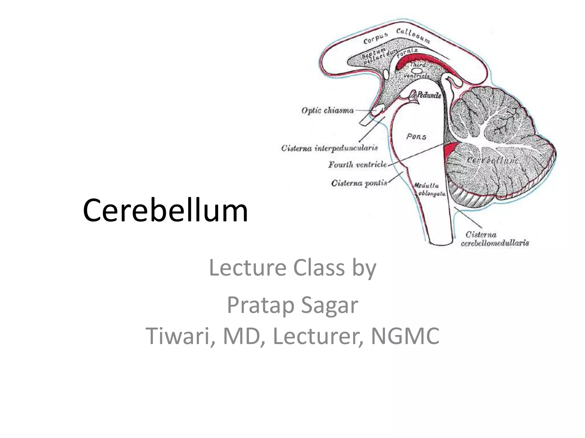
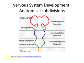
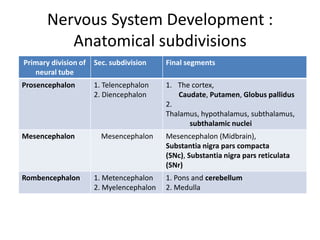
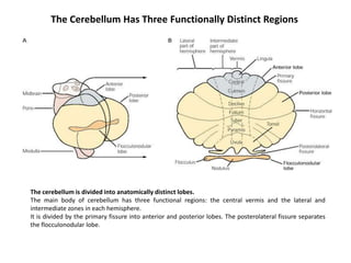
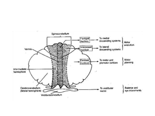
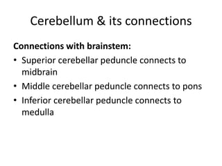
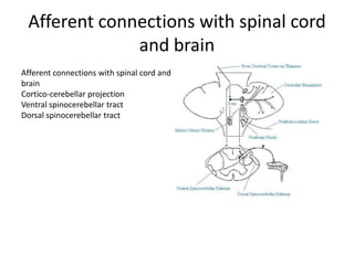
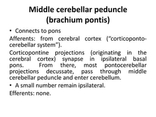
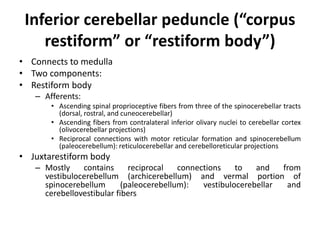
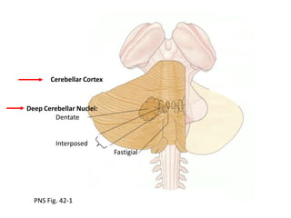
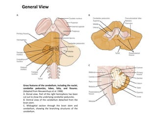
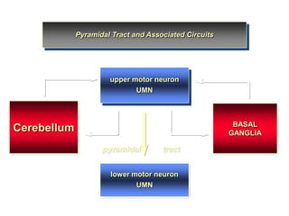
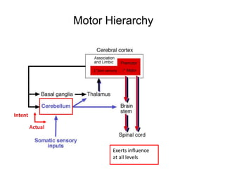
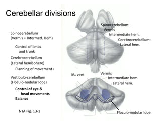
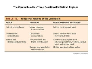
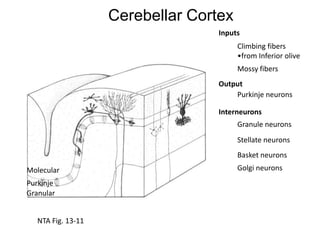
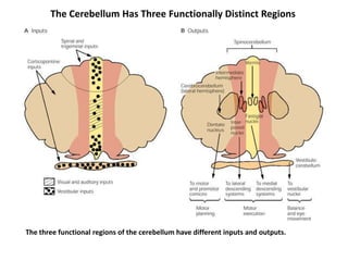
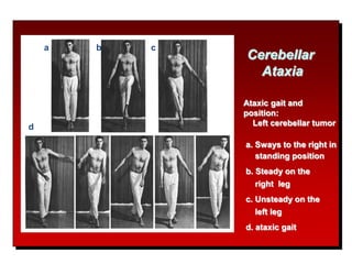
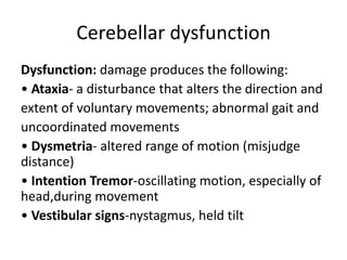
The document discusses the anatomy and functions of the cerebellum. It begins by outlining the embryonic development and subdivision of the nervous system. It then describes the three functionally distinct regions of the cerebellum - the vestibulocerebellum, spinocerebellum, and cerebrocerebellum - and their different inputs and outputs. The document also discusses the cerebellar peduncles, cortex, and nuclei. It outlines the roles of the cerebellum in motor control and coordination and describes clinical signs of cerebellar dysfunction such as ataxia.
enlists and the description of the different descending tracts of the CNS. cortico spinal tract, cortico bulbar tract, extra pyramidal and pyramiddal tracts, homunculus, vestibulospinal tract, reticulo spinal tracts, tectospinal tract, autonomic tract, uppermotor neuron lesion, lower motor neuron lesion, spinal cord injury, brown sequard syndrome. spinal cord infection, degenerative disorders of spinal cord,
Pyramidal tract by Sunita.M.Tiwale,Prof. Dept of physiology,D.Y.Patil Medical...Physiology Dept
Specific Learning Objectives:
At the end of session the students should be able to :
Enumerate the descending tracts.
Describe the origin, course, termination, collaterals of Pyramidal tract.
Describe the functions of the pyramidal tract.
Largest part of hind brain.
Called “ silent area/Little Brain ”
Weight- 150 gms.
Cerebellar cortex is a large folded sheet, each fold is called Folium.
Connected to brain stem by 3 pairs of peduncles- Superior (Brachium conjunctiva), Middle (Brachium Pontis) & Inferior (Restiform body) peduncle.
Anatomy & functions of the Brainstem & CerebellumRafid Rashid
Provides a good description of the anatomy of the brainstem & cerebellum; their parts, structure, blood supply & a brief description of their functions.
This is a slide presentation for MBBS students. a brief overview of hemochromatosis, an iron overload condition. overview of hemochromatosis, pathophysiology, clinical features, approach, and management
Liver transplantation; notes of DM/DNB/SpecialistsPratap Tiwari
Liver transplantation; extensive notes of DM/DNB/Specialists. This was my notes for my exam compiled from several sources, credit goes to original authors. This is just for quick revision
This is a lecture note for 5th-semester MBBS students. Lecture notes on hepatology, liver disease, and liver abscess. Introduction to a liver abscess, pyogenic liver abscess, causes, approach and management of liver abscess.
This is a lecture note for 5th semester MBBS students. Lecture notes on hepatology, liver disease, alcoholic liver disease, alcohol-related liver disease, portal hypertension, hepatic encephalopathy, and acute liver failure. Introduction to acute liver failure, causes, approach, and management of acute liver failure .
This is a lecture note for 5th semester MBBS students. Lecture notes on hepatology, liver disease, alcoholic liver disease, alcohol-related liver disease, portal hypertension, and hepatic encephalopathy. Introduction to hepatic encephalopathy, causes, differentials, approach, and management of hepatic encephalopathy .
This is a lecture note for 5th semester MBBS students. Lecture notes on hepatology, liver disease, alcoholic liver disease, alcohol-related liver disease, alcoholic hepatitis, portal hypertension, ascites. Introduction to ascites and management of ascites.
This is a lecture note for 5th semester MBBS students. Lecture notes on hepatology, liver disease, alcoholic liver disease, alcohol-related liver disease, portal hypertension, ascites. Introduction to ascites and management of ascites.
brief lecture notes for 5th sem MBBS, on portal hypertension and varices. Introduction to portal hypertension and esophageal and gastric varices and management of variceal bleeding.
Chronic liver disease, lecture presentation for 5th sem MBBS students. Introduction to chronic liver disease, notes on liver fibrosis, alcoholic hepatitis, liver histology and overview.
- Video recording of this lecture in English language: https://youtu.be/lK81BzxMqdo
- Video recording of this lecture in Arabic language: https://youtu.be/Ve4P0COk9OI
- Link to download the book free: https://nephrotube.blogspot.com/p/nephrotube-nephrology-books.html
- Link to NephroTube website: www.NephroTube.com
- Link to NephroTube social media accounts: https://nephrotube.blogspot.com/p/join-nephrotube-on-social-media.html
The prostate is an exocrine gland of the male mammalian reproductive system
It is a walnut-sized gland that forms part of the male reproductive system and is located in front of the rectum and just below the urinary bladder
Function is to store and secrete a clear, slightly alkaline fluid that constitutes 10-30% of the volume of the seminal fluid that along with the spermatozoa, constitutes semen
A healthy human prostate measures (4cm-vertical, by 3cm-horizontal, 2cm ant-post ).
It surrounds the urethra just below the urinary bladder. It has anterior, median, posterior and two lateral lobes
It’s work is regulated by androgens which are responsible for male sex characteristics
Generalised disease of the prostate due to hormonal derangement which leads to non malignant enlargement of the gland (increase in the number of epithelial cells and stromal tissue)to cause compression of the urethra leading to symptoms (LUTS
New Directions in Targeted Therapeutic Approaches for Older Adults With Mantl...i3 Health
i3 Health is pleased to make the speaker slides from this activity available for use as a non-accredited self-study or teaching resource.
This slide deck presented by Dr. Kami Maddocks, Professor-Clinical in the Division of Hematology and
Associate Division Director for Ambulatory Operations
The Ohio State University Comprehensive Cancer Center, will provide insight into new directions in targeted therapeutic approaches for older adults with mantle cell lymphoma.
STATEMENT OF NEED
Mantle cell lymphoma (MCL) is a rare, aggressive B-cell non-Hodgkin lymphoma (NHL) accounting for 5% to 7% of all lymphomas. Its prognosis ranges from indolent disease that does not require treatment for years to very aggressive disease, which is associated with poor survival (Silkenstedt et al, 2021). Typically, MCL is diagnosed at advanced stage and in older patients who cannot tolerate intensive therapy (NCCN, 2022). Although recent advances have slightly increased remission rates, recurrence and relapse remain very common, leading to a median overall survival between 3 and 6 years (LLS, 2021). Though there are several effective options, progress is still needed towards establishing an accepted frontline approach for MCL (Castellino et al, 2022). Treatment selection and management of MCL are complicated by the heterogeneity of prognosis, advanced age and comorbidities of patients, and lack of an established standard approach for treatment, making it vital that clinicians be familiar with the latest research and advances in this area. In this activity chaired by Michael Wang, MD, Professor in the Department of Lymphoma & Myeloma at MD Anderson Cancer Center, expert faculty will discuss prognostic factors informing treatment, the promising results of recent trials in new therapeutic approaches, and the implications of treatment resistance in therapeutic selection for MCL.
Target Audience
Hematology/oncology fellows, attending faculty, and other health care professionals involved in the treatment of patients with mantle cell lymphoma (MCL).
Learning Objectives
1.) Identify clinical and biological prognostic factors that can guide treatment decision making for older adults with MCL
2.) Evaluate emerging data on targeted therapeutic approaches for treatment-naive and relapsed/refractory MCL and their applicability to older adults
3.) Assess mechanisms of resistance to targeted therapies for MCL and their implications for treatment selection
micro teaching on communication m.sc nursing.pdfAnurag Sharma
Microteaching is a unique model of practice teaching. It is a viable instrument for the. desired change in the teaching behavior or the behavior potential which, in specified types of real. classroom situations, tends to facilitate the achievement of specified types of objectives.
Pulmonary Thromboembolism - etilogy, types, medical- Surgical and nursing man...VarunMahajani
Disruption of blood supply to lung alveoli due to blockage of one or more pulmonary blood vessels is called as Pulmonary thromboembolism. In this presentation we will discuss its causes, types and its management in depth.
Title: Sense of Taste
Presenter: Dr. Faiza, Assistant Professor of Physiology
Qualifications:
MBBS (Best Graduate, AIMC Lahore)
FCPS Physiology
ICMT, CHPE, DHPE (STMU)
MPH (GC University, Faisalabad)
MBA (Virtual University of Pakistan)
Learning Objectives:
Describe the structure and function of taste buds.
Describe the relationship between the taste threshold and taste index of common substances.
Explain the chemical basis and signal transduction of taste perception for each type of primary taste sensation.
Recognize different abnormalities of taste perception and their causes.
Key Topics:
Significance of Taste Sensation:
Differentiation between pleasant and harmful food
Influence on behavior
Selection of food based on metabolic needs
Receptors of Taste:
Taste buds on the tongue
Influence of sense of smell, texture of food, and pain stimulation (e.g., by pepper)
Primary and Secondary Taste Sensations:
Primary taste sensations: Sweet, Sour, Salty, Bitter, Umami
Chemical basis and signal transduction mechanisms for each taste
Taste Threshold and Index:
Taste threshold values for Sweet (sucrose), Salty (NaCl), Sour (HCl), and Bitter (Quinine)
Taste index relationship: Inversely proportional to taste threshold
Taste Blindness:
Inability to taste certain substances, particularly thiourea compounds
Example: Phenylthiocarbamide
Structure and Function of Taste Buds:
Composition: Epithelial cells, Sustentacular/Supporting cells, Taste cells, Basal cells
Features: Taste pores, Taste hairs/microvilli, and Taste nerve fibers
Location of Taste Buds:
Found in papillae of the tongue (Fungiform, Circumvallate, Foliate)
Also present on the palate, tonsillar pillars, epiglottis, and proximal esophagus
Mechanism of Taste Stimulation:
Interaction of taste substances with receptors on microvilli
Signal transduction pathways for Umami, Sweet, Bitter, Sour, and Salty tastes
Taste Sensitivity and Adaptation:
Decrease in sensitivity with age
Rapid adaptation of taste sensation
Role of Saliva in Taste:
Dissolution of tastants to reach receptors
Washing away the stimulus
Taste Preferences and Aversions:
Mechanisms behind taste preference and aversion
Influence of receptors and neural pathways
Impact of Sensory Nerve Damage:
Degeneration of taste buds if the sensory nerve fiber is cut
Abnormalities of Taste Detection:
Conditions: Ageusia, Hypogeusia, Dysgeusia (parageusia)
Causes: Nerve damage, neurological disorders, infections, poor oral hygiene, adverse drug effects, deficiencies, aging, tobacco use, altered neurotransmitter levels
Neurotransmitters and Taste Threshold:
Effects of serotonin (5-HT) and norepinephrine (NE) on taste sensitivity
Supertasters:
25% of the population with heightened sensitivity to taste, especially bitterness
Increased number of fungiform papillae
These lecture slides, by Dr Sidra Arshad, offer a quick overview of physiological basis of a normal electrocardiogram.
Learning objectives:
1. Define an electrocardiogram (ECG) and electrocardiography
2. Describe how dipoles generated by the heart produce the waveforms of the ECG
3. Describe the components of a normal electrocardiogram of a typical bipolar leads (limb II)
4. Differentiate between intervals and segments
5. Enlist some common indications for obtaining an ECG
Study Resources:
1. Chapter 11, Guyton and Hall Textbook of Medical Physiology, 14th edition
2. Chapter 9, Human Physiology - From Cells to Systems, Lauralee Sherwood, 9th edition
3. Chapter 29, Ganong’s Review of Medical Physiology, 26th edition
4. Electrocardiogram, StatPearls - https://www.ncbi.nlm.nih.gov/books/NBK549803/
5. ECG in Medical Practice by ABM Abdullah, 4th edition
6. ECG Basics, http://www.nataliescasebook.com/tag/e-c-g-basics
These simplified slides by Dr. Sidra Arshad present an overview of the non-respiratory functions of the respiratory tract.
Learning objectives:
1. Enlist the non-respiratory functions of the respiratory tract
2. Briefly explain how these functions are carried out
3. Discuss the significance of dead space
4. Differentiate between minute ventilation and alveolar ventilation
5. Describe the cough and sneeze reflexes
Study Resources:
1. Chapter 39, Guyton and Hall Textbook of Medical Physiology, 14th edition
2. Chapter 34, Ganong’s Review of Medical Physiology, 26th edition
3. Chapter 17, Human Physiology by Lauralee Sherwood, 9th edition
4. Non-respiratory functions of the lungs https://academic.oup.com/bjaed/article/13/3/98/278874
2. Nervous System Development :
Anatomical subdivisions
Source : http://en.wikipedia.org/wiki/File:EmbryonicBrain.svg
Reference: Gray’s Anatomy
3. Nervous System Development :
Anatomical subdivisions
Primary division of
neural tube
Sec. subdivision
Final segments
Prosencephalon
1. Telencephalon
2. Diencephalon
1. The cortex,
Caudate, Putamen, Globus pallidus
2.
Thalamus, hypothalamus, subthalamus,
subthalamic nuclei
Mesencephalon
Rombencephalon
Mesencephalon
1. Metencephalon
2. Myelencephalon
Mesencephalon (Midbrain),
Substantia nigra pars compacta
(SNc), Substantia nigra pars reticulata
(SNr)
1. Pons and cerebellum
2. Medulla
4. The Cerebellum Has Three Functionally Distinct Regions
The cerebellum is divided into anatomically distinct lobes.
The main body of cerebellum has three functional regions: the central vermis and the lateral and
intermediate zones in each hemisphere.
It is divided by the primary fissure into anterior and posterior lobes. The posterolateral fissure separates
the flocculonodular lobe.
5.
6. Cerebellum & its connections
Connections with brainstem:
• Superior cerebellar peduncle connects to
midbrain
• Middle cerebellar peduncle connects to pons
• Inferior cerebellar peduncle connects to
medulla
7. Afferent connections with spinal cord
and brain
Afferent connections with spinal cord and
brain
Cortico-cerebellar projection
Ventral spinocerebellar tract
Dorsal spinocerebellar tract
9. Sections of the Cerebellum
Vestibulocerebellum or archicerebellum
• Comprises the flocculonodular lobe
• Extensive connections with the vestibular system
Spinocerebellum or paleocerebellum
• Comprises the vermis (medial) & paravermal (intermediate)
region
• Extensive connections with the spinal cord & brainstem
Cerebrocerebellum or neocerebellum
• Comprises lateral portions of cerebellar hemispheres
(excluding paravermal regions)
• Extensive connections with cerebral cortex through relay
stations in cerebellar nuclei and dorsal thalamus
11. Superior cerebellar peduncle
(brachium conjunctivum)
• Connects to midbrain
Afferents: only ventral spinocerebellar tract
Efferents:
• Most of the efferents from the cerebellum
• All of the efferents from three (out of four)
pairs of nuclei: dentate, emboliform, and
globose
12. Middle cerebellar peduncle
(brachium pontis)
• Connects to pons
Afferents: from cerebral cortex (“corticopontocerebellar system”).
Corticopontine projections (originating in the
cerebral cortex) synapse in ipsilateral basal
pons.
From there, most pontocerebellar
projections decussate, pass through middle
cerebellar peduncle and enter cerebellum.
• A small number remain ipsilateral.
Efferents: none.
13. Inferior cerebellar peduncle (“corpus
restiform” or “restiform body”)
• Connects to medulla
• Two components:
• Restiform body
– Afferents:
• Ascending spinal proprioceptive fibers from three of the spinocerebellar tracts
(dorsal, rostral, and cuneocerebellar)
• Ascending fibers from contralateral inferior olivary nuclei to cerebellar cortex
(olivocerebellar projections)
• Reciprocal connections with motor reticular formation and spinocerebellum
(paleocerebellum): reticulocerebellar and cerebelloreticular projections
• Juxtarestiform body
– Mostly
contains
reciprocal
connections
to
and
from
vestibulocerebellum (archicerebellum) and vermal portion of
spinocerebellum
(paleocerebellum):
vestibulocerebellar
and
cerebellovestibular fibers
15. General View
Gross features of the cerebellum, including the nuclei,
cerebellar peduncles, lobes, folia, and fissures.
(Adapted from Nieuwenhuys et al. 1988)
A. Dorsal view. Part of the right hemisphere has been
cut out to show the underlying cerebellar peduncles.
B. Ventral view of the cerebellum detached from the
brain stem.
C. Midsagittal section through the brain stem and
cerebellum, showing the branching structures of the
cerebellum.
16. Pyramidal Tract and Associated Circuits
upper motor neuron
UMN
BASAL
GANGLIA
Cerebellum
pyramidal
tract
lower motor neuron
UMN
24. Cerebellar dysfunction
Dysfunction: damage produces the following:
• Ataxia- a disturbance that alters the direction and
extent of voluntary movements; abnormal gait and
uncoordinated movements
• Dysmetria- altered range of motion (misjudge
distance)
• Intention Tremor-oscillating motion, especially of
head,during movement
• Vestibular signs-nystagmus, held tilt
25. Cerebellar dysfunction
1. Small lesions produce no signs or only transient
symptoms; small deficits are compensated for by
other parts of the brain
2. Lesions of the cerebellar hemispheres result in
loss of muscular coordination and jerky puppet-like
movements of the limbs on the ipsilateral side
(same side as lesion)
3. Lesions of the vermis result in truncal tremor
and gait ataxia (splayed stance and swaying of
the body while walking)[
In terms of development, the human nervous system is often classified based on the original 3 primitive vesicles from which it develops: These primary vesicles form in the normal development of the neural tube (time ??)of the human fetus and initially include prosencephalon,mesencephalon, and rhombencephalon,. Later in development of the nervous system each section itself turns into smaller components and form the final segments of the brain as shown in the table and the structures that are relevant to the basal ganglia are shown in bold):like the caudate,putamenglobuspallidus,stn and the substantianigra.