SlideShare a Scribd company logo
Ankle Joint and the Joints of Foot
Dr. Komal Parmar
9th of March, 2021
Part 1- Tibiofibular Articulation
Tibiofibular
Joints
Before we begin, let’s revise….
• Superior/ Proximal
• Interosseus
• Inferior/ Distal
Superior Tibiofibular
Joint
• Plane type synovial joint
• The primary function of the PTFJ is
dissipation of torsional stresses applied at the
ankle and the lateral tibial bending moments.
• Joint capsule attached along the margins of
articular surfaces.
• Anterior and Posterior proximal tibiofibular
ligaments
• The synovial membrane lines the articular
capsule which may communicate with the knee
joint via the subpopliteal recess in about 10% of
the population.
• The fibular facet is usually elliptical or
circular and nearly flat or slightly grooved. The
surfaces are covered with hyaline cartilage
underlining the synovial nature of this joint.
• The tunnel
entrance is a
musculo-
aponeurotic arch
derived from the
peroneus longus
muscle.
• Unyielding
musculo-
aponeurotic
fibular arch.
Fibular Tunnel
Interosseous joint
• Syndesmosis
• Run downward and
lateralward.
• The membrane is continuous
distally with the interosseous
ligament of the distal
tibiofibular joint.
• The interosseous membrane
acts as a conduit for stress
transmission to the fibula. An
intact membrane keeps the
fibula active during the loads
and motions of normal gait.
Remember that there are Tibiofibular and
Talofibular ligaments near the formation of
ankle joint. Don’t get them confused.
The PITFL is made up a superficial ligament and deep ligament (often called the
inferior transverse tibiofibular ligament)
Part 2- Ankle Joint
ANKLE (TALOCRURAL)
JOINT
• Hinge, uniaxial
• Mortise for Talus-
The lower end of
the tibia, malleolus
of the fibula and
inferior transverse
tibiofibular
ligament
Range of Motion
• Hinge joint with a dynamic axis of rotation
• Normal range of dorsiflexion is 10º when
the knee is straight, and 30º with the knee
flexed (when the calcaneal tendon will be
relaxed).
• The range of normal plantar flexion is 30º.
• Dorsiflexion results in the joint adopting the
‘closepacked’ position, with maximal
congruence and ligamentous tension
• From this position, all major thrusting
movements are exerted, in walking,
running and jumping.
• The superior talar surface is broader in
front, and in dorsiflexion the malleolar gap
is increased by slight lateral rotation of
the fibula, by ‘give’ at the inferior
tibiofibular syndesmosis and gliding at the
superior tibiofibular joint
Articulating Surfaces
• covered by hyaline cartilage
Joint Capsule
attachment
• It is attached proximally to the borders of the
tibial and malleolar articular surfaces, and
distally to the talus near the margins of its
trochlear surface, except in front where it
reaches the dorsum of the talar neck.
• Amaha, K., Nimura, A., Yamaguchi, R. et al. Anatomic study of the medial side of the
ankle base on the joint capsule: an alternative description of the deltoid and spring
ligament. J EXP ORTOP 6, 2 (2019). https://doi.org/10.1186/s40634-019-0171-y
Ligaments
Medial (Deltoid)
Ligament
• TIBIA: Attached to the
apex and the anterior
and posterior borders of
the medial malleolus.
• Superficial fibres:
• Anterior (tibionavicular):
navicular tuberosity,
medial margin of the
plantar calcaneonavicular
ligament
• Intermediate
(tibiocalcaneal): entire
length of the
sustentaculum tali;
• Posterior fibres
(posterior tibiotalar):
medial side of the talus
and its medial tubercle.
• Deep fibres (anterior
tibiotalar): tip of the
medial malleolus to
the non-articular part
of the medial talar
surface.
• Relations: The
ligament is crossed by
the tendons of
• tibialis posterior
• Flexor digitorum
longus.
Medial (Deltoid)
Ligament
Lateral Ligament Complex
• The lateral ligament has three
discrete parts.
• Anterior talofibular ligament:
the anterior margin of the fibula
to front of the lateral articular
facet of Talus and to the lateral
aspect of its neck
• The posterior talofibular
ligament: The distal part of the
lateral malleolar fossa to the
lateral tubercle of the posterior
talar process; a ‘tibial slip’ of
fibres connects it to the medial
malleolus.
• The calcaneofibular ligament:
From a depression anterior to
the apex of the fibular malleolus
to a tubercle on the lateral
calcaneal surface
• crossed by the tendons of
fibularis longus and brevis
• The lateral ligament complex is
injured most commonly with
inversion sprains, often during
sport; the posterior talofibular
ligament is almost always
spared.
• Synovial membrane The joint is lined by
synovial membrane which projects into the
inferior (distal) tibiofibular joint.
• Vascular supply and lymphatic drainage
The talocrural joint is supplied by malleolar
branches of the anterior and posterior tibial
and fibular arteries. Lymphatic drainage is
via vessels accompanying the arteries and
via the long and short saphenous veins
superficially.
• Innervation The talocrural joint is
innervated by branches from the deep
fibular, saphenous, sural and tibial nerves
(or medial and lateral plantar nerves,
depending on the level of division of the
tibial nerve).
Relations
• The long saphenous vein and
saphenous nerve cross the ankle
joint medial to the tendon of tibialis
anterior and anterior to the medial
malleolus, the nerve lying posterior
to the vein.
•Factors maintaining stability
• Passive stability is conferred upon the ankle
mainly by the medial and lateral ligament
complexes, the distal tibiofibular ligaments, the
tendons crossing the joint, the bony contours
and the capsular attachments.
• Dynamic stability is conferred by gravity,
muscle action and ground reaction forces.
• Stability requires the continuous action of soleus
assisted by gastrocnemius: it increases when
leaning forward, and decreases when leaning
backwards.
• If backward sway takes the projection of the
centre of gravity (‘weight line’) posterior to the
transverse axes of the ankle joints, the plantar
flexors relax and the dorsiflexors contract.
Part 3- Small Joints of Foot
1. Inferior Tibiofibular
2. Talocalcaneal (Subtalar Joint)
3. Talocalcaneonavicular joint (Subtalar Joint)
4. Calcaneocuboid joint
5. Naviculocuneiform joints
6. Cuboideonavicular joint
7. Intercuneiform and cuneocuboid joints
8. Tarsometatarsal articulations
9. Intermetatarsal joints
10. Metatarsophalangeal articulations
11. Interphalangeal articulations
Talocalcaneal Joint
• Anterior and posterior
articulations between the
calcaneus and talus form a
functional unit often termed the
‘subtalar joint’.
• The posterior articulation is
referred to as the
talocalcaneal joint and the
anterior articulation is regarded
as part of the
talocalcaneonavicular
joint.
• The bones are connected by a
fibrous capsule, and by lateral,
medial, interosseous
talocalcaneal and cervical
Talocalcaneal Joint
Talocalcaneal Joint
Joint Capsule
• Attached to the margins of articular surfaces
Talocalcaneal Joint
Ligaments
• Lateral talocalcanal eligaments
• medial talocalcaneal ligaments
• interosseous talocalcaneal ligaments
• Cervical ligament.
• Lateral talocalcaneal ligament- lateral
talar process to the lateral calcaneal
surface.
• Medial talocalcaneal ligament- medial
talar tubercle to the back of the
sustentaculum tali and adjacent medial
surface of the calcaneus.
• Its fibres blend with the medial (deltoid)
ligament of the ankle joint.
• Interosseous talocalcaneal ligament:
flat, bilaminar transverse band in the
sinus tarsi
• Sulcus tali to the calcaneal sulcus.
• The posterior lamina of the ligament is
associated with the talocalcaneal joint,
and the anterior lamina with the
talocalcaneonavicular joint.
• Cervical ligament : On talar Neck
lateral to the tarsal sinus and attached to
the superior calcaneal surface.
Talocalcaneal Joint
Talocalcaneal Joint
Talocalcaneal Joint
Talocalcaneal Joint
Talocalcaneal Joint
• Synovial membrane The synovial cavity of the talocalcaneal joint is
usually quite separate and does not communicate with those of other
tarsal joints. However, direct communication with the ankle joint has been
observed in rare instances.
• Innervation The talocalcaneal joint is innervated by branches of the
posterior tibial, medial plantar and sural nerves.
• Relations Posteromedially, posterior tibial artery and vein, the tibial nerve
and the tendon of flexor hallucis longus.
• These neurovascular structures are at risk in posteromedial approaches to
the ankle and talocalcaneal joints.
• On the lateral side, the tendon of fibularis brevis lies anterior to the tendon
of fibularis longus, both passing behind the fibular malleolus.
• The sural nerve lies just posterior to the fibular tendons and is at risk
during lateral exposure of the joint.
• Muscles producing movement Heel inversion is controlled by
tibialis anterior, tibialis posterior and the gastrocnemius–soleus complex
via the calcaneal tendon; the long flexors of the toes also contribute.
Heel eversion results from the pull of fibularis longus, brevis and
tertius in addition to the long extensors of the toes.
Talocalcaneal Joint
Talocalcaneonavicular joint
• Two articulations, i.e. the anterior ‘subtalar’ joint and the talonavicular
joint.
• Compound, multiaxial articulation.
Talocalcaneonavicular Joint
• The ovoid talar
head is continuous
with the triple-
faceted anterior
area of its inferior
surface.
• The whole head
fits the concavity
formed collectively
by the posterior
surface of the
navicular, the
middle and anterior
talar facets of the
calcaneus, and the
superior
fibrocartilaginous
surface of the
plantar
calcaneonavicular
ligament (spring
ligament).
Talocalcaneonavicular Joint
• Fibrous Capsule- Thin and poorly developed anteriorly,
posteriorly blends with the anterior part of the
interosseous ligament filling the tarsal sinus.
• Ligaments:
• Talonavicular ligament: connects the dorsal surfaces of the neck of the talus and
the navicular
• Plantar calcaneonavicular (spring) ligament: Spring Ligament thick band
connecting the anterior margin of the sustentaculum tali to the plantar surface of
the navicular.
• Sustains the medial longitudinal arch of the foot
• The dorsal surface of the ligament
has a triangular fibrocartilaginous
facet on which part of the talar head
rests.
• Its plantar surface is supported
medially by the tendon of tibialis
posterior and laterally by the tendons
of flexors hallucis longus and
digitorum longus; its medial border is
blended with the anterior superficial
fibres of the medial (deltoid)
Talocalcaneonavicular Joint
Sinus Tarsi
Calcaneocuboid Joint
• same level as the talonavicular joint and, together, they represent the
transverse tarsal joint.
• Saddle (sellar) or biaxial joint with concavo-convex surfaces.
• 2 cm proximal to the tubercle on the fifth metatarsal base
Calcaneocuboid Joint
• Fibrous capsule: Thickened dorsally as the
dorsal calcaneocuboid ligament.
• The synovial cavity of this joint is
separate, and does not communicate with
those of other tarsal articulations
• Ligaments The ligaments of the
calcaneocuboid joint are the
• Bifurcate ligament
• long plantar ligament
• plantar calcaneocuboid ligaments
Calcaneocuboid Joint
Bifurcate Ligament:
• strong Y-shaped
band
• attached by its stem
proximally to the
anterior part of the
upper calcaneal
surface, and distally
it divides into
calcaneocuboid and
calcaneonavicular
parts.
• Calcaneocuboid
part forms the main
bond between the
two rows of tarsal
bones
• The (medial)
calcaneonavicular
ligament is attached
to the dorsolateral
aspect of the
navicular.
Calcaneocuboid Joint
• Long plantar ligament
• Longest ligament associated
with the tarsus
• Attachment:
• calcaneus: anterior to the
processes of its tuberosity and
from its anterior tubercle
• Cuboid: the ridge and
tuberosity on the plantar surface
of the and continue to the bases
of the second to fourth, and
sometimes fifth,
metatarsals.
• This ligament, together with the
groove on the plantar surface of
the cuboid, makes a tunnel
for the tendon of
fibularis longus. It is a
most powerful factor limiting
depression of the lateral
longitudinal arch.
Calcaneocuboid Joint
• Plantar
calcaneocuboid
ligament
• Aka Short Planter
Ligament
• Deeper than the
long plantar
ligament
•Attachment:
Anterior calcaneal
tubercle and the
depression anterior
to it, to the
adjoining part of the
plantar surface of
Calcaneocuboid Joint
Naviculocuneiform joints
• Articulating surfaces
• the distal navicular surface: transversely convex and divided into three
facets
• Fibrous capsule The fibrous capsule is continuous with those of the
intercuneiform and cuneocuboid joints and it is also connected to the
second and third cuneometatarsal joints and intermetatarsal joints
between the second to fourth metatarsal bones.
• Ligaments The ligaments
of the naviculocuneiform
joint are the
• dorsal and plantar
ligaments.
• Dorsal and plantar
(Cuneionavicular)
ligaments
• connect the navicular to
each cuneiform; of the
three dorsal ligaments, one
is attached to each
cuneiform.
• The fasciculus from the
navicular to the medial
cuneiform is continued as
the capsule of the joint
around its medial aspect,
and then blends medially
with the plantar ligament.
• Plantar ligaments have
similar attachments and
receive slips from the
tendon of tibialis posterior.
Cuboideonavicular joint
• The cuboideonavicular joint is
usually a fibrous joint.
Syndesmosis
• dorsal, plantar and
interosseous
ligaments
• articular capsule and synovial lining
are continuous with
naviculocuneiform joint.
• The interosseous
ligament is made of strong
transverse fibres and connects
nonarticular parts of adjacent
surfaces to the two bones
Intercuneiform and cuneocuboid
joints
• all synovial and approximately plane or slightly curved.
• Their articular capsules and synovial linings are continuous with those
of the naviculocuneiform joints.
Tarsometatarsal Joints
• Plane, Synovial
• Articulating
surfaces
• First metatarsal:
medial cuneiform
• Second: between
the medial and lateral
cuneiforms and
articulates with the
intermediate
cuneiform
• Third: lateral
cuneiform
• Fourth: lateral
cuneiform and the
cuboid
• Fibrous capsule The
hallucal joint has its
own capsule.
• The articular capsules
and cavities of the
second and third are
continuous with those
of the intercuneiform
and
naviculocuneiform
joints, but are
separated from the
fourth and fifth joints
by an interosseous
ligament between the
lateral cuneiform and
fourth metatarsal
base.
• Ligaments The
bones are connected
by dorsal and plantar
tarsometatarsal and
interosseous
cuneometatarsal
ligaments.
• Interosseous
cuneometatarsal
ligaments
• three
• One (the strongest) passes
from the lateral surface of
the medial cuneiform to the
adjacent angle of the
second metatarsal
• Known as Lisfranc’s
ligament.
• A second ligament
connects the lateral
cuneiform to the adjacent
angle of the second
metatarsal
• A third ligament connects
the lateral angle of the
lateral cuneiform to the
adjacent fourth metatarsal
base.
• Dorsal, Plantar and
Interosseus Ligaments
• The intermetatarsal
interosseous ligaments
are very strong and are
present between all the
lateral four metatarsals
• they are absent between
the first and second
metatarsals.
• The base of the second
metatarsal is joined to the
first tarsometatarsal joint
by the medial
interosseous ligament
(Lisfranc’s ligament)
which connects the
plantar aspect of the
second metatarsal to the
medial cuneiform.
Intermetatarsal
joints
Metatarsophalangeal Joints
• Articulating
surfaces Articular
surfaces cover the
distal and plantar,
but not the dorsal,
aspects of the
metatarsal heads.
• The plantar aspect
of the first
metatarsal head
has two
longitudinal
grooves separated
by a ridge (the
crista).
• Articular areas on
the proximal
phalangeal bases
are concave.
• Plantar ligaments.
• Deep transverse metatarsal ligaments The deep transverse metatarsal ligaments are four
short, wide, flat bands that unite the plantar ligaments of adjoining metatarsophalangeal
joints.
• Collateral ligaments They are attached to the dorsal tubercles on the metatarsal heads and
the corresponding side of the phalangeal bases and they slope downwards and forwards.
• The first metatarsophalangeal joint also contains metatarso-sesamoid ligaments.
• Each collateral ligament consists of the phalangeal collateral ligament, which inserts into the
base of the proximal phalanx, and the accessory collateral ligament, which inserts into the
plantar plate.
movements
• the range of active extension
(50–60º)
• flexion (30–40º
• When the foot is on the
ground, metatarsophalangeal
joints are already extended to
at least 25º
• The range of passive
movements in these joints is
• 90º (extension) and 45º
(flexion)
Interphalangeal
articulations
• pure hinge joints
• Each has an articular capsule
• two collateral ligaments.
• The plantar surface of the capsule is a
thickened fibrous plate, like the plantar
metatarsophalangeal ligaments, and is often
termed the plantar ligament.
Part 4- Postures and Movements
of Foot
Foot Posture
• Foot posture is generally characterized by the
contour of the medial longitudinal arch, and is
typically divided into
• normal (rectus)
• low-arched (planus)
• highly-arched (cavus)
• The term “pronated” is used to indicate a foot that
undergoes greater lowering of the medial
longitudinal arch and more medial distribution of
plantar loading during gait and
• “supinated” to indicate a foot that undergoes
greater elevation of the medial longitudinal arch and
more lateral distribution of plantar loading during
gait.
• As the foot is loaded, eversion of the subtalar
joint, dorsiflexion of the ankle,
and abduction of the forefoot occur
Talipes Equinovarus
Midtarsal Joints
Talus Anatomy
• The junction of the head and body of the talus
subtends an angle approximately measuring 120◦
when measured between the axis of the head and
a transverse coronal plane passing across the
superior articular surface of the talus.
• The transverse or long axis of the articular surface
of the head appears to be medially rotated with
respect to the transverse plane drawn across the
superior articular surface of the talus,
corresponding roughly with the mid-points of the
tibial and fibular articulating surfaces on either side
of the body of the talus
average inclination of 42° in the sagittal plane and 23° medial deviation in the axial
plane when relating to the long axis of the foot.
Ankle joint and joints of foot

More Related Content

What's hot

Muscle biomechanics
Muscle biomechanicsMuscle biomechanics
Muscle biomechanics
Meghan Phutane
 
The hip complex
The hip complexThe hip complex
The hip complex
Dr.Rajal Sukhiyaji
 
Kinesiology of the Shoulder
Kinesiology of the ShoulderKinesiology of the Shoulder
Kinesiology of the Shoulder
Sado Anatomist
 
Biomechanics of hip complex 3
Biomechanics of hip complex 3Biomechanics of hip complex 3
Biomechanics of hip complex 3
Dibyendunarayan Bid
 
Q angle (Quadriceps angle) Assessment
Q angle (Quadriceps angle) Assessment   Q angle (Quadriceps angle) Assessment
Q angle (Quadriceps angle) Assessment
Syed Adil
 
Arches of foot
Arches of footArches of foot
Arches of foot
Idris Siddiqui
 
Subtalar joint
Subtalar jointSubtalar joint
Subtalar joint
Idris Siddiqui
 
The ankle joint
The  ankle jointThe  ankle joint
The ankle joint
Idris Siddiqui
 
BIOMECHANICS OF HIP JOINT BY Dr. VIKRAM
BIOMECHANICS OF HIP JOINT BY Dr. VIKRAMBIOMECHANICS OF HIP JOINT BY Dr. VIKRAM
BIOMECHANICS OF HIP JOINT BY Dr. VIKRAM
Vicky Vikram
 
Knee Joint Anatomy
Knee Joint AnatomyKnee Joint Anatomy
Knee Joint Anatomy
Kommireddy Kumar
 
Shoulder joint, sterno clavicular joint, acromio-clavicular joint (2)
Shoulder joint,  sterno clavicular joint, acromio-clavicular joint (2)Shoulder joint,  sterno clavicular joint, acromio-clavicular joint (2)
Shoulder joint, sterno clavicular joint, acromio-clavicular joint (2)
Dr. Mohammad Mahmoud
 
Median nerve
Median nerveMedian nerve
Median nerve
Idris Siddiqui
 
ANATOMICAL PULLEYS.ppt
ANATOMICAL PULLEYS.pptANATOMICAL PULLEYS.ppt
ANATOMICAL PULLEYS.ppt
SYED MASOOD
 
Biomechanics of HIP
Biomechanics of HIPBiomechanics of HIP
Biomechanics of HIP
Muhammadasif909
 
Biomechanics of shoulder complex
Biomechanics of shoulder complexBiomechanics of shoulder complex
Biomechanics of shoulder complex
debashree roy
 
The cervical spine
The cervical spineThe cervical spine
The cervical spine
Vibhuti Nautiyal
 
Ankle joint Anatomy
Ankle joint AnatomyAnkle joint Anatomy
Ankle joint Anatomy
dr.supriti verma bhatnagar
 
Anatomy of ankle and foot
Anatomy of ankle and footAnatomy of ankle and foot
Anatomy of ankle and foot
Dr VAIBHAV KALYANJI VIRA
 
Elbow complex (Biomechanics)
Elbow complex (Biomechanics)Elbow complex (Biomechanics)
Elbow complex (Biomechanics)
Ajith lolita
 

What's hot (20)

Muscle biomechanics
Muscle biomechanicsMuscle biomechanics
Muscle biomechanics
 
Anatomy of the Knee Joint
Anatomy of the Knee JointAnatomy of the Knee Joint
Anatomy of the Knee Joint
 
The hip complex
The hip complexThe hip complex
The hip complex
 
Kinesiology of the Shoulder
Kinesiology of the ShoulderKinesiology of the Shoulder
Kinesiology of the Shoulder
 
Biomechanics of hip complex 3
Biomechanics of hip complex 3Biomechanics of hip complex 3
Biomechanics of hip complex 3
 
Q angle (Quadriceps angle) Assessment
Q angle (Quadriceps angle) Assessment   Q angle (Quadriceps angle) Assessment
Q angle (Quadriceps angle) Assessment
 
Arches of foot
Arches of footArches of foot
Arches of foot
 
Subtalar joint
Subtalar jointSubtalar joint
Subtalar joint
 
The ankle joint
The  ankle jointThe  ankle joint
The ankle joint
 
BIOMECHANICS OF HIP JOINT BY Dr. VIKRAM
BIOMECHANICS OF HIP JOINT BY Dr. VIKRAMBIOMECHANICS OF HIP JOINT BY Dr. VIKRAM
BIOMECHANICS OF HIP JOINT BY Dr. VIKRAM
 
Knee Joint Anatomy
Knee Joint AnatomyKnee Joint Anatomy
Knee Joint Anatomy
 
Shoulder joint, sterno clavicular joint, acromio-clavicular joint (2)
Shoulder joint,  sterno clavicular joint, acromio-clavicular joint (2)Shoulder joint,  sterno clavicular joint, acromio-clavicular joint (2)
Shoulder joint, sterno clavicular joint, acromio-clavicular joint (2)
 
Median nerve
Median nerveMedian nerve
Median nerve
 
ANATOMICAL PULLEYS.ppt
ANATOMICAL PULLEYS.pptANATOMICAL PULLEYS.ppt
ANATOMICAL PULLEYS.ppt
 
Biomechanics of HIP
Biomechanics of HIPBiomechanics of HIP
Biomechanics of HIP
 
Biomechanics of shoulder complex
Biomechanics of shoulder complexBiomechanics of shoulder complex
Biomechanics of shoulder complex
 
The cervical spine
The cervical spineThe cervical spine
The cervical spine
 
Ankle joint Anatomy
Ankle joint AnatomyAnkle joint Anatomy
Ankle joint Anatomy
 
Anatomy of ankle and foot
Anatomy of ankle and footAnatomy of ankle and foot
Anatomy of ankle and foot
 
Elbow complex (Biomechanics)
Elbow complex (Biomechanics)Elbow complex (Biomechanics)
Elbow complex (Biomechanics)
 

Similar to Ankle joint and joints of foot

Ankle Joint.pptx
Ankle Joint.pptxAnkle Joint.pptx
Ankle Joint.pptx
AkinsolaAyomidotun
 
Lower limb joints
Lower limb jointsLower limb joints
Lower limb joints
mohammed Qazzaz
 
Ankle joint
Ankle  jointAnkle  joint
KNEE JOINT GK.pptx
KNEE JOINT GK.pptxKNEE JOINT GK.pptx
KNEE JOINT GK.pptx
RifaRahmalia2
 
Ankel joint
Ankel jointAnkel joint
The Ankle Joint.pptx Dr Haki Selaj Residency in Orthopedic and Traumatology i...
The Ankle Joint.pptx Dr Haki Selaj Residency in Orthopedic and Traumatology i...The Ankle Joint.pptx Dr Haki Selaj Residency in Orthopedic and Traumatology i...
The Ankle Joint.pptx Dr Haki Selaj Residency in Orthopedic and Traumatology i...
HakiSelaj1
 
KNEE JOINT GK.pptx
KNEE JOINT GK.pptxKNEE JOINT GK.pptx
KNEE JOINT GK.pptx
MrSHAH18
 
Bursae around knee joint
Bursae around knee jointBursae around knee joint
Bursae around knee joint
Dr Sushant S. Sonarkar
 
Anatomy of knee
Anatomy of kneeAnatomy of knee
Anatomy of knee
DibinKThomas3
 
anatomyofthekneejoint-170329185745.pdf
anatomyofthekneejoint-170329185745.pdfanatomyofthekneejoint-170329185745.pdf
anatomyofthekneejoint-170329185745.pdf
UmaMaheshwariJ3
 
Anatomy of the knee joint
Anatomy of the knee jointAnatomy of the knee joint
Anatomy of the knee joint
Rajesh Raj
 
Knee joint.pptx
Knee joint.pptxKnee joint.pptx
Knee joint.pptx
ApoorvGarg28
 
knee ..pptx
knee ..pptxknee ..pptx
knee ..pptx
Vivekchanda4
 
Knee joint.pptx
Knee joint.pptxKnee joint.pptx
Knee joint.pptx
Sundip Charmode
 
Joints of lower limb
Joints of lower limbJoints of lower limb
Joints of lower limb
Prashanth Kumar
 
anklejointsupriti-181110140613.pdf
anklejointsupriti-181110140613.pdfanklejointsupriti-181110140613.pdf
anklejointsupriti-181110140613.pdf
UmaMaheshwariJ3
 
8. hip joint
8. hip joint8. hip joint
8. hip joint
Dr. Mohammad Mahmoud
 
Biomechanics of ankle_joint
Biomechanics of ankle_jointBiomechanics of ankle_joint
Biomechanics of ankle_jointNityal Kumar
 
Knee joint ps3
Knee joint  ps3Knee joint  ps3
Knee joint ps3
DR. SHRUTI SINGH
 

Similar to Ankle joint and joints of foot (20)

Ankle Joint.pptx
Ankle Joint.pptxAnkle Joint.pptx
Ankle Joint.pptx
 
Lower limb joints
Lower limb jointsLower limb joints
Lower limb joints
 
Ankle joint
Ankle  jointAnkle  joint
Ankle joint
 
KNEE JOINT GK.pptx
KNEE JOINT GK.pptxKNEE JOINT GK.pptx
KNEE JOINT GK.pptx
 
joint GK .pptx
joint GK .pptxjoint GK .pptx
joint GK .pptx
 
Ankel joint
Ankel jointAnkel joint
Ankel joint
 
The Ankle Joint.pptx Dr Haki Selaj Residency in Orthopedic and Traumatology i...
The Ankle Joint.pptx Dr Haki Selaj Residency in Orthopedic and Traumatology i...The Ankle Joint.pptx Dr Haki Selaj Residency in Orthopedic and Traumatology i...
The Ankle Joint.pptx Dr Haki Selaj Residency in Orthopedic and Traumatology i...
 
KNEE JOINT GK.pptx
KNEE JOINT GK.pptxKNEE JOINT GK.pptx
KNEE JOINT GK.pptx
 
Bursae around knee joint
Bursae around knee jointBursae around knee joint
Bursae around knee joint
 
Anatomy of knee
Anatomy of kneeAnatomy of knee
Anatomy of knee
 
anatomyofthekneejoint-170329185745.pdf
anatomyofthekneejoint-170329185745.pdfanatomyofthekneejoint-170329185745.pdf
anatomyofthekneejoint-170329185745.pdf
 
Anatomy of the knee joint
Anatomy of the knee jointAnatomy of the knee joint
Anatomy of the knee joint
 
Knee joint.pptx
Knee joint.pptxKnee joint.pptx
Knee joint.pptx
 
knee ..pptx
knee ..pptxknee ..pptx
knee ..pptx
 
Knee joint.pptx
Knee joint.pptxKnee joint.pptx
Knee joint.pptx
 
Joints of lower limb
Joints of lower limbJoints of lower limb
Joints of lower limb
 
anklejointsupriti-181110140613.pdf
anklejointsupriti-181110140613.pdfanklejointsupriti-181110140613.pdf
anklejointsupriti-181110140613.pdf
 
8. hip joint
8. hip joint8. hip joint
8. hip joint
 
Biomechanics of ankle_joint
Biomechanics of ankle_jointBiomechanics of ankle_joint
Biomechanics of ankle_joint
 
Knee joint ps3
Knee joint  ps3Knee joint  ps3
Knee joint ps3
 

More from Komal Parmar

Perineum- 2
Perineum- 2Perineum- 2
Perineum- 2
Komal Parmar
 
Sole of foot soft tissue and muscles
Sole of foot  soft tissue and musclesSole of foot  soft tissue and muscles
Sole of foot soft tissue and muscles
Komal Parmar
 
Abdomen surface marking and radiology
Abdomen  surface marking and radiologyAbdomen  surface marking and radiology
Abdomen surface marking and radiology
Komal Parmar
 
Radiological anatomy of lower limb
Radiological anatomy of lower limbRadiological anatomy of lower limb
Radiological anatomy of lower limb
Komal Parmar
 
Mid brain 2
Mid brain 2Mid brain 2
Mid brain 2
Komal Parmar
 
Cerebrum
CerebrumCerebrum
Cerebrum
Komal Parmar
 
Pelvic Skeleton
Pelvic SkeletonPelvic Skeleton
Pelvic Skeleton
Komal Parmar
 
Fetal circulation
Fetal circulation Fetal circulation
Fetal circulation
Komal Parmar
 
Development of Arteries
Development of ArteriesDevelopment of Arteries
Development of Arteries
Komal Parmar
 
Muscle Microanatomy
Muscle MicroanatomyMuscle Microanatomy
Muscle Microanatomy
Komal Parmar
 
Cell Menbrane
Cell MenbraneCell Menbrane
Cell Menbrane
Komal Parmar
 
Perineum
PerineumPerineum
Perineum
Komal Parmar
 
Pelvic Diaphragm
Pelvic DiaphragmPelvic Diaphragm
Pelvic Diaphragm
Komal Parmar
 
Microtomy
MicrotomyMicrotomy
Microtomy
Komal Parmar
 
Tissue Processing for Histopathological Analysis
Tissue Processing for Histopathological AnalysisTissue Processing for Histopathological Analysis
Tissue Processing for Histopathological Analysis
Komal Parmar
 
GIT blood supply
GIT blood supplyGIT blood supply
GIT blood supply
Komal Parmar
 
Skin
SkinSkin
Lymphatic system- Thymus and MALT
Lymphatic system- Thymus and MALTLymphatic system- Thymus and MALT
Lymphatic system- Thymus and MALT
Komal Parmar
 
Lymphatic system- Lymph nodes and Spleen
Lymphatic system- Lymph nodes and SpleenLymphatic system- Lymph nodes and Spleen
Lymphatic system- Lymph nodes and Spleen
Komal Parmar
 
Blastulation
Blastulation Blastulation
Blastulation
Komal Parmar
 

More from Komal Parmar (20)

Perineum- 2
Perineum- 2Perineum- 2
Perineum- 2
 
Sole of foot soft tissue and muscles
Sole of foot  soft tissue and musclesSole of foot  soft tissue and muscles
Sole of foot soft tissue and muscles
 
Abdomen surface marking and radiology
Abdomen  surface marking and radiologyAbdomen  surface marking and radiology
Abdomen surface marking and radiology
 
Radiological anatomy of lower limb
Radiological anatomy of lower limbRadiological anatomy of lower limb
Radiological anatomy of lower limb
 
Mid brain 2
Mid brain 2Mid brain 2
Mid brain 2
 
Cerebrum
CerebrumCerebrum
Cerebrum
 
Pelvic Skeleton
Pelvic SkeletonPelvic Skeleton
Pelvic Skeleton
 
Fetal circulation
Fetal circulation Fetal circulation
Fetal circulation
 
Development of Arteries
Development of ArteriesDevelopment of Arteries
Development of Arteries
 
Muscle Microanatomy
Muscle MicroanatomyMuscle Microanatomy
Muscle Microanatomy
 
Cell Menbrane
Cell MenbraneCell Menbrane
Cell Menbrane
 
Perineum
PerineumPerineum
Perineum
 
Pelvic Diaphragm
Pelvic DiaphragmPelvic Diaphragm
Pelvic Diaphragm
 
Microtomy
MicrotomyMicrotomy
Microtomy
 
Tissue Processing for Histopathological Analysis
Tissue Processing for Histopathological AnalysisTissue Processing for Histopathological Analysis
Tissue Processing for Histopathological Analysis
 
GIT blood supply
GIT blood supplyGIT blood supply
GIT blood supply
 
Skin
SkinSkin
Skin
 
Lymphatic system- Thymus and MALT
Lymphatic system- Thymus and MALTLymphatic system- Thymus and MALT
Lymphatic system- Thymus and MALT
 
Lymphatic system- Lymph nodes and Spleen
Lymphatic system- Lymph nodes and SpleenLymphatic system- Lymph nodes and Spleen
Lymphatic system- Lymph nodes and Spleen
 
Blastulation
Blastulation Blastulation
Blastulation
 

Recently uploaded

Ozempic: Preoperative Management of Patients on GLP-1 Receptor Agonists
Ozempic: Preoperative Management of Patients on GLP-1 Receptor Agonists  Ozempic: Preoperative Management of Patients on GLP-1 Receptor Agonists
Ozempic: Preoperative Management of Patients on GLP-1 Receptor Agonists
Saeid Safari
 
Ocular injury ppt Upendra pal optometrist upums saifai etawah
Ocular injury  ppt  Upendra pal  optometrist upums saifai etawahOcular injury  ppt  Upendra pal  optometrist upums saifai etawah
Ocular injury ppt Upendra pal optometrist upums saifai etawah
pal078100
 
Tom Selleck Health: A Comprehensive Look at the Iconic Actor’s Wellness Journey
Tom Selleck Health: A Comprehensive Look at the Iconic Actor’s Wellness JourneyTom Selleck Health: A Comprehensive Look at the Iconic Actor’s Wellness Journey
Tom Selleck Health: A Comprehensive Look at the Iconic Actor’s Wellness Journey
greendigital
 
Charaka Samhita Sutra sthana Chapter 15 Upakalpaniyaadhyaya
Charaka Samhita Sutra sthana Chapter 15 UpakalpaniyaadhyayaCharaka Samhita Sutra sthana Chapter 15 Upakalpaniyaadhyaya
Charaka Samhita Sutra sthana Chapter 15 Upakalpaniyaadhyaya
Dr KHALID B.M
 
New Directions in Targeted Therapeutic Approaches for Older Adults With Mantl...
New Directions in Targeted Therapeutic Approaches for Older Adults With Mantl...New Directions in Targeted Therapeutic Approaches for Older Adults With Mantl...
New Directions in Targeted Therapeutic Approaches for Older Adults With Mantl...
i3 Health
 
Report Back from SGO 2024: What’s the Latest in Cervical Cancer?
Report Back from SGO 2024: What’s the Latest in Cervical Cancer?Report Back from SGO 2024: What’s the Latest in Cervical Cancer?
Report Back from SGO 2024: What’s the Latest in Cervical Cancer?
bkling
 
Phone Us ❤85270-49040❤ #ℂall #gIRLS In Surat By Surat @ℂall @Girls Hotel With...
Phone Us ❤85270-49040❤ #ℂall #gIRLS In Surat By Surat @ℂall @Girls Hotel With...Phone Us ❤85270-49040❤ #ℂall #gIRLS In Surat By Surat @ℂall @Girls Hotel With...
Phone Us ❤85270-49040❤ #ℂall #gIRLS In Surat By Surat @ℂall @Girls Hotel With...
Savita Shen $i11
 
TEST BANK for Operations Management, 14th Edition by William J. Stevenson, Ve...
TEST BANK for Operations Management, 14th Edition by William J. Stevenson, Ve...TEST BANK for Operations Management, 14th Edition by William J. Stevenson, Ve...
TEST BANK for Operations Management, 14th Edition by William J. Stevenson, Ve...
kevinkariuki227
 
Knee anatomy and clinical tests 2024.pdf
Knee anatomy and clinical tests 2024.pdfKnee anatomy and clinical tests 2024.pdf
Knee anatomy and clinical tests 2024.pdf
vimalpl1234
 
HOT NEW PRODUCT! BIG SALES FAST SHIPPING NOW FROM CHINA!! EU KU DB BK substit...
HOT NEW PRODUCT! BIG SALES FAST SHIPPING NOW FROM CHINA!! EU KU DB BK substit...HOT NEW PRODUCT! BIG SALES FAST SHIPPING NOW FROM CHINA!! EU KU DB BK substit...
HOT NEW PRODUCT! BIG SALES FAST SHIPPING NOW FROM CHINA!! EU KU DB BK substit...
GL Anaacs
 
ANATOMY AND PHYSIOLOGY OF URINARY SYSTEM.pptx
ANATOMY AND PHYSIOLOGY OF URINARY SYSTEM.pptxANATOMY AND PHYSIOLOGY OF URINARY SYSTEM.pptx
ANATOMY AND PHYSIOLOGY OF URINARY SYSTEM.pptx
Swetaba Besh
 
micro teaching on communication m.sc nursing.pdf
micro teaching on communication m.sc nursing.pdfmicro teaching on communication m.sc nursing.pdf
micro teaching on communication m.sc nursing.pdf
Anurag Sharma
 
Flu Vaccine Alert in Bangalore Karnataka
Flu Vaccine Alert in Bangalore KarnatakaFlu Vaccine Alert in Bangalore Karnataka
Flu Vaccine Alert in Bangalore Karnataka
addon Scans
 
ACUTE SCROTUM.....pdf. ACUTE SCROTAL CONDITIOND
ACUTE SCROTUM.....pdf. ACUTE SCROTAL CONDITIONDACUTE SCROTUM.....pdf. ACUTE SCROTAL CONDITIOND
ACUTE SCROTUM.....pdf. ACUTE SCROTAL CONDITIOND
DR SETH JOTHAM
 
Surat @ℂall @Girls ꧁❤8527049040❤꧂@ℂall @Girls Service Vip Top Model Safe
Surat @ℂall @Girls ꧁❤8527049040❤꧂@ℂall @Girls Service Vip Top Model SafeSurat @ℂall @Girls ꧁❤8527049040❤꧂@ℂall @Girls Service Vip Top Model Safe
Surat @ℂall @Girls ꧁❤8527049040❤꧂@ℂall @Girls Service Vip Top Model Safe
Savita Shen $i11
 
BRACHYTHERAPY OVERVIEW AND APPLICATORS
BRACHYTHERAPY OVERVIEW  AND  APPLICATORSBRACHYTHERAPY OVERVIEW  AND  APPLICATORS
BRACHYTHERAPY OVERVIEW AND APPLICATORS
Krishan Murari
 
New Drug Discovery and Development .....
New Drug Discovery and Development .....New Drug Discovery and Development .....
New Drug Discovery and Development .....
NEHA GUPTA
 
How STIs Influence the Development of Pelvic Inflammatory Disease.pptx
How STIs Influence the Development of Pelvic Inflammatory Disease.pptxHow STIs Influence the Development of Pelvic Inflammatory Disease.pptx
How STIs Influence the Development of Pelvic Inflammatory Disease.pptx
FFragrant
 
Non-respiratory Functions of the Lungs.pdf
Non-respiratory Functions of the Lungs.pdfNon-respiratory Functions of the Lungs.pdf
Non-respiratory Functions of the Lungs.pdf
MedicoseAcademics
 
Superficial & Deep Fascia of the NECK.pptx
Superficial & Deep Fascia of the NECK.pptxSuperficial & Deep Fascia of the NECK.pptx
Superficial & Deep Fascia of the NECK.pptx
Dr. Rabia Inam Gandapore
 

Recently uploaded (20)

Ozempic: Preoperative Management of Patients on GLP-1 Receptor Agonists
Ozempic: Preoperative Management of Patients on GLP-1 Receptor Agonists  Ozempic: Preoperative Management of Patients on GLP-1 Receptor Agonists
Ozempic: Preoperative Management of Patients on GLP-1 Receptor Agonists
 
Ocular injury ppt Upendra pal optometrist upums saifai etawah
Ocular injury  ppt  Upendra pal  optometrist upums saifai etawahOcular injury  ppt  Upendra pal  optometrist upums saifai etawah
Ocular injury ppt Upendra pal optometrist upums saifai etawah
 
Tom Selleck Health: A Comprehensive Look at the Iconic Actor’s Wellness Journey
Tom Selleck Health: A Comprehensive Look at the Iconic Actor’s Wellness JourneyTom Selleck Health: A Comprehensive Look at the Iconic Actor’s Wellness Journey
Tom Selleck Health: A Comprehensive Look at the Iconic Actor’s Wellness Journey
 
Charaka Samhita Sutra sthana Chapter 15 Upakalpaniyaadhyaya
Charaka Samhita Sutra sthana Chapter 15 UpakalpaniyaadhyayaCharaka Samhita Sutra sthana Chapter 15 Upakalpaniyaadhyaya
Charaka Samhita Sutra sthana Chapter 15 Upakalpaniyaadhyaya
 
New Directions in Targeted Therapeutic Approaches for Older Adults With Mantl...
New Directions in Targeted Therapeutic Approaches for Older Adults With Mantl...New Directions in Targeted Therapeutic Approaches for Older Adults With Mantl...
New Directions in Targeted Therapeutic Approaches for Older Adults With Mantl...
 
Report Back from SGO 2024: What’s the Latest in Cervical Cancer?
Report Back from SGO 2024: What’s the Latest in Cervical Cancer?Report Back from SGO 2024: What’s the Latest in Cervical Cancer?
Report Back from SGO 2024: What’s the Latest in Cervical Cancer?
 
Phone Us ❤85270-49040❤ #ℂall #gIRLS In Surat By Surat @ℂall @Girls Hotel With...
Phone Us ❤85270-49040❤ #ℂall #gIRLS In Surat By Surat @ℂall @Girls Hotel With...Phone Us ❤85270-49040❤ #ℂall #gIRLS In Surat By Surat @ℂall @Girls Hotel With...
Phone Us ❤85270-49040❤ #ℂall #gIRLS In Surat By Surat @ℂall @Girls Hotel With...
 
TEST BANK for Operations Management, 14th Edition by William J. Stevenson, Ve...
TEST BANK for Operations Management, 14th Edition by William J. Stevenson, Ve...TEST BANK for Operations Management, 14th Edition by William J. Stevenson, Ve...
TEST BANK for Operations Management, 14th Edition by William J. Stevenson, Ve...
 
Knee anatomy and clinical tests 2024.pdf
Knee anatomy and clinical tests 2024.pdfKnee anatomy and clinical tests 2024.pdf
Knee anatomy and clinical tests 2024.pdf
 
HOT NEW PRODUCT! BIG SALES FAST SHIPPING NOW FROM CHINA!! EU KU DB BK substit...
HOT NEW PRODUCT! BIG SALES FAST SHIPPING NOW FROM CHINA!! EU KU DB BK substit...HOT NEW PRODUCT! BIG SALES FAST SHIPPING NOW FROM CHINA!! EU KU DB BK substit...
HOT NEW PRODUCT! BIG SALES FAST SHIPPING NOW FROM CHINA!! EU KU DB BK substit...
 
ANATOMY AND PHYSIOLOGY OF URINARY SYSTEM.pptx
ANATOMY AND PHYSIOLOGY OF URINARY SYSTEM.pptxANATOMY AND PHYSIOLOGY OF URINARY SYSTEM.pptx
ANATOMY AND PHYSIOLOGY OF URINARY SYSTEM.pptx
 
micro teaching on communication m.sc nursing.pdf
micro teaching on communication m.sc nursing.pdfmicro teaching on communication m.sc nursing.pdf
micro teaching on communication m.sc nursing.pdf
 
Flu Vaccine Alert in Bangalore Karnataka
Flu Vaccine Alert in Bangalore KarnatakaFlu Vaccine Alert in Bangalore Karnataka
Flu Vaccine Alert in Bangalore Karnataka
 
ACUTE SCROTUM.....pdf. ACUTE SCROTAL CONDITIOND
ACUTE SCROTUM.....pdf. ACUTE SCROTAL CONDITIONDACUTE SCROTUM.....pdf. ACUTE SCROTAL CONDITIOND
ACUTE SCROTUM.....pdf. ACUTE SCROTAL CONDITIOND
 
Surat @ℂall @Girls ꧁❤8527049040❤꧂@ℂall @Girls Service Vip Top Model Safe
Surat @ℂall @Girls ꧁❤8527049040❤꧂@ℂall @Girls Service Vip Top Model SafeSurat @ℂall @Girls ꧁❤8527049040❤꧂@ℂall @Girls Service Vip Top Model Safe
Surat @ℂall @Girls ꧁❤8527049040❤꧂@ℂall @Girls Service Vip Top Model Safe
 
BRACHYTHERAPY OVERVIEW AND APPLICATORS
BRACHYTHERAPY OVERVIEW  AND  APPLICATORSBRACHYTHERAPY OVERVIEW  AND  APPLICATORS
BRACHYTHERAPY OVERVIEW AND APPLICATORS
 
New Drug Discovery and Development .....
New Drug Discovery and Development .....New Drug Discovery and Development .....
New Drug Discovery and Development .....
 
How STIs Influence the Development of Pelvic Inflammatory Disease.pptx
How STIs Influence the Development of Pelvic Inflammatory Disease.pptxHow STIs Influence the Development of Pelvic Inflammatory Disease.pptx
How STIs Influence the Development of Pelvic Inflammatory Disease.pptx
 
Non-respiratory Functions of the Lungs.pdf
Non-respiratory Functions of the Lungs.pdfNon-respiratory Functions of the Lungs.pdf
Non-respiratory Functions of the Lungs.pdf
 
Superficial & Deep Fascia of the NECK.pptx
Superficial & Deep Fascia of the NECK.pptxSuperficial & Deep Fascia of the NECK.pptx
Superficial & Deep Fascia of the NECK.pptx
 

Ankle joint and joints of foot

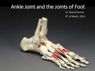
  • 1. Ankle Joint and the Joints of Foot Dr. Komal Parmar 9th of March, 2021
  • 2. Part 1- Tibiofibular Articulation
  • 3. Tibiofibular Joints Before we begin, let’s revise…. • Superior/ Proximal • Interosseus • Inferior/ Distal
  • 4. Superior Tibiofibular Joint • Plane type synovial joint • The primary function of the PTFJ is dissipation of torsional stresses applied at the ankle and the lateral tibial bending moments. • Joint capsule attached along the margins of articular surfaces. • Anterior and Posterior proximal tibiofibular ligaments • The synovial membrane lines the articular capsule which may communicate with the knee joint via the subpopliteal recess in about 10% of the population. • The fibular facet is usually elliptical or circular and nearly flat or slightly grooved. The surfaces are covered with hyaline cartilage underlining the synovial nature of this joint.
  • 5.
  • 6.
  • 7. • The tunnel entrance is a musculo- aponeurotic arch derived from the peroneus longus muscle. • Unyielding musculo- aponeurotic fibular arch. Fibular Tunnel
  • 8.
  • 9.
  • 10. Interosseous joint • Syndesmosis • Run downward and lateralward. • The membrane is continuous distally with the interosseous ligament of the distal tibiofibular joint. • The interosseous membrane acts as a conduit for stress transmission to the fibula. An intact membrane keeps the fibula active during the loads and motions of normal gait.
  • 11.
  • 12. Remember that there are Tibiofibular and Talofibular ligaments near the formation of ankle joint. Don’t get them confused.
  • 13.
  • 14.
  • 15. The PITFL is made up a superficial ligament and deep ligament (often called the inferior transverse tibiofibular ligament)
  • 16.
  • 17.
  • 18. Part 2- Ankle Joint
  • 19.
  • 20.
  • 21. ANKLE (TALOCRURAL) JOINT • Hinge, uniaxial • Mortise for Talus- The lower end of the tibia, malleolus of the fibula and inferior transverse tibiofibular ligament
  • 22. Range of Motion • Hinge joint with a dynamic axis of rotation • Normal range of dorsiflexion is 10º when the knee is straight, and 30º with the knee flexed (when the calcaneal tendon will be relaxed). • The range of normal plantar flexion is 30º. • Dorsiflexion results in the joint adopting the ‘closepacked’ position, with maximal congruence and ligamentous tension • From this position, all major thrusting movements are exerted, in walking, running and jumping. • The superior talar surface is broader in front, and in dorsiflexion the malleolar gap is increased by slight lateral rotation of the fibula, by ‘give’ at the inferior tibiofibular syndesmosis and gliding at the superior tibiofibular joint
  • 23. Articulating Surfaces • covered by hyaline cartilage
  • 24.
  • 25.
  • 26. Joint Capsule attachment • It is attached proximally to the borders of the tibial and malleolar articular surfaces, and distally to the talus near the margins of its trochlear surface, except in front where it reaches the dorsum of the talar neck.
  • 27. • Amaha, K., Nimura, A., Yamaguchi, R. et al. Anatomic study of the medial side of the ankle base on the joint capsule: an alternative description of the deltoid and spring ligament. J EXP ORTOP 6, 2 (2019). https://doi.org/10.1186/s40634-019-0171-y
  • 28.
  • 30. Medial (Deltoid) Ligament • TIBIA: Attached to the apex and the anterior and posterior borders of the medial malleolus. • Superficial fibres: • Anterior (tibionavicular): navicular tuberosity, medial margin of the plantar calcaneonavicular ligament • Intermediate (tibiocalcaneal): entire length of the sustentaculum tali; • Posterior fibres (posterior tibiotalar): medial side of the talus and its medial tubercle.
  • 31. • Deep fibres (anterior tibiotalar): tip of the medial malleolus to the non-articular part of the medial talar surface. • Relations: The ligament is crossed by the tendons of • tibialis posterior • Flexor digitorum longus. Medial (Deltoid) Ligament
  • 33. • The lateral ligament has three discrete parts. • Anterior talofibular ligament: the anterior margin of the fibula to front of the lateral articular facet of Talus and to the lateral aspect of its neck • The posterior talofibular ligament: The distal part of the lateral malleolar fossa to the lateral tubercle of the posterior talar process; a ‘tibial slip’ of fibres connects it to the medial malleolus. • The calcaneofibular ligament: From a depression anterior to the apex of the fibular malleolus to a tubercle on the lateral calcaneal surface • crossed by the tendons of fibularis longus and brevis • The lateral ligament complex is injured most commonly with inversion sprains, often during sport; the posterior talofibular ligament is almost always spared.
  • 34. • Synovial membrane The joint is lined by synovial membrane which projects into the inferior (distal) tibiofibular joint. • Vascular supply and lymphatic drainage The talocrural joint is supplied by malleolar branches of the anterior and posterior tibial and fibular arteries. Lymphatic drainage is via vessels accompanying the arteries and via the long and short saphenous veins superficially. • Innervation The talocrural joint is innervated by branches from the deep fibular, saphenous, sural and tibial nerves (or medial and lateral plantar nerves, depending on the level of division of the tibial nerve).
  • 36. • The long saphenous vein and saphenous nerve cross the ankle joint medial to the tendon of tibialis anterior and anterior to the medial malleolus, the nerve lying posterior to the vein.
  • 37. •Factors maintaining stability • Passive stability is conferred upon the ankle mainly by the medial and lateral ligament complexes, the distal tibiofibular ligaments, the tendons crossing the joint, the bony contours and the capsular attachments. • Dynamic stability is conferred by gravity, muscle action and ground reaction forces. • Stability requires the continuous action of soleus assisted by gastrocnemius: it increases when leaning forward, and decreases when leaning backwards. • If backward sway takes the projection of the centre of gravity (‘weight line’) posterior to the transverse axes of the ankle joints, the plantar flexors relax and the dorsiflexors contract.
  • 38.
  • 39.
  • 40. Part 3- Small Joints of Foot 1. Inferior Tibiofibular 2. Talocalcaneal (Subtalar Joint) 3. Talocalcaneonavicular joint (Subtalar Joint) 4. Calcaneocuboid joint 5. Naviculocuneiform joints 6. Cuboideonavicular joint 7. Intercuneiform and cuneocuboid joints 8. Tarsometatarsal articulations 9. Intermetatarsal joints 10. Metatarsophalangeal articulations 11. Interphalangeal articulations
  • 41. Talocalcaneal Joint • Anterior and posterior articulations between the calcaneus and talus form a functional unit often termed the ‘subtalar joint’. • The posterior articulation is referred to as the talocalcaneal joint and the anterior articulation is regarded as part of the talocalcaneonavicular joint. • The bones are connected by a fibrous capsule, and by lateral, medial, interosseous talocalcaneal and cervical
  • 44. Joint Capsule • Attached to the margins of articular surfaces Talocalcaneal Joint Ligaments • Lateral talocalcanal eligaments • medial talocalcaneal ligaments • interosseous talocalcaneal ligaments • Cervical ligament.
  • 45. • Lateral talocalcaneal ligament- lateral talar process to the lateral calcaneal surface. • Medial talocalcaneal ligament- medial talar tubercle to the back of the sustentaculum tali and adjacent medial surface of the calcaneus. • Its fibres blend with the medial (deltoid) ligament of the ankle joint. • Interosseous talocalcaneal ligament: flat, bilaminar transverse band in the sinus tarsi • Sulcus tali to the calcaneal sulcus. • The posterior lamina of the ligament is associated with the talocalcaneal joint, and the anterior lamina with the talocalcaneonavicular joint. • Cervical ligament : On talar Neck lateral to the tarsal sinus and attached to the superior calcaneal surface. Talocalcaneal Joint
  • 50. • Synovial membrane The synovial cavity of the talocalcaneal joint is usually quite separate and does not communicate with those of other tarsal joints. However, direct communication with the ankle joint has been observed in rare instances. • Innervation The talocalcaneal joint is innervated by branches of the posterior tibial, medial plantar and sural nerves. • Relations Posteromedially, posterior tibial artery and vein, the tibial nerve and the tendon of flexor hallucis longus. • These neurovascular structures are at risk in posteromedial approaches to the ankle and talocalcaneal joints. • On the lateral side, the tendon of fibularis brevis lies anterior to the tendon of fibularis longus, both passing behind the fibular malleolus. • The sural nerve lies just posterior to the fibular tendons and is at risk during lateral exposure of the joint. • Muscles producing movement Heel inversion is controlled by tibialis anterior, tibialis posterior and the gastrocnemius–soleus complex via the calcaneal tendon; the long flexors of the toes also contribute. Heel eversion results from the pull of fibularis longus, brevis and tertius in addition to the long extensors of the toes. Talocalcaneal Joint
  • 51. Talocalcaneonavicular joint • Two articulations, i.e. the anterior ‘subtalar’ joint and the talonavicular joint. • Compound, multiaxial articulation. Talocalcaneonavicular Joint
  • 53. • The ovoid talar head is continuous with the triple- faceted anterior area of its inferior surface. • The whole head fits the concavity formed collectively by the posterior surface of the navicular, the middle and anterior talar facets of the calcaneus, and the superior fibrocartilaginous surface of the plantar calcaneonavicular ligament (spring ligament). Talocalcaneonavicular Joint
  • 54. • Fibrous Capsule- Thin and poorly developed anteriorly, posteriorly blends with the anterior part of the interosseous ligament filling the tarsal sinus. • Ligaments: • Talonavicular ligament: connects the dorsal surfaces of the neck of the talus and the navicular • Plantar calcaneonavicular (spring) ligament: Spring Ligament thick band connecting the anterior margin of the sustentaculum tali to the plantar surface of the navicular. • Sustains the medial longitudinal arch of the foot • The dorsal surface of the ligament has a triangular fibrocartilaginous facet on which part of the talar head rests. • Its plantar surface is supported medially by the tendon of tibialis posterior and laterally by the tendons of flexors hallucis longus and digitorum longus; its medial border is blended with the anterior superficial fibres of the medial (deltoid) Talocalcaneonavicular Joint
  • 57.
  • 58.
  • 59.
  • 60.
  • 61. Calcaneocuboid Joint • same level as the talonavicular joint and, together, they represent the transverse tarsal joint. • Saddle (sellar) or biaxial joint with concavo-convex surfaces. • 2 cm proximal to the tubercle on the fifth metatarsal base Calcaneocuboid Joint
  • 62. • Fibrous capsule: Thickened dorsally as the dorsal calcaneocuboid ligament. • The synovial cavity of this joint is separate, and does not communicate with those of other tarsal articulations • Ligaments The ligaments of the calcaneocuboid joint are the • Bifurcate ligament • long plantar ligament • plantar calcaneocuboid ligaments Calcaneocuboid Joint
  • 63. Bifurcate Ligament: • strong Y-shaped band • attached by its stem proximally to the anterior part of the upper calcaneal surface, and distally it divides into calcaneocuboid and calcaneonavicular parts. • Calcaneocuboid part forms the main bond between the two rows of tarsal bones • The (medial) calcaneonavicular ligament is attached to the dorsolateral aspect of the navicular. Calcaneocuboid Joint
  • 64. • Long plantar ligament • Longest ligament associated with the tarsus • Attachment: • calcaneus: anterior to the processes of its tuberosity and from its anterior tubercle • Cuboid: the ridge and tuberosity on the plantar surface of the and continue to the bases of the second to fourth, and sometimes fifth, metatarsals. • This ligament, together with the groove on the plantar surface of the cuboid, makes a tunnel for the tendon of fibularis longus. It is a most powerful factor limiting depression of the lateral longitudinal arch. Calcaneocuboid Joint
  • 66. • Plantar calcaneocuboid ligament • Aka Short Planter Ligament • Deeper than the long plantar ligament •Attachment: Anterior calcaneal tubercle and the depression anterior to it, to the adjoining part of the plantar surface of Calcaneocuboid Joint
  • 70. Naviculocuneiform joints • Articulating surfaces • the distal navicular surface: transversely convex and divided into three facets • Fibrous capsule The fibrous capsule is continuous with those of the intercuneiform and cuneocuboid joints and it is also connected to the second and third cuneometatarsal joints and intermetatarsal joints between the second to fourth metatarsal bones.
  • 71.
  • 72.
  • 73.
  • 74. • Ligaments The ligaments of the naviculocuneiform joint are the • dorsal and plantar ligaments. • Dorsal and plantar (Cuneionavicular) ligaments • connect the navicular to each cuneiform; of the three dorsal ligaments, one is attached to each cuneiform. • The fasciculus from the navicular to the medial cuneiform is continued as the capsule of the joint around its medial aspect, and then blends medially with the plantar ligament. • Plantar ligaments have similar attachments and receive slips from the tendon of tibialis posterior.
  • 75.
  • 76.
  • 77.
  • 78. Cuboideonavicular joint • The cuboideonavicular joint is usually a fibrous joint. Syndesmosis • dorsal, plantar and interosseous ligaments • articular capsule and synovial lining are continuous with naviculocuneiform joint. • The interosseous ligament is made of strong transverse fibres and connects nonarticular parts of adjacent surfaces to the two bones
  • 79. Intercuneiform and cuneocuboid joints • all synovial and approximately plane or slightly curved. • Their articular capsules and synovial linings are continuous with those of the naviculocuneiform joints.
  • 80.
  • 81.
  • 82.
  • 83. Tarsometatarsal Joints • Plane, Synovial • Articulating surfaces • First metatarsal: medial cuneiform • Second: between the medial and lateral cuneiforms and articulates with the intermediate cuneiform • Third: lateral cuneiform • Fourth: lateral cuneiform and the cuboid
  • 84. • Fibrous capsule The hallucal joint has its own capsule. • The articular capsules and cavities of the second and third are continuous with those of the intercuneiform and naviculocuneiform joints, but are separated from the fourth and fifth joints by an interosseous ligament between the lateral cuneiform and fourth metatarsal base. • Ligaments The bones are connected by dorsal and plantar tarsometatarsal and interosseous cuneometatarsal ligaments.
  • 85. • Interosseous cuneometatarsal ligaments • three • One (the strongest) passes from the lateral surface of the medial cuneiform to the adjacent angle of the second metatarsal • Known as Lisfranc’s ligament. • A second ligament connects the lateral cuneiform to the adjacent angle of the second metatarsal • A third ligament connects the lateral angle of the lateral cuneiform to the adjacent fourth metatarsal base.
  • 86.
  • 87.
  • 88. • Dorsal, Plantar and Interosseus Ligaments • The intermetatarsal interosseous ligaments are very strong and are present between all the lateral four metatarsals • they are absent between the first and second metatarsals. • The base of the second metatarsal is joined to the first tarsometatarsal joint by the medial interosseous ligament (Lisfranc’s ligament) which connects the plantar aspect of the second metatarsal to the medial cuneiform. Intermetatarsal joints
  • 89.
  • 90. Metatarsophalangeal Joints • Articulating surfaces Articular surfaces cover the distal and plantar, but not the dorsal, aspects of the metatarsal heads. • The plantar aspect of the first metatarsal head has two longitudinal grooves separated by a ridge (the crista). • Articular areas on the proximal phalangeal bases are concave.
  • 91.
  • 92.
  • 93. • Plantar ligaments. • Deep transverse metatarsal ligaments The deep transverse metatarsal ligaments are four short, wide, flat bands that unite the plantar ligaments of adjoining metatarsophalangeal joints. • Collateral ligaments They are attached to the dorsal tubercles on the metatarsal heads and the corresponding side of the phalangeal bases and they slope downwards and forwards. • The first metatarsophalangeal joint also contains metatarso-sesamoid ligaments. • Each collateral ligament consists of the phalangeal collateral ligament, which inserts into the base of the proximal phalanx, and the accessory collateral ligament, which inserts into the plantar plate.
  • 94. movements • the range of active extension (50–60º) • flexion (30–40º • When the foot is on the ground, metatarsophalangeal joints are already extended to at least 25º • The range of passive movements in these joints is • 90º (extension) and 45º (flexion)
  • 95. Interphalangeal articulations • pure hinge joints • Each has an articular capsule • two collateral ligaments. • The plantar surface of the capsule is a thickened fibrous plate, like the plantar metatarsophalangeal ligaments, and is often termed the plantar ligament.
  • 96. Part 4- Postures and Movements of Foot
  • 97. Foot Posture • Foot posture is generally characterized by the contour of the medial longitudinal arch, and is typically divided into • normal (rectus) • low-arched (planus) • highly-arched (cavus)
  • 98. • The term “pronated” is used to indicate a foot that undergoes greater lowering of the medial longitudinal arch and more medial distribution of plantar loading during gait and • “supinated” to indicate a foot that undergoes greater elevation of the medial longitudinal arch and more lateral distribution of plantar loading during gait. • As the foot is loaded, eversion of the subtalar joint, dorsiflexion of the ankle, and abduction of the forefoot occur
  • 99.
  • 100.
  • 102.
  • 103.
  • 104.
  • 105.
  • 106.
  • 108.
  • 109. Talus Anatomy • The junction of the head and body of the talus subtends an angle approximately measuring 120◦ when measured between the axis of the head and a transverse coronal plane passing across the superior articular surface of the talus. • The transverse or long axis of the articular surface of the head appears to be medially rotated with respect to the transverse plane drawn across the superior articular surface of the talus, corresponding roughly with the mid-points of the tibial and fibular articulating surfaces on either side of the body of the talus
  • 110.
  • 111.
  • 112. average inclination of 42° in the sagittal plane and 23° medial deviation in the axial plane when relating to the long axis of the foot.