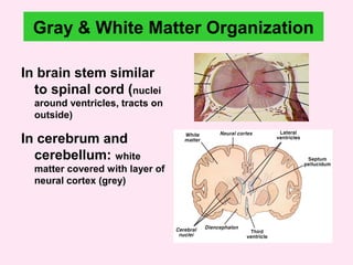
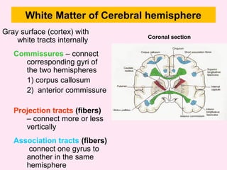
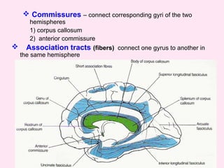
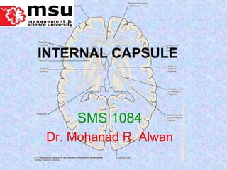
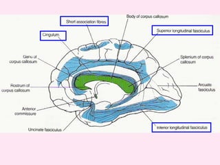
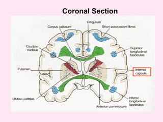
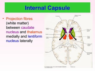
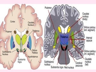
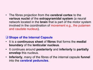
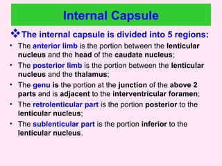
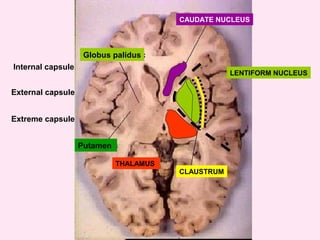
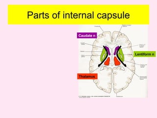
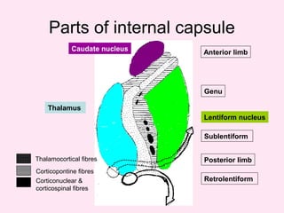
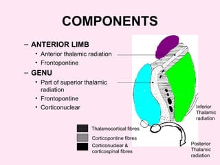
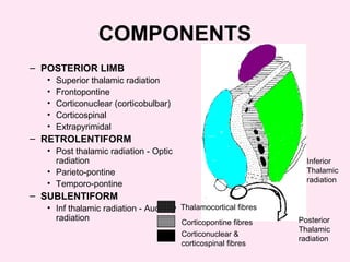
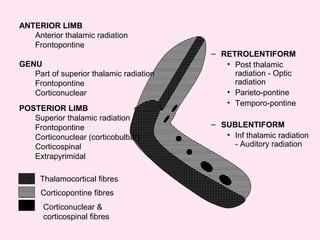
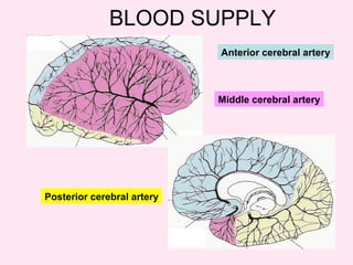
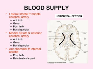
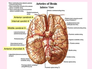
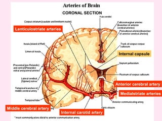
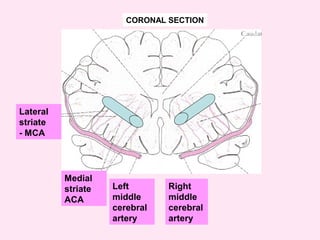
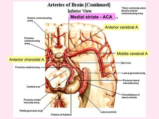
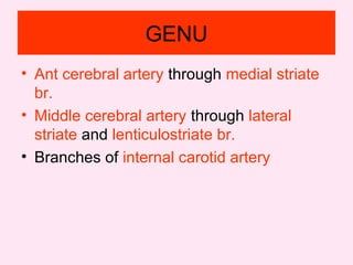
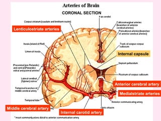
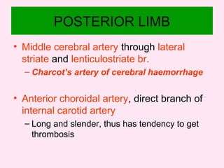
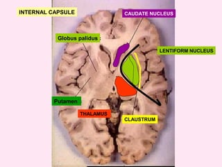
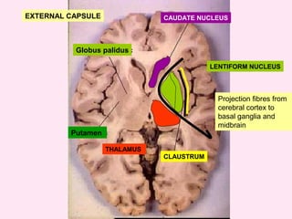
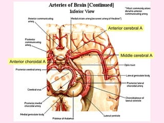
The internal capsule is a central white matter structure that contains important projection fibers connecting the cerebral cortex with deep gray matter structures. It is divided into the anterior limb, genu, posterior limb, retrolenticular and sublenticular parts. The internal capsule receives its blood supply from the middle cerebral artery, anterior cerebral artery, and anterior choroidal artery. Strokes or lesions involving the internal capsule can cause motor or sensory deficits depending on the fibers affected.
2. • Define internal capsule and enumerate the
parts
• Illustrate the internal capsule and describe
the components of each part
• Describe the blood supply and its clinical
importance
OBJECTIVES
3. Gray & White Matter Organization
In brain stem similar
to spinal cord (nuclei
around ventricles, tracts on
outside)
In cerebrum and
cerebellum: white
matter covered with layer of
neural cortex (grey)
4. White Matter of Cerebral hemisphere
Gray surface (cortex) with
white tracts internally
Commissures – connect
corresponding gyri of
the two hemispheres
1) corpus callosum
2) anterior commissure
Projection tracts (fibers)
– connect more or less
vertically
Association tracts (fibers)
connect one gyrus to
another in the same
hemisphere
Coronal section
5. Commissures – connect corresponding gyri of the two
hemispheres
1) corpus callosum
2) anterior commissure
Association tracts (fibers) connect one gyrus to another in
the same hemisphere
12. • Projection fibres
(white matter)
between caudate
nucleus and thalamus
medially and lentiform
nucleus laterally
Internal Capsule
13.
14. • The fibres projection from the cerebral cortex to the
various nuclei of the extrapyramidal system (a neural
network located in the brain that is part of the motor system
involved in the coordination of movement e.g., the putamen
and caudate nucleus).
Shape of the Internal Capsule
• It is a continuous sheet of fibres that forms the medial
boundary of the lenticular nucleus.
• It continues around posteriorly and inferiorly to partially
envelop this nucleus.
• Inferiorly, many of the fibres of the internal capsule funnel
into the cerebral peduncles.
15. Superiorly, the fibres fan out into the corona radiata.
Here, they travel in the cerebral white matter to reach
their cortical origins or destinations.
The entire fibre system is shaped like a trumpet with a
large notch cut out of its bell.
The notch corresponds to the location of where this sheet
of fibres is interrupted by the lateral sulcus.
The lenticular nucleus sits where a mute would sit in a
trumpet.
16. Internal Capsule
The internal capsule is divided into 5 regions:
• The anterior limb is the portion between the lenticular
nucleus and the head of the caudate nucleus;
• The posterior limb is the portion between the lenticular
nucleus and the thalamus;
• The genu is the portion at the junction of the above 2
parts and is adjacent to the interventricular foramen;
• The retrolenticular part is the portion posterior to the
lenticular nucleus;
• The sublenticular part is the portion inferior to the
lenticular nucleus.
17. Portion Description Origin Destination
Anterior Limb
Anterior thalamic
radiation
• Anterior nucleus
• DM
• Cingulate gyrus
• Prefrontal cortex
Genu Relays to motor areas
• VA
• VL
• Premotor cortex
• Primary motor
cortex
Posterior Limb
1. Motor pathways:
o Corticospinal
tract
o Corticobulbar
tract
2. Somatosensory
relays
1. Motor cortex
2. VPL/VPM
1. Motor pathways:
o Spinal cord
o Brainstem
2. Somatosensory
relays:
o Primary
somatosentory
cortex
Retrolenticular
• Association relay
• Optic radiation
• Pulvinar
• LGN
• Association cortex
• Visual cortex
Sublenticular
• Optic radiation
• Auditory radiation
• LGN
• MGN
• Visual cortex
• Auditory cortex
33. ANTERIOR LIMB
• Ant cerebral artery through medial striate
br.
• Middle cerebral artery through lateral
striate and lenticulostriate br.
- pass through the lentiform N to supply the striate
34. GENU
• Ant cerebral artery through medial striate
br.
• Middle cerebral artery through lateral
striate and lenticulostriate br.
• Branches of internal carotid artery
36. POSTERIOR LIMB
• Middle cerebral artery through lateral
striate and lenticulostriate br.
– Charcot’s artery of cerebral haemorrhage
• Anterior choroidal artery, direct branch of
internal carotid artery
– Long and slender, thus has tendency to get
thrombosis