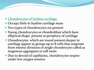
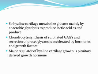
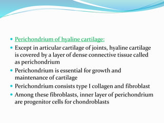
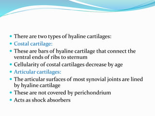
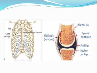
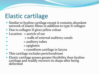
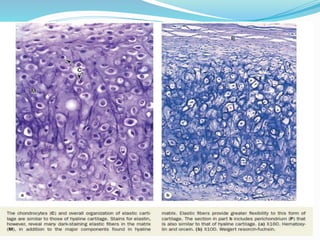
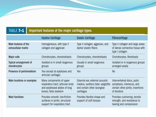
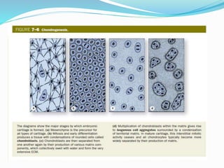
- Ladi Anudeep
ISM-IUK
 Cartilage is a tough and flexible form of connective
tissue and can be also considered as modified
connective tissue
 Cartilage is characterised by ECM and high
concentrations of GAG’s, proteoglycans
 Cartilage differs from typical connective tissue mainly
in nature of ground substance
Functions of cartilage
 Due to ECM, it allows the tissue to bear mechanical
stress
 In respiratory tracts, ears, nose, cartilage forms
framework supporting soft tissues
 Cartilage provides shock absorbing and sliding regions
with in joints and facilitate bone movements due to
smooth lubricated surface and resilency
Components of cartilage
 Cells- chondrocytes
 Ground substance/ matrix
 Fibres- collagen fibres
 Cells- chondrocytes
 cartilage consists of cells called as chondrocytes (
chondros: cartilage ; kytos: cell)
 These are embedded in matrix
 Chondrocytes synthesize and maintain ECM components
 Chondrocytes located in matrix cavities called as lacunae
 Nucleus is euchromatic
 Mitochondria, Golgi apparatus, endoplasmic
reticulum are prominent
 As cartilage cells matures, nuclei become
heterochromatic and cells organelles become less
prominent
 Cytoplasm contains glycogen and lipids
S TEM OF CHONDROCYTE
 Ground substance / matrix
 it is made up of complex molecules containing proteins and
carbohydrates( proteoglycans)
 Principal macromolecules present in cartilage matrix are:
 >collagen
 >hyaluronic acid
 >proteoglycans
 >glycoprotein
 Semi rigid nature of cartilage is due to water bound to negatively
charged sulphated GAG chains extending from proteoglycan core
proteins
 Collagen fibres
 type II collagen is present
 However, fibro cartilage and perichondrium consists of type I collagen
Classification of cartilage
 There are three types of cartilges that can be recognised
depending on number and variety of fibres in the matrix:
 Hyaline cartilage: ( hyalos: glass)
 Most common form
 Homogenous
 Semi-transparent in fresh state
 Principle collagen is type II collagen
 Location: > articular surfaces of movable joints
 > respiratory passages ( nose, larynx, trachea,
bronchi)
 > ventral ends of ribs
 > epiphyseal plates of long bones
 In embryo, hyaline cartilage forms temporary skeleton
that is replaced by bone
 Osteo-arthritis occurs during ageing which involves
gradual loss or changed physical properties of hyaline
cartilage that lines articular ends of bones on joints
 Joints that are weight bearing, heavily used are more
prone to cartilage degeneration
 Fragments released by wear and tear to articular
cartilage trigger secretion of matrix metallo –proteases
from macrophages which cause damage, pain and
inflammation with the joint
 Matrix of hyaline cartilage:
 Dry weight of hyaline cartilage is 40% which is embedded
in hydrated gel of proteoglycans and structural
glycoproteins
 proteoglycans cause matrix to be basophilic
 Most of the collagen in hyaline cartilage is type II collagen
 Most abundant proteoglycan of hyaline cartilage is
aggrecan( core protein) which consists of 150 GAG’s side
chains of chondroitin sulphate, keratin sulphate
 Proteoglycans non-covalently bound by link proteins to
long polymers of hyaluronic acid
 Proteoglycan complexes bind to type II collagen
 Water bound to GAG’s in proteoglycans constitute 60-80%
of fresh weight of fresh hyaline cartilage
 Another important component of matrix is chondronectin
which is a multi adhesive protein that binds specifically to
GAG’s, collagen II and integrins
 Two types of matrix:
 Territorial matrix: immediately around lacunae housing
chondrocytes, matrix stains deeper giving capsule like
appearance
 Interterritorial matrix: pale staining matrix seperatig cell
nests
 Chondrocytes of hyaline cartilage:
 Occupy little in hyaline cartilage mass
 Two types of chondrocytes are present
 Young chondrocytes or chondroblast which have
elliptical shape present at periphery of cartilage
 Chondrocytes which are round present deeper in
cartilage appear in groups up to 8 cells that originate
from mitotic divisions of single chondrocyte called as
isogenous aggregates or cell nests
 Due to devoid of capillaries, chondrocytes respire
under low oxygen tension
 So hyaline cartilage metabolize glucose mainly by
anaerobic glycolysis to produce lactic acid as end
product
 Chondrocyte synthesis of sulphated GAG’s and
secretion of proteoglycans is accelerated by hormones
and growth factors
 Major regulator of hyaline cartilage growth is pituitary
derived growth hormone
 Perichondrium of hyaline cartilage:
 Except in articular cartilage of joints, hyaline cartilage
is covered by a layer of dense connective tissue called
as perichondrium
 Perichondrium is essential for growth and
maintenance of cartilage
 Perichondrium consists type I collagen and fibroblast
 Among these fibroblasts, inner layer of perichondrium
are progenitor cells for chondroblasts
 There are two types of hyaline cartilages:
 Costal cartilage:
 These are bars of hyaline cartilage that connect the
ventral ends of ribs to sternum
 Cellularity of costal cartilages decrease by age
 Articular cartilages:
 The articular surfaces of most synovial joints are lined
by hyaline cartilage
 These are not covered by perichondrium
 Acts as shock absorbers
Elastic cartilage
 Similar to hyaline cartilage except it contains abundant
network of elastic fibres in addition to type II collagen
 Due to collagen II gives yellow colour
 Location: > auricle of ear
 > walls of external auditory canals
 > auditory tubes
 > epiglottis
 > cuneiform cartilage in larynx
 This cartilage includes perichondrium
 Elastic cartilage posses greater flexibility than hyaline
cartilage and readily recovers its shape after being
deformed
Fibrocartilage
 It is a combination of hyaline cartilage and dense
connective tissue
 Location: > intervertebral discs
 > attachments of ligaments
 > pubic symphysis
 Chondrocytes are single and are aligned isogenous
aggregates which produce type II collagen
 Scarcity of proteoglycans make matrix of fibro
cartilage more acidophilic
 In this type of cartilage perichondrium is absent
 Intervertebral discs of spinal column primarily consist
of fibro cartilage, lubricated cushions and shock
absorbers
 Intervertebral discs has two components:
 Peripheral annulus fibrosus which is rich in type I
collagen
 Central nucleus pulposus which is gel like matrix and
is rich in hyaluronic acid
summary
chondrogenesis
 Cartilage forms from embryonic mesenchyme by the
process called as chondrogenesis
 First indication of this process is rounding up of
mesenchymal cells which have extensions and divide
rapidly called as chondroblast and chondrocytes
 When proliferation is ceased, these cells have
basophilic cytoplasm, rich in RER for collagen
synthesis
 Production of ECM encloses these cells in cavities
called lacunae
 During embryonic development, differentiation of cartilage
takes place from the centre outward
 So more central cells have characteristics of chondrocytes
and peripheral cells have characteristics of chondroblast
 Superficial mesenchyme forms perichondrium
 In cartilage two types of growth is seen
 > interstitial growth: which is a result from mitotic
division of pre existing chondrobalst
 This type of growth Is important in increasing the length of
long bones
 > appositional growth: which involves differentiation of
new chondroblasts from perichondrium
 Damaged cartilage undergoes slow and incomplete
repair, primarily by the activity of cells in the
perichondrium which invade the injured area and
produce new cartilage and this character is not seen in
young children
 In extensively damaged areas, perichondrium
produces a scar of dense connective tissue instead of
ne cartilage
 The poor capacity of cartilage for repair or
regeneration is due to its avascular in nature
CARTILAGE

CARTILAGE

  • 1.
  • 2.
     Cartilage isa tough and flexible form of connective tissue and can be also considered as modified connective tissue  Cartilage is characterised by ECM and high concentrations of GAG’s, proteoglycans  Cartilage differs from typical connective tissue mainly in nature of ground substance
  • 3.
    Functions of cartilage Due to ECM, it allows the tissue to bear mechanical stress  In respiratory tracts, ears, nose, cartilage forms framework supporting soft tissues  Cartilage provides shock absorbing and sliding regions with in joints and facilitate bone movements due to smooth lubricated surface and resilency
  • 4.
    Components of cartilage Cells- chondrocytes  Ground substance/ matrix  Fibres- collagen fibres  Cells- chondrocytes  cartilage consists of cells called as chondrocytes ( chondros: cartilage ; kytos: cell)  These are embedded in matrix  Chondrocytes synthesize and maintain ECM components  Chondrocytes located in matrix cavities called as lacunae  Nucleus is euchromatic
  • 5.
     Mitochondria, Golgiapparatus, endoplasmic reticulum are prominent  As cartilage cells matures, nuclei become heterochromatic and cells organelles become less prominent  Cytoplasm contains glycogen and lipids
  • 6.
    S TEM OFCHONDROCYTE
  • 7.
     Ground substance/ matrix  it is made up of complex molecules containing proteins and carbohydrates( proteoglycans)  Principal macromolecules present in cartilage matrix are:  >collagen  >hyaluronic acid  >proteoglycans  >glycoprotein  Semi rigid nature of cartilage is due to water bound to negatively charged sulphated GAG chains extending from proteoglycan core proteins  Collagen fibres  type II collagen is present  However, fibro cartilage and perichondrium consists of type I collagen
  • 8.
    Classification of cartilage There are three types of cartilges that can be recognised depending on number and variety of fibres in the matrix:  Hyaline cartilage: ( hyalos: glass)  Most common form  Homogenous  Semi-transparent in fresh state  Principle collagen is type II collagen  Location: > articular surfaces of movable joints  > respiratory passages ( nose, larynx, trachea, bronchi)  > ventral ends of ribs  > epiphyseal plates of long bones
  • 9.
     In embryo,hyaline cartilage forms temporary skeleton that is replaced by bone  Osteo-arthritis occurs during ageing which involves gradual loss or changed physical properties of hyaline cartilage that lines articular ends of bones on joints  Joints that are weight bearing, heavily used are more prone to cartilage degeneration  Fragments released by wear and tear to articular cartilage trigger secretion of matrix metallo –proteases from macrophages which cause damage, pain and inflammation with the joint
  • 10.
     Matrix ofhyaline cartilage:  Dry weight of hyaline cartilage is 40% which is embedded in hydrated gel of proteoglycans and structural glycoproteins  proteoglycans cause matrix to be basophilic  Most of the collagen in hyaline cartilage is type II collagen  Most abundant proteoglycan of hyaline cartilage is aggrecan( core protein) which consists of 150 GAG’s side chains of chondroitin sulphate, keratin sulphate  Proteoglycans non-covalently bound by link proteins to long polymers of hyaluronic acid
  • 12.
     Proteoglycan complexesbind to type II collagen  Water bound to GAG’s in proteoglycans constitute 60-80% of fresh weight of fresh hyaline cartilage  Another important component of matrix is chondronectin which is a multi adhesive protein that binds specifically to GAG’s, collagen II and integrins  Two types of matrix:  Territorial matrix: immediately around lacunae housing chondrocytes, matrix stains deeper giving capsule like appearance  Interterritorial matrix: pale staining matrix seperatig cell nests
  • 13.
     Chondrocytes ofhyaline cartilage:  Occupy little in hyaline cartilage mass  Two types of chondrocytes are present  Young chondrocytes or chondroblast which have elliptical shape present at periphery of cartilage  Chondrocytes which are round present deeper in cartilage appear in groups up to 8 cells that originate from mitotic divisions of single chondrocyte called as isogenous aggregates or cell nests  Due to devoid of capillaries, chondrocytes respire under low oxygen tension
  • 14.
     So hyalinecartilage metabolize glucose mainly by anaerobic glycolysis to produce lactic acid as end product  Chondrocyte synthesis of sulphated GAG’s and secretion of proteoglycans is accelerated by hormones and growth factors  Major regulator of hyaline cartilage growth is pituitary derived growth hormone
  • 15.
     Perichondrium ofhyaline cartilage:  Except in articular cartilage of joints, hyaline cartilage is covered by a layer of dense connective tissue called as perichondrium  Perichondrium is essential for growth and maintenance of cartilage  Perichondrium consists type I collagen and fibroblast  Among these fibroblasts, inner layer of perichondrium are progenitor cells for chondroblasts
  • 17.
     There aretwo types of hyaline cartilages:  Costal cartilage:  These are bars of hyaline cartilage that connect the ventral ends of ribs to sternum  Cellularity of costal cartilages decrease by age  Articular cartilages:  The articular surfaces of most synovial joints are lined by hyaline cartilage  These are not covered by perichondrium  Acts as shock absorbers
  • 19.
    Elastic cartilage  Similarto hyaline cartilage except it contains abundant network of elastic fibres in addition to type II collagen  Due to collagen II gives yellow colour  Location: > auricle of ear  > walls of external auditory canals  > auditory tubes  > epiglottis  > cuneiform cartilage in larynx  This cartilage includes perichondrium  Elastic cartilage posses greater flexibility than hyaline cartilage and readily recovers its shape after being deformed
  • 21.
    Fibrocartilage  It isa combination of hyaline cartilage and dense connective tissue  Location: > intervertebral discs  > attachments of ligaments  > pubic symphysis  Chondrocytes are single and are aligned isogenous aggregates which produce type II collagen  Scarcity of proteoglycans make matrix of fibro cartilage more acidophilic  In this type of cartilage perichondrium is absent
  • 23.
     Intervertebral discsof spinal column primarily consist of fibro cartilage, lubricated cushions and shock absorbers  Intervertebral discs has two components:  Peripheral annulus fibrosus which is rich in type I collagen  Central nucleus pulposus which is gel like matrix and is rich in hyaluronic acid
  • 25.
  • 26.
    chondrogenesis  Cartilage formsfrom embryonic mesenchyme by the process called as chondrogenesis  First indication of this process is rounding up of mesenchymal cells which have extensions and divide rapidly called as chondroblast and chondrocytes  When proliferation is ceased, these cells have basophilic cytoplasm, rich in RER for collagen synthesis  Production of ECM encloses these cells in cavities called lacunae
  • 28.
     During embryonicdevelopment, differentiation of cartilage takes place from the centre outward  So more central cells have characteristics of chondrocytes and peripheral cells have characteristics of chondroblast  Superficial mesenchyme forms perichondrium  In cartilage two types of growth is seen  > interstitial growth: which is a result from mitotic division of pre existing chondrobalst  This type of growth Is important in increasing the length of long bones  > appositional growth: which involves differentiation of new chondroblasts from perichondrium
  • 30.
     Damaged cartilageundergoes slow and incomplete repair, primarily by the activity of cells in the perichondrium which invade the injured area and produce new cartilage and this character is not seen in young children  In extensively damaged areas, perichondrium produces a scar of dense connective tissue instead of ne cartilage  The poor capacity of cartilage for repair or regeneration is due to its avascular in nature