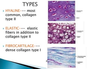
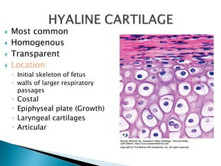
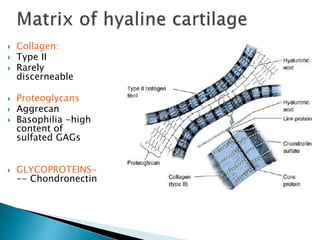
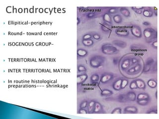
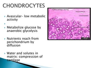
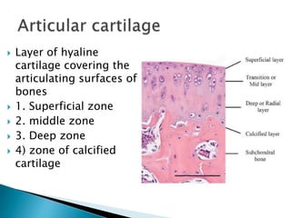
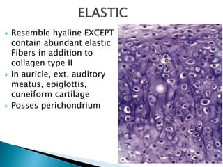
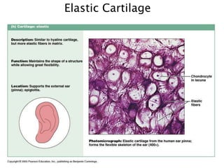
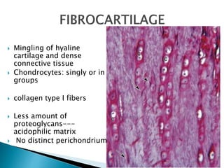
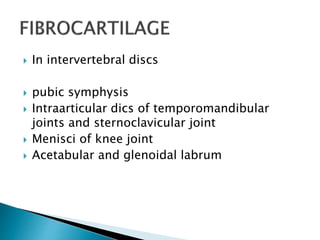
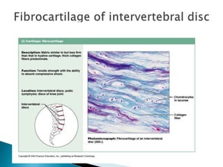
 Describe the histological features of cartilage.
 Describe the histological changes in cartilage
in degenerative diseases
 Special type of connective tissue, in which
firm consistency of ECM allows it to bear
mechanical stress, without permanent
distortion
 Protects soft tissue
 Shock absorbance
 Facilitates bone movements
 Growth of bones
 Semi rigid-solvation water
 Avascular
 Composed of Chondrocytes & ECM----fibers
& ground substance
 Chondrocytes: synthesize and secrete matrix
component
 ECM: Collagen, proteoglycans, hyaluronic
acid, glycoprotein
 Dense connective
tissue surrounds
cartilage in most
places except articular
cartilage
 Outer Fibrous layer:
Type I collagen fibers,
fibroblasts, Vascular
supply, nerves and
lymphatic vessels
 Inner cellular layer:
 Chondroblasts
 HYALINE--- most
common, collagen
type II
 ELASTIC--- elastic
fibers in addition to
collagen type II
 FIBROCARTILAGE---
dense collagen type I
 Most common
 Homogenous
 Transparent
 Location:
◦ Initial skeleton of fetus
◦ walls of larger respiratory
passages
◦ Costal
◦ Epiphyseal plate (Growth)
◦ Laryngeal cartilages
◦ Articular
 Collagen:
 Type II
 Rarely
discerneable
 Proteoglycans
 Aggrecan
 Basophilia -high
content of
sulfated GAGs
 GLYCOPROTEINS-
-- Chondronectin
 Ellipitical-periphery
 Round- toward center
 ISOGENOUS GROUP-
 TERRITORIAL MATRIX
 INTER TERRITORIAL MATRIX
 In routine histological
preparations--- shrinkage
 Avascular– low metabolic
activity

 Metabolize glucose by
anaerobic glycolysis
 Nutrients reach from
perichondrium by
diffusion
 Water and solutes in
matrix: compression of
cartilage
 Layer of hyaline
cartilage covering the
articulating surfaces of
bones
 1. Superficial zone
 2. middle zone
 3. Deep zone
 4) zone of calcified
cartilage
 Osteoarthritis
 Aging
 Weight bearing joints
 Gradual loss of cartilage
 Change in physical properties of
cartilage
 Resemble hyaline EXCEPT
contain abundant elastic
Fibers in addition to
collagen type II
 In auricle, ext. auditory
meatus, epiglottis,
cuneiform cartilage
 Posses perichondrium
Elastic Cartilage
 Mingling of hyaline
cartilage and dense
connective tissue
 Chondrocytes: singly or in
groups
 collagen type I fibers
 Less amount of
proteoglycans---
acidophilic matrix
 No distinct perichondrium
 In intervertebral discs
 pubic symphysis
 Intraarticular dics of temporomandibular
joints and sternoclavicular joint
 Menisci of knee joint
 Acetabular and glenoidal labrum
cartilage histology (2).pptx

cartilage histology (2).pptx

  • 2.
     Describe thehistological features of cartilage.  Describe the histological changes in cartilage in degenerative diseases
  • 3.
     Special typeof connective tissue, in which firm consistency of ECM allows it to bear mechanical stress, without permanent distortion  Protects soft tissue  Shock absorbance  Facilitates bone movements  Growth of bones  Semi rigid-solvation water  Avascular
  • 4.
     Composed ofChondrocytes & ECM----fibers & ground substance  Chondrocytes: synthesize and secrete matrix component  ECM: Collagen, proteoglycans, hyaluronic acid, glycoprotein
  • 5.
     Dense connective tissuesurrounds cartilage in most places except articular cartilage  Outer Fibrous layer: Type I collagen fibers, fibroblasts, Vascular supply, nerves and lymphatic vessels  Inner cellular layer:  Chondroblasts
  • 6.
     HYALINE--- most common,collagen type II  ELASTIC--- elastic fibers in addition to collagen type II  FIBROCARTILAGE--- dense collagen type I
  • 7.
     Most common Homogenous  Transparent  Location: ◦ Initial skeleton of fetus ◦ walls of larger respiratory passages ◦ Costal ◦ Epiphyseal plate (Growth) ◦ Laryngeal cartilages ◦ Articular
  • 8.
     Collagen:  TypeII  Rarely discerneable  Proteoglycans  Aggrecan  Basophilia -high content of sulfated GAGs  GLYCOPROTEINS- -- Chondronectin
  • 9.
     Ellipitical-periphery  Round-toward center  ISOGENOUS GROUP-  TERRITORIAL MATRIX  INTER TERRITORIAL MATRIX  In routine histological preparations--- shrinkage
  • 10.
     Avascular– lowmetabolic activity   Metabolize glucose by anaerobic glycolysis  Nutrients reach from perichondrium by diffusion  Water and solutes in matrix: compression of cartilage
  • 11.
     Layer ofhyaline cartilage covering the articulating surfaces of bones  1. Superficial zone  2. middle zone  3. Deep zone  4) zone of calcified cartilage
  • 12.
     Osteoarthritis  Aging Weight bearing joints  Gradual loss of cartilage  Change in physical properties of cartilage
  • 13.
     Resemble hyalineEXCEPT contain abundant elastic Fibers in addition to collagen type II  In auricle, ext. auditory meatus, epiglottis, cuneiform cartilage  Posses perichondrium
  • 14.
  • 15.
     Mingling ofhyaline cartilage and dense connective tissue  Chondrocytes: singly or in groups  collagen type I fibers  Less amount of proteoglycans--- acidophilic matrix  No distinct perichondrium
  • 16.
     In intervertebraldiscs  pubic symphysis  Intraarticular dics of temporomandibular joints and sternoclavicular joint  Menisci of knee joint  Acetabular and glenoidal labrum

Editor's Notes

  • #4 Cartilage is a specialized form of connective tissue in which the firm consistency of the ECM allows the tissue to bear mechanical stresses without permanent distortion. In the respiratory system cartilage forms a framework supporting soft tissues. Because it is smooth-surfaced and resilient, cartilage provides a shock-absorbing and sliding area for joints and facilitates bone movements. Cartilage is also essential for the development and growth of long bones, both before and after birth. Semi rigid : water bound to negatively charged sulfated GAGs.
  • #5 Cartilage consists of cells called chondrocytes (Gr. chondros, cartilage + kytos, cell) and an extensive extracellular matrix composed of fibers and ground substance. Chondrocytes synthesize and secrete the ECM and the cells themselves are located in matrix cavities called lacunae.  Collagen, hyaluronic acid, proteoglycans, and small amounts of several glycoproteins are the principal macromolecules present in all types of cartilage matrix.
  • #6 Except in the articular cartilage of joints, all hyaline cartilage is covered by a layer of dense connective tissue, the perichondrium, which is essential for the growth and maintenance of cartilage (Figure 7–2). It consists largely of collagen type I fibers and contains numerous fibroblasts. Although cells in the inner layer of the perichondrium resemble fibroblasts, they are precursors for chondroblasts which divide and differentiate into chondrocytes.
  • #7 As a consequence of different functional requirements, three forms of cartilage have evolved, each exhibiting variation in matrix composition. In the matrix of hyaline cartilage, the most common form, type II collagen is the principal collagen type. The more pliable and distensible elastic cartilage possesses, in addition to collagen type II, an abundance of elastic fibers within its matrix. Fibrocartilage, present in regions of the body subjected to pulling forces, is characterized by a matrix containing a dense network of coarse type I collagen fibers.
  • #8 Hyaline cartilage is the most common and best studied of the three forms. Fresh hyaline cartilage is bluish-white and translucent. In the embryo, it serves as a temporary skeleton until it is gradually replaced by bone. In adult mammals, hyaline cartilage is located in the articular surfaces of the movable joints, in the walls of larger respiratory passages (nose, larynx, trachea, bronchi), in the ventral ends of ribs, where they articulate with the sternum, and in the epiphyseal plate, where it is responsible for the longitudinal growth of bone
  • #9 Cartilage proteoglycans contain chondroitin 4-sulfate, chondroitin 6-sulfate, and keratan sulfate, covalently linked to core proteins. Hundreds of these proteoglycans are bound non-covalently to long molecules of hyaluronic acid by link proteins, forming very large proteoglycan aggregates such as aggrecan that interact with collagen. Structurally, proteoglycans resemble bottle brushes, the protein core being the stem and the radiating glycosaminoglycan (GAG) chains the bristles. The high content of solvation water bound to the negative charges of the GAGs acts as a shock absorber or biomechanical spring; this is of great functional importance, especially in articular cartilages. In addition to type II collagen and proteoglycan, an important component of cartilage matrix is the structural multiadhesive glycoprotein chondronectin. Like fibronectin in connective tissue, this macromolecule binds specifically to GAGs, collagen type II and integrins, mediating the adherence of chondrocytes to the ECM. Cartilage matrix is generally basophilic due to the high concentration of sulfated GAGs and staining variations within the matrix reflect differences in the molecular composition.
  • #10 Immediately surrounding each chondrocyte the ECM is richer in GAGs and poor in collagen. These areas comprise the territorial matrix and usually stain differently from the rest of the matrix At the periphery of hyaline cartilage, young chondrocytes have an elliptic shape, with the long axis parallel to the surface. Farther in, they are round and may appear in groups of up to eight cells originating from mitotic divisions of a single chondrocyte. These groups are called isogenous aggregates (Gr. isos, equal, + genos, family). Chondrocytes synthesize collagens and the other matrix molecules. As matrix is produced, cells in the aggregates are moved apart and occupy separate lacunae.
  • #11 Because cartilage is devoid of blood capillaries, chondrocytes respire under low oxygen tension. Hyaline cartilage cells metabolize glucose mainly by anaerobic glycolysis to produce lactic acid as the end product. Nutrients from the blood diffuse through the perichondrium to reach the more deeply placed cartilage cells. Transport of water and solutes is promoted by the pumping action of intermittent cartilage compression and decompression. Because of the limits of diffusion, the maximum width of the cartilage is limited and cartilage usually is found as small, thin plates of tissue. Chondrocyte function is hormone dependent. Secretion of proteoglycans is accelerated by hormones and growth factor.
  • #12 Superficial zone: Tangential zone: Type II collagen fibers arranged parallel to surface. Elongated chondrocytes are also arranged are also organized parallel to surface. There is abudance of collagen fibils, concentration of proteoglycans is less; this layer is pressure resistant which protects the deeper layers. Middle zone: spherical chondrocytes in ECM. It contain aggrecan, thicker type II collagen fibrils. Collagen fibrils are in oblique direction. Deep zone: spherical chondrocytes in short columns which are perpendicular to the articular surface. Large amount of aggrecan and very thick collagen fibers. Collagen fibers are perpendicular to the free surface. Due to high content of aggrecan and perpendicular arrangement of collagen fibrils, it provides greatest resistant against compressive forces. Zone of calcified cartilage: calcified extracellular matrix in which small chondorocytes are embeded,. It serves to bind the cartilage with the bone.
  • #13 Osteoarthritis, a chronic condition that commonly occurs during aging, involves the gradual loss or changed physical properties of the hyaline cartilage that lines the articular ends of bones in joints. Joints that are weight-bearing (knees, hips) or heavily used (wrist, fingers) are most prone to cartilage degeneration. Fragments released by wear-and-tear to the articular cartilage trigger secretion of matrix metalloproteinases and other factors from macrophages in adjacent tissues, which exacerbate damage and cause within the joint  pain and inflammation
  • #14 Elastic cartilage is essentially very similar to hyaline cartilage except that it contains an abundant network of fine elastic fibers in addition to collagen type II fibrils (Figure 7–4). Fresh elastic cartilage has a yellowish color owing to the presence of elastin in the elastic fibers. Elastic cartilage is frequently found to be gradually continuous with hyaline cartilage. Like hyaline cartilage, elastic cartilage possesses a perichondrium. Elastic cartilage is found in the auricle of the ear, the walls of the external auditory canals, the auditory (eustachian) tubes, the epiglottis, and the cuneiform cartilage in the larynx.  
  • #16 Fibrocartilage takes various forms in different structures but is essentially a mingling of hyaline cartilage and dense connective tissue (Figures 7–5 and 7–1d). Chondrocytes of fibrocartilage occur singly and often in aligned isogenous aggregates, producing type II collagen and other ECM components, although the matrix around these chondrocytes is typically sparse. Areas with chondrocytes and hyaline matrix are separated by other regions with fibroblasts and dense bundles of type I collagen which confer extra tensile strength to this tissue (Figure 7–5). The relative scarcity of proteoglycans overall makes fibrocartilage matrix more acidophilic than that of hyaline or elastic cartilage. There is no distinct surrounding perichondrium in fibrocartilage. Held in place by ligaments, intervertebral discs are discussed further with joints in Chapter 8.
  • #17 It is found in intervertebral discs, in attachments of certain ligaments, and in the pubic symphysis—all places where it serves as very tough, yet cushioning support tissue for bone.
  • #18 Intervertebral discs of the spinal column are composed primarily of fibrocartilage and act as lubricated cushions and shock absorbers preventing damage to adjacent vertebrae from abrasive forces or impacts. Intervertebral disks are composed of fibrocartilage primarily. They are situated between the vertebrae and are held to them by ligaments. Each disk has two major histological components: the peripheral annulus fibrosus rich in bundles of type I collagen and the central nucleus pulposus with a gel-like matrix rich in hyaluronic acid. Intervertebral disks act as lubricated cushions and shock absorbers preventing adjacent vertebrae from being damaged by abrasive forces or impact during movement of the spinal column.