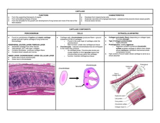
;
CARTILAGE
FUNCTIONS
● Form the supporting framework of organs
● Form articulating surfaces of bones
● Form the template for the growth and development of long bones and most of the rest of the
fetal skeleton
CHARACTERISTICS
● Develops from mesenchymal cells
● Avascular (secretes anti-angiogenesis factor - substance that prevents blood vessel growth)
● Aneural (lacks nerves)
CARTILAGE COMPONENTS
PERICHONDRIUM CELLS EXTRACELLULAR MATRIX
● Found on peripheries of hyaline and elastic cartilage
except articular hyaline cartilage of long bones and
fibrocartilage
PERIPHERAL (OUTER) LAYER/ FIBROUS LAYER
● Separates cartilage from other tissues
● Vascularized, DICT with type I collagen
● Contains fibroblasts - produces collagen fibers
● Contains blood vessels and nerves
INNER LAYER/ CHONDROGENIC LAYER/ CELLULAR LAYER
● Gives rise to future chondrocytes
● Gives rise to chondroblasts
● Cartilage cells: chondroblasts (produces fibers + ground
substance (ECM) of cartilage)
o Found in the outer layer of cartilage under the
perichondrium
o Main function: create new cartilaginous tissue
● Chondrocytes - matured chondroblasts that are entrapped
to the matrix they produce
o As cartilage grows. Chondrocytes divide and
cluster together to form lacunae (space that
chondrocytes occupy within the cartilage)
o function: maintain cartilaginous tissue
● Collagen and elastic fibers (depending on collagen type)
o Provides strength
● Type 2 Collagen predominates
o Highly refractile
● Proteoglycans, notably aggrecan
o Aggrecan contains numerous chondroitin
sulfate (enables cartilage to deform their shape;
allows resilience; withstand mechanical stress
compression forces)
● High content of bound water allows cartilage to serve as a
shock absorber
CARTILAGE TYPES
PARAMETER HYALINE ELASTIC FIBROCARTILAGE
Important
information
● Derived from the greek word hayalus = glass
● Most common
● Provides a firm structural and flexible support
● In embryo - forms the temporary skeleton that is
gradually replaced by bone
● Territorial matrix - darker matrix immediately
around chondrocytes
● Interterritorial matrix - less intensely stained area
between the isogenous groups of chondrocytes
● Lacunae - contains mature chondrocytes
● Better flexibility than hyaline ● type 1 collagen fibers - strongest and toughest
type
Main features of the
extracellular matrix
● Homogeneous (because the type 2 collagen fibrils
present have the same refractive index as that of
ground substance), with type II collagen and
aggrecan
● Rich in ground substance
● Bluish, basophilic
● Few elastic fibers
● Type II collagen, aggrecan, and darker elastic
fibers
● Numerous elastic fibers - visualized using silver
stain
● Type II collagen and large areas of dense regular
connective tissue with type I collagen
● Thick type 1 collagen fibers predominate, hence
hyaline cartilage matrix not that visible
Major cells ● Chondrocytes, chondroblasts ● Chondrocytes, chondroblasts ● Chondrocytes, fibroblasts
Typical arrangement
of chondrocytes
● random/Isolated or in abundant isogenous groups
● Small chondrocytes
● Usually in less isogenous groups
● Big chondrocytes
● Isolated or in isogenous groups arranged axially
● Small chondrocytes usually distributed in rows
Presences of
perichondrium
● Yes (except at epiphysis and articular cartilage) ● Yes ● No Perichondrium
Main locations of
examples
● Trachea, ribs, nose larynx
● Many components of upper respiratory tract;
articular ends and epiphyseal plates of long bones;
fetal skeleton
● EEEE: External ear, external acoustic meatus,
eustachian tubes, auditory tube, epiglottis and
certain other laryngeal cartilages
● PIA: Pubic symphysis, Intervertebral discs,
attachments of certain ligaments, meniscus, and
certain other joints; insertions of tendons
Main functions ● Provides smooth, low-friction surfaces in
joints; structural support for respiratory tract,
resilient cushion, resists compressive stress
● Provides flexible shape and support of soft
tissues
● Provides cushioning, tensile strength, and
resistance to tearing and compression
Laboratory Slides
HISTOLOGY CARTILAGE
HISTOLOGY CARTILAGE

HISTOLOGY CARTILAGE

  • 1.
    ; CARTILAGE FUNCTIONS ● Form thesupporting framework of organs ● Form articulating surfaces of bones ● Form the template for the growth and development of long bones and most of the rest of the fetal skeleton CHARACTERISTICS ● Develops from mesenchymal cells ● Avascular (secretes anti-angiogenesis factor - substance that prevents blood vessel growth) ● Aneural (lacks nerves) CARTILAGE COMPONENTS PERICHONDRIUM CELLS EXTRACELLULAR MATRIX ● Found on peripheries of hyaline and elastic cartilage except articular hyaline cartilage of long bones and fibrocartilage PERIPHERAL (OUTER) LAYER/ FIBROUS LAYER ● Separates cartilage from other tissues ● Vascularized, DICT with type I collagen ● Contains fibroblasts - produces collagen fibers ● Contains blood vessels and nerves INNER LAYER/ CHONDROGENIC LAYER/ CELLULAR LAYER ● Gives rise to future chondrocytes ● Gives rise to chondroblasts ● Cartilage cells: chondroblasts (produces fibers + ground substance (ECM) of cartilage) o Found in the outer layer of cartilage under the perichondrium o Main function: create new cartilaginous tissue ● Chondrocytes - matured chondroblasts that are entrapped to the matrix they produce o As cartilage grows. Chondrocytes divide and cluster together to form lacunae (space that chondrocytes occupy within the cartilage) o function: maintain cartilaginous tissue ● Collagen and elastic fibers (depending on collagen type) o Provides strength ● Type 2 Collagen predominates o Highly refractile ● Proteoglycans, notably aggrecan o Aggrecan contains numerous chondroitin sulfate (enables cartilage to deform their shape; allows resilience; withstand mechanical stress compression forces) ● High content of bound water allows cartilage to serve as a shock absorber
  • 2.
    CARTILAGE TYPES PARAMETER HYALINEELASTIC FIBROCARTILAGE Important information ● Derived from the greek word hayalus = glass ● Most common ● Provides a firm structural and flexible support ● In embryo - forms the temporary skeleton that is gradually replaced by bone ● Territorial matrix - darker matrix immediately around chondrocytes ● Interterritorial matrix - less intensely stained area between the isogenous groups of chondrocytes ● Lacunae - contains mature chondrocytes ● Better flexibility than hyaline ● type 1 collagen fibers - strongest and toughest type Main features of the extracellular matrix ● Homogeneous (because the type 2 collagen fibrils present have the same refractive index as that of ground substance), with type II collagen and aggrecan ● Rich in ground substance ● Bluish, basophilic ● Few elastic fibers ● Type II collagen, aggrecan, and darker elastic fibers ● Numerous elastic fibers - visualized using silver stain ● Type II collagen and large areas of dense regular connective tissue with type I collagen ● Thick type 1 collagen fibers predominate, hence hyaline cartilage matrix not that visible Major cells ● Chondrocytes, chondroblasts ● Chondrocytes, chondroblasts ● Chondrocytes, fibroblasts Typical arrangement of chondrocytes ● random/Isolated or in abundant isogenous groups ● Small chondrocytes ● Usually in less isogenous groups ● Big chondrocytes ● Isolated or in isogenous groups arranged axially ● Small chondrocytes usually distributed in rows Presences of perichondrium ● Yes (except at epiphysis and articular cartilage) ● Yes ● No Perichondrium Main locations of examples ● Trachea, ribs, nose larynx ● Many components of upper respiratory tract; articular ends and epiphyseal plates of long bones; fetal skeleton ● EEEE: External ear, external acoustic meatus, eustachian tubes, auditory tube, epiglottis and certain other laryngeal cartilages ● PIA: Pubic symphysis, Intervertebral discs, attachments of certain ligaments, meniscus, and certain other joints; insertions of tendons Main functions ● Provides smooth, low-friction surfaces in joints; structural support for respiratory tract, resilient cushion, resists compressive stress ● Provides flexible shape and support of soft tissues ● Provides cushioning, tensile strength, and resistance to tearing and compression Laboratory Slides