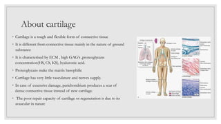
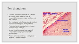
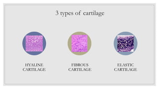
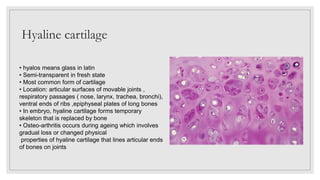
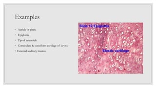
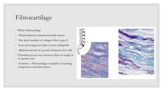
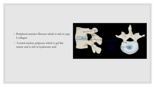
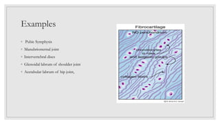
CLASSIFICATION OF
CARTILAGE
Aditi Biswas-1056
About cartilage
◦ Cartilage is a tough and flexible form of connective tissue
◦ It is different from connective tissue mainly in the nature of ground
substance
◦ It is characterised by ECM , high GAG’s ,proteoglycans
concentration(HS, CS, KS), hyaluronic acid.
◦ Proteoglycans make the matrix basophilic
◦ Cartilage has very little vasculature and nerves supply.
◦ In case of extensive damage, perichondrium produces a scar of
dense connective tissue instead of new cartilage.
◦ The poor repair capacity of cartilage or regeneration is due to its
avascular in nature
Perichondrium
◦ Cartilage is covered externally by a dense
connective tissue sheath known as
perichondrium except articular cartilage and
fibro cartilage.
◦ Has two layers- outer fibrous layer (vascular)
and inner chondrogenic layer (cellular)
◦ Perichondrium is essential for growth and
maintenance of cartilage
◦ Among these fibroblasts, inner layer of
perichondrium are progenitor cells for
chondroblasts
◦ Perichondrium consists type I collagen and
fibroblast ( more acidophilic)
3 types of cartilage
HYALINE
CARTILAGE
FIBROUS
CARTILAGE
ELASTIC
CARTILAGE
Hyaline cartilage
• hyalos means glass in latin
• Semi-transparent in fresh state
• Most common form of cartilage
• Location: articular surfaces of movable joints ,
respiratory passages ( nose, larynx, trachea, bronchi),
ventral ends of ribs ,epiphyseal plates of long bones
• In embryo, hyaline cartilage forms temporary
skeleton that is replaced by bone
• Osteo-arthritis occurs during ageing which involves
gradual loss or changed physical
properties of hyaline cartilage that lines articular ends
of bones on joints
Cells
1.They are derived from undifferentiated mesenchymal cells,
these have cytoplasmic processes.
2.Young cells(elliptical shaped) are small known as
chondroblasts are present at the periphery, they multiply to
chondrocytes
3.Older and mature cells are known as chondrocytes
4.Chondrocytes are bigger in size and are found in spaces
called as lacunae
5. They are found either groups of 2-4 cells together known
as cell nest or individual cells
6.They are responsible for production of fibres and ground
substance of the cartilage . Old mature cells are incapable
of multiplication
Chondrocyte
◦ Occupy in lacunae
◦ Responsible for the synthesis of collagen
fibers and ground substances
◦ Isogenous groups of chondrocyte
◦ Nucleus is euchromatic initially
◦ Mitochondria, Golgi apparatus, endoplasmic
reticulum are prominent .
◦ As cartilage cells matures, nuclei become
heterochromatic and cells organelles
become less prominent
Ground substance
◦ Type II collagen fibers
◦ Collagen- stability & strength
◦ Fibers constitute about the 40% of dry weight of cartilage
◦ 150 Keratan sulphate and chondroitin sulphate side chains
attach to ACAN ( aggrecan) or cartilage
specific proteoglycan core protein.
◦ This aggregate attaches (non-covalently) to the hyaluronic acid
backbone via Linker protein
◦ This structure then binds to collagen fibre II
◦ Semi rigid nature of cartilage is due to water bound to negatively charged sulphated GAG chains extending from proteoglycan core
proteins
◦ Another important component of matrix is chondronectin which is a multi adhesive protein that binds specifically to GAG’s, collagen II
and integrins
• Highest concentration present around the lacuna
• Matrix around the cells is brighter and deep blue in
colour than other areas, this matrix is known as
capsule / territorial matrix. (newly formed)
• Two groups of cells are separated by a lightly
coloured matrix known inter-territorial matrix
• Due to devoid of capillaries, chondrocytes respire
under low oxygen conditions
• So hyaline cartilage metabolize glucose mainly by
anaerobic glycolysis to produce lactic acid as end
product
• Chondrocyte synthesis of sulphated GAG’s and
secretion of proteoglycans is accelerated by
hormones and growth factors
• Major regulator of hyaline cartilage growth is
pituitary derived growth hormone
Examples
Function of hyaline cartilage
◦ Articular cartilage provides smooth surface for movement at joints
◦ Though it is flexible, it provides support because of its firmness
◦ Firmness of the cartilage keeps the lumen of trachea and bronchi patent
◦ It facts as the base for formation of skeletal bones in the embryological growth period.
◦ Epiphyseal plates of long bones
Elastic cartilage
◦ Yellow fibrocartilage
◦ Perichondrium present
◦ Characterized by the presence of elastic fibers in abundance
which branch and anastomose
◦ Chondrocytes are larger than those of hyaline cartilage
◦ Chondrocytes are found singly or group two in lacuna.
◦ Though the matrix of hyaline cartilage calcifies in old age, the
matrix of elastic cartilage doesn't calcify with age
◦ Function : It provides shape and support to the organ with
elasticity
Examples
◦ Auricle or pinna
◦ Epiglottis
◦ Tip of artenoids
◦ Corniculate & cuneiform cartilage of larynx
◦ External auditory meatus
Fibrocartilage
◦ White Fibrocartilage
◦ Perichondrium is characteristically absent
◦ Has thick bundles of collagen fibers (type I)
◦ lesser proteoglycans make it more acidophilic
◦ Minimal amount of ground substance, few cells
◦ Chondrocytes are seen between fibers in single or
in narrow rows
◦ Function : Fibrocartilage is capable of resisting
compressive and shear forces
◦ Peripheral annulus fibrosus which is rich in type
I collagen
◦ Central nucleus pulposus which is gel like
matrix and is rich in hyaluronic acid
Examples
◦ Pubic Symphysis
◦ Manubriosternal joint
◦ Intervertebral discs
◦ Glenoidal labrum of shoulder joint
◦ Acetabular labrum of hip joint,
Thank you for listening !

Classification of cartilage.pptx

  • 1.
  • 2.
    About cartilage ◦ Cartilageis a tough and flexible form of connective tissue ◦ It is different from connective tissue mainly in the nature of ground substance ◦ It is characterised by ECM , high GAG’s ,proteoglycans concentration(HS, CS, KS), hyaluronic acid. ◦ Proteoglycans make the matrix basophilic ◦ Cartilage has very little vasculature and nerves supply. ◦ In case of extensive damage, perichondrium produces a scar of dense connective tissue instead of new cartilage. ◦ The poor repair capacity of cartilage or regeneration is due to its avascular in nature
  • 3.
    Perichondrium ◦ Cartilage iscovered externally by a dense connective tissue sheath known as perichondrium except articular cartilage and fibro cartilage. ◦ Has two layers- outer fibrous layer (vascular) and inner chondrogenic layer (cellular) ◦ Perichondrium is essential for growth and maintenance of cartilage ◦ Among these fibroblasts, inner layer of perichondrium are progenitor cells for chondroblasts ◦ Perichondrium consists type I collagen and fibroblast ( more acidophilic)
  • 4.
    3 types ofcartilage HYALINE CARTILAGE FIBROUS CARTILAGE ELASTIC CARTILAGE
  • 5.
    Hyaline cartilage • hyalosmeans glass in latin • Semi-transparent in fresh state • Most common form of cartilage • Location: articular surfaces of movable joints , respiratory passages ( nose, larynx, trachea, bronchi), ventral ends of ribs ,epiphyseal plates of long bones • In embryo, hyaline cartilage forms temporary skeleton that is replaced by bone • Osteo-arthritis occurs during ageing which involves gradual loss or changed physical properties of hyaline cartilage that lines articular ends of bones on joints
  • 6.
    Cells 1.They are derivedfrom undifferentiated mesenchymal cells, these have cytoplasmic processes. 2.Young cells(elliptical shaped) are small known as chondroblasts are present at the periphery, they multiply to chondrocytes 3.Older and mature cells are known as chondrocytes 4.Chondrocytes are bigger in size and are found in spaces called as lacunae 5. They are found either groups of 2-4 cells together known as cell nest or individual cells 6.They are responsible for production of fibres and ground substance of the cartilage . Old mature cells are incapable of multiplication
  • 7.
    Chondrocyte ◦ Occupy inlacunae ◦ Responsible for the synthesis of collagen fibers and ground substances ◦ Isogenous groups of chondrocyte ◦ Nucleus is euchromatic initially ◦ Mitochondria, Golgi apparatus, endoplasmic reticulum are prominent . ◦ As cartilage cells matures, nuclei become heterochromatic and cells organelles become less prominent
  • 8.
    Ground substance ◦ TypeII collagen fibers ◦ Collagen- stability & strength ◦ Fibers constitute about the 40% of dry weight of cartilage ◦ 150 Keratan sulphate and chondroitin sulphate side chains attach to ACAN ( aggrecan) or cartilage specific proteoglycan core protein. ◦ This aggregate attaches (non-covalently) to the hyaluronic acid backbone via Linker protein ◦ This structure then binds to collagen fibre II ◦ Semi rigid nature of cartilage is due to water bound to negatively charged sulphated GAG chains extending from proteoglycan core proteins ◦ Another important component of matrix is chondronectin which is a multi adhesive protein that binds specifically to GAG’s, collagen II and integrins
  • 9.
    • Highest concentrationpresent around the lacuna • Matrix around the cells is brighter and deep blue in colour than other areas, this matrix is known as capsule / territorial matrix. (newly formed) • Two groups of cells are separated by a lightly coloured matrix known inter-territorial matrix • Due to devoid of capillaries, chondrocytes respire under low oxygen conditions • So hyaline cartilage metabolize glucose mainly by anaerobic glycolysis to produce lactic acid as end product • Chondrocyte synthesis of sulphated GAG’s and secretion of proteoglycans is accelerated by hormones and growth factors • Major regulator of hyaline cartilage growth is pituitary derived growth hormone
  • 10.
  • 11.
    Function of hyalinecartilage ◦ Articular cartilage provides smooth surface for movement at joints ◦ Though it is flexible, it provides support because of its firmness ◦ Firmness of the cartilage keeps the lumen of trachea and bronchi patent ◦ It facts as the base for formation of skeletal bones in the embryological growth period. ◦ Epiphyseal plates of long bones
  • 12.
    Elastic cartilage ◦ Yellowfibrocartilage ◦ Perichondrium present ◦ Characterized by the presence of elastic fibers in abundance which branch and anastomose ◦ Chondrocytes are larger than those of hyaline cartilage ◦ Chondrocytes are found singly or group two in lacuna. ◦ Though the matrix of hyaline cartilage calcifies in old age, the matrix of elastic cartilage doesn't calcify with age ◦ Function : It provides shape and support to the organ with elasticity
  • 13.
    Examples ◦ Auricle orpinna ◦ Epiglottis ◦ Tip of artenoids ◦ Corniculate & cuneiform cartilage of larynx ◦ External auditory meatus
  • 14.
    Fibrocartilage ◦ White Fibrocartilage ◦Perichondrium is characteristically absent ◦ Has thick bundles of collagen fibers (type I) ◦ lesser proteoglycans make it more acidophilic ◦ Minimal amount of ground substance, few cells ◦ Chondrocytes are seen between fibers in single or in narrow rows ◦ Function : Fibrocartilage is capable of resisting compressive and shear forces
  • 15.
    ◦ Peripheral annulusfibrosus which is rich in type I collagen ◦ Central nucleus pulposus which is gel like matrix and is rich in hyaluronic acid
  • 16.
    Examples ◦ Pubic Symphysis ◦Manubriosternal joint ◦ Intervertebral discs ◦ Glenoidal labrum of shoulder joint ◦ Acetabular labrum of hip joint,
  • 17.
    Thank you forlistening !