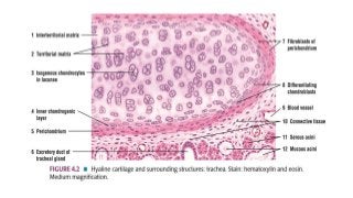
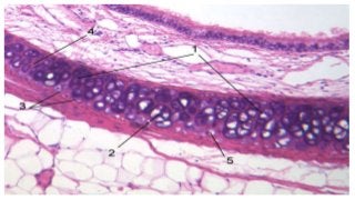
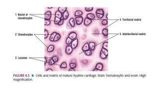
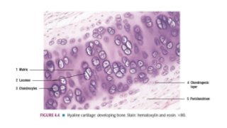
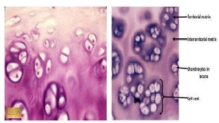
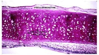
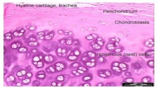
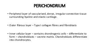
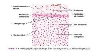
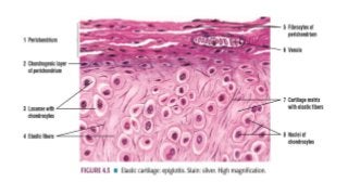
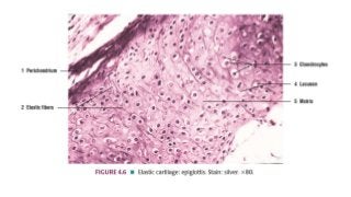
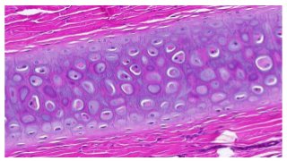
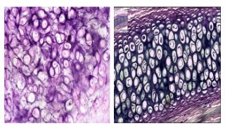
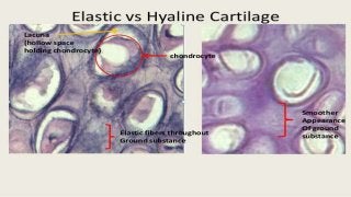
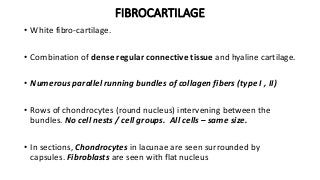
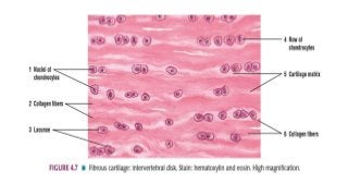
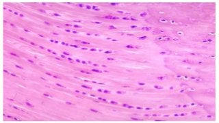
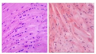
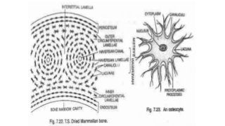
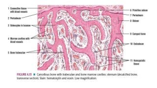
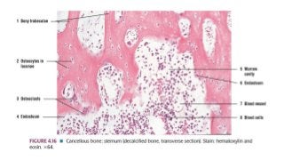
CARTILAGE
DR. SUNDIP CHARMODE
ASSOCIATE PROFESSOR
DEPARTMENT OF ANATOMY
AIIMS RAJKOT
INTRODUCTION
• It is a specialized connective tissue.
CHARATERISTIC FEATURES
• Chondroblasts – cartilage forming cells differentiate from
mesenchymal cells. It has very limited ability for regeneration.
• During fetal life, cartilage is much more widely distributed than in
adult. Hence, the greater part of skeleton is cartilaginous in early fetal
life.
• Presence of Cartilage canals in the cartilage is well established.
• Cartilage canals – small artery surrounded with numerous venules
and capillaries.
CHARATERISTIC FEATURES
• Replacement of cartilage by bone is Endochondral ossification.
• Cartilage canals also play a role in ossification of cartilage by carrying
bone forming cells into it.
• Calcification is the process in which matrix hardens because of
deposition of calcium salts in it.
• It is an avascular tissue. Nutrition is via diffusion from vessels in the
perichondrium or in cartilage canals.
GROWTH OF CARTILAGE
• Interstitial growth -
• Appositional growth -
COMPONENTS OF CARTILAGE
 Cells – Chondrocytes
 Ground substance / Inter-cellular / Extra-cellular matrix
 Fibres – Collagen fibers and elastic fibers
CARTILAGE CELLS
• Cells of cartilage – Chondrocytes which lie in spaces / Lacunae present in
matrix
• Chondroblasts: Mesenchymal progenitor cells- Small, immature and
metabolically active.
• Chondrocytes: Mature, large (dia. – 40 um) and produced from
chondroblasts. Cytoplasm of chondrocytes contains glycogen and lipids.
• Chondroblasts – secrete components of extracellular matrix
• Chondrocytes – nourishment and maintenance of cartilage
GROUND SUBSTANCE
• It is rich in proteins and carbohydrates i.e. Proteo-gylcans and
Glycoproteins (chondronectin)
• Proteogylcan – Complex formed by protein and long chain
polysaccharides (glycosaminoglycans).
• Glycosaminoglycans – Chondroitin sulphate,, Hyaluronic acid,
Keratan sulphate
• The core protein - Aggrecan
GROUND SUBSTANCE - PROTEOGYLCANS
• Tightly bound - negatively charged – attracts water and sodium ions -
firm gel like consistency.
High water content confers following abilities:
1. Maintains hydration of matrix
2. Allows diffusion of molecules to and from the chondrocytes
3. Confers cartilage the ability to resist compression
GROUND SUBSTANCE - GLYCOPROTEINS
• Glycoproteins (Chondronectin) – Macromolecules which bind to GAG
and collagen fibres.
• Facilitate adhesion of Chondroblasts and Chondrocytes to collagen
fibres of matrix.
GROUND SUBSTANCE – GENERAL FUNCTIONS
• Facilitate adhesion between various elements of connective tissue.
• Fibroblasts - Intercellular ground substance
• Osteoblasts, Chondrocytes, Chondroblasts and smooth muscle cells
also produce ground substance.
FIBRES
• Collagen fibres are chemically distinct from the usual fibres – Type II
collagen
• Fibrocartilage and Perichondrium – Type I collagen
• The collagen /elastin fibres give cartilage its firmness and resilience.
• Collagen fibres not seen in routine stains – same RI to that of GS.
HYALINE CARTILAGE
• Hyalos – glass – transparent
• Inter-cellular substance appears homogenous (basophilic).
• Special stains – can visualise Collagen Fibers – type II
• Fibres – resist tensile strength , Matrix – resist compressive forces
• H and E stain:
I. Matrix – blue (basophilic)
II. Matrix just beneath perichondrium – acidophilic
HYALINE CARTILAGE
• Towards the centre – chondrocytes are large, in groups (2-4)
• The cells tend to remain together due to surrounding dense matrix.
• Groups of cells – Cell nests / Iso-genous cell groups.
• Cell nests are formed by division of a single parent cell.
• Towards the periphery, the cells become small and flattened, parallel
to the perichondrium. (Differentiating Chondrobasts)
HYALINE CARTILAGE
• Lacunae
• Territorial matrix / Lacunar capsule – deeper stained
• Inter-territorial matrix – pale stained
• Calcification of hyaline cartilage occurs in old age.
• With advancing age, Collagen fibres become much more prominent in
hyaline cartilage
HYALINE CARTILAGE
• Conversion of Hyaline cartilage to Fibro-cartilage – sign of ageing in
body.
• E.g. Costal cartilages, articular cartilages, thyroid, cricoid, arytenoid
cartilage, tracheal rings, large bronchi, epiphyseal plate, parts of nasal
septum, lateral wall of nose.
PERICHONDRIUM
• Peripheral layer of vascularised, dense, irregular connective tissue
surrounding hyaline and elastic cartilage.
• Outer fibrous layer – Type I collagen fibres and fibroblasts
• Inner cellular layer – contains chondrogenic cells – differentiate to
form – chondroblasts – secrete matrix. Chondroblasts differentiate
into chondrocytes.
ELASTIC CARTILAGE
• Yellow fibro-cartilage
• In addition to Collagen fibres (type –II), numerous elastic fibres are present
which branch and anastomose.
• Elastic fibres difficult to visualise in H and E stains.
• Special stains used are Resorcin – fuschin, Orcein, Verhoeff’s method –
bluish black
• Surface is covered by perichondrium containing fibrocytes, venules.
• Large chondrocytes in the lacunae – centre, small and flat cells – periphery.
ELASTIC CARTILAGE
• Chondrocytes larger than those in hyaline cartilage. Found in single
or in twos in lacunae.
• Other features similar to hyaline cartilage. Do not undergo
calcification. Presence of perichondrium
• It has greater flexibility than hyaline cartilage.
• Examples : Auricle, lateral part of external acoustic meatus, wall of
medial part of auditory tube, epiglottis, corniculate and cuneiform
cartilage.
FIBROCARTILAGE
• White fibro-cartilage.
• Combination of dense regular connective tissue and hyaline cartilage.
• Numerous parallel running bundles of collagen fibers (type I , II)
• Rows of chondrocytes (round nucleus) intervening between the
bundles. No cell nests / cell groups. All cells – same size.
• In sections, Chondrocytes in lacunae are seen surrounded by
capsules. Fibroblasts are seen with flat nucleus
FIBROCARTILAGE
• Chondrocytes are similar to that of hyaline, but they have
considerably less cartilage matrix material.
• Collagen fibres – Type I, II
• Fibro-cartilage unlike Hyaline cartilage does not undergo
calcification
• Absent perichondrium
FIBROCARTILAGE
• Examples :
i. Secondary cartilaginous joints, articular disc, menisci
ii. Disc of Temporo-mandibular jt, sterno-clavicular jt, knee joint
iii. Glenoidal labrum, acetabular labrum
iv. Bony Grooves lodging tendons are lined by fibro-cartilage
v. Sites where tendons insert into bone are covered by fibro-cartilage.
BONE
DR. SUNDIP CHARMODE
ASSOCIATE PROFESSOR
DEPARTMENT OF ANATOMY
AIIMS RAJKOT
SLIDES
1. Compact bone -
• Transverse section
• Longitudinal section
2. Cancellous bone – T.S.
TYPES OF BONE
 Based of histology:
• Compact bone
• Cancellous / Spongy bone
 Base on maturity:
• Mature/ Lamellar bone
• Immature / Woven bone
 Based on manner of development:
• Cartilage bone
• Membranous bone
COMPACT BONE
• Made up of Lamellae
• Lacunae
• Canaliculi
• Types of lamellae
• Haversian canal
• Osteon / Haversian
system
• Volkmann’s canal
• Osteocytes and its
processes
COMPACT, DRIED BONE –
TRANSVERSE SECTION
COMPACT, DRIED BONE –
LONGITUDINAL SECTION
CANCELLOUS BONE – TRANSVERSE
SECTION
 Made up of layers / lamellae
SUMMARY
THANK YOU

Histology - Bone and Cartilage.pptx

  • 1.
    CARTILAGE DR. SUNDIP CHARMODE ASSOCIATEPROFESSOR DEPARTMENT OF ANATOMY AIIMS RAJKOT
  • 2.
    INTRODUCTION • It isa specialized connective tissue.
  • 3.
    CHARATERISTIC FEATURES • Chondroblasts– cartilage forming cells differentiate from mesenchymal cells. It has very limited ability for regeneration. • During fetal life, cartilage is much more widely distributed than in adult. Hence, the greater part of skeleton is cartilaginous in early fetal life. • Presence of Cartilage canals in the cartilage is well established. • Cartilage canals – small artery surrounded with numerous venules and capillaries.
  • 4.
    CHARATERISTIC FEATURES • Replacementof cartilage by bone is Endochondral ossification. • Cartilage canals also play a role in ossification of cartilage by carrying bone forming cells into it. • Calcification is the process in which matrix hardens because of deposition of calcium salts in it. • It is an avascular tissue. Nutrition is via diffusion from vessels in the perichondrium or in cartilage canals.
  • 5.
    GROWTH OF CARTILAGE •Interstitial growth - • Appositional growth -
  • 6.
    COMPONENTS OF CARTILAGE Cells – Chondrocytes  Ground substance / Inter-cellular / Extra-cellular matrix  Fibres – Collagen fibers and elastic fibers
  • 7.
    CARTILAGE CELLS • Cellsof cartilage – Chondrocytes which lie in spaces / Lacunae present in matrix • Chondroblasts: Mesenchymal progenitor cells- Small, immature and metabolically active. • Chondrocytes: Mature, large (dia. – 40 um) and produced from chondroblasts. Cytoplasm of chondrocytes contains glycogen and lipids. • Chondroblasts – secrete components of extracellular matrix • Chondrocytes – nourishment and maintenance of cartilage
  • 8.
    GROUND SUBSTANCE • Itis rich in proteins and carbohydrates i.e. Proteo-gylcans and Glycoproteins (chondronectin) • Proteogylcan – Complex formed by protein and long chain polysaccharides (glycosaminoglycans). • Glycosaminoglycans – Chondroitin sulphate,, Hyaluronic acid, Keratan sulphate • The core protein - Aggrecan
  • 9.
    GROUND SUBSTANCE -PROTEOGYLCANS • Tightly bound - negatively charged – attracts water and sodium ions - firm gel like consistency. High water content confers following abilities: 1. Maintains hydration of matrix 2. Allows diffusion of molecules to and from the chondrocytes 3. Confers cartilage the ability to resist compression
  • 10.
    GROUND SUBSTANCE -GLYCOPROTEINS • Glycoproteins (Chondronectin) – Macromolecules which bind to GAG and collagen fibres. • Facilitate adhesion of Chondroblasts and Chondrocytes to collagen fibres of matrix.
  • 11.
    GROUND SUBSTANCE –GENERAL FUNCTIONS • Facilitate adhesion between various elements of connective tissue. • Fibroblasts - Intercellular ground substance • Osteoblasts, Chondrocytes, Chondroblasts and smooth muscle cells also produce ground substance.
  • 12.
    FIBRES • Collagen fibresare chemically distinct from the usual fibres – Type II collagen • Fibrocartilage and Perichondrium – Type I collagen • The collagen /elastin fibres give cartilage its firmness and resilience. • Collagen fibres not seen in routine stains – same RI to that of GS.
  • 13.
    HYALINE CARTILAGE • Hyalos– glass – transparent • Inter-cellular substance appears homogenous (basophilic). • Special stains – can visualise Collagen Fibers – type II • Fibres – resist tensile strength , Matrix – resist compressive forces • H and E stain: I. Matrix – blue (basophilic) II. Matrix just beneath perichondrium – acidophilic
  • 14.
    HYALINE CARTILAGE • Towardsthe centre – chondrocytes are large, in groups (2-4) • The cells tend to remain together due to surrounding dense matrix. • Groups of cells – Cell nests / Iso-genous cell groups. • Cell nests are formed by division of a single parent cell. • Towards the periphery, the cells become small and flattened, parallel to the perichondrium. (Differentiating Chondrobasts)
  • 15.
    HYALINE CARTILAGE • Lacunae •Territorial matrix / Lacunar capsule – deeper stained • Inter-territorial matrix – pale stained • Calcification of hyaline cartilage occurs in old age. • With advancing age, Collagen fibres become much more prominent in hyaline cartilage
  • 16.
    HYALINE CARTILAGE • Conversionof Hyaline cartilage to Fibro-cartilage – sign of ageing in body. • E.g. Costal cartilages, articular cartilages, thyroid, cricoid, arytenoid cartilage, tracheal rings, large bronchi, epiphyseal plate, parts of nasal septum, lateral wall of nose.
  • 24.
    PERICHONDRIUM • Peripheral layerof vascularised, dense, irregular connective tissue surrounding hyaline and elastic cartilage. • Outer fibrous layer – Type I collagen fibres and fibroblasts • Inner cellular layer – contains chondrogenic cells – differentiate to form – chondroblasts – secrete matrix. Chondroblasts differentiate into chondrocytes.
  • 26.
    ELASTIC CARTILAGE • Yellowfibro-cartilage • In addition to Collagen fibres (type –II), numerous elastic fibres are present which branch and anastomose. • Elastic fibres difficult to visualise in H and E stains. • Special stains used are Resorcin – fuschin, Orcein, Verhoeff’s method – bluish black • Surface is covered by perichondrium containing fibrocytes, venules. • Large chondrocytes in the lacunae – centre, small and flat cells – periphery.
  • 27.
    ELASTIC CARTILAGE • Chondrocyteslarger than those in hyaline cartilage. Found in single or in twos in lacunae. • Other features similar to hyaline cartilage. Do not undergo calcification. Presence of perichondrium • It has greater flexibility than hyaline cartilage. • Examples : Auricle, lateral part of external acoustic meatus, wall of medial part of auditory tube, epiglottis, corniculate and cuneiform cartilage.
  • 34.
    FIBROCARTILAGE • White fibro-cartilage. •Combination of dense regular connective tissue and hyaline cartilage. • Numerous parallel running bundles of collagen fibers (type I , II) • Rows of chondrocytes (round nucleus) intervening between the bundles. No cell nests / cell groups. All cells – same size. • In sections, Chondrocytes in lacunae are seen surrounded by capsules. Fibroblasts are seen with flat nucleus
  • 35.
    FIBROCARTILAGE • Chondrocytes aresimilar to that of hyaline, but they have considerably less cartilage matrix material. • Collagen fibres – Type I, II • Fibro-cartilage unlike Hyaline cartilage does not undergo calcification • Absent perichondrium
  • 36.
    FIBROCARTILAGE • Examples : i.Secondary cartilaginous joints, articular disc, menisci ii. Disc of Temporo-mandibular jt, sterno-clavicular jt, knee joint iii. Glenoidal labrum, acetabular labrum iv. Bony Grooves lodging tendons are lined by fibro-cartilage v. Sites where tendons insert into bone are covered by fibro-cartilage.
  • 40.
    BONE DR. SUNDIP CHARMODE ASSOCIATEPROFESSOR DEPARTMENT OF ANATOMY AIIMS RAJKOT
  • 41.
    SLIDES 1. Compact bone- • Transverse section • Longitudinal section 2. Cancellous bone – T.S.
  • 42.
    TYPES OF BONE Based of histology: • Compact bone • Cancellous / Spongy bone  Base on maturity: • Mature/ Lamellar bone • Immature / Woven bone  Based on manner of development: • Cartilage bone • Membranous bone
  • 43.
    COMPACT BONE • Madeup of Lamellae • Lacunae • Canaliculi • Types of lamellae • Haversian canal • Osteon / Haversian system • Volkmann’s canal • Osteocytes and its processes
  • 45.
    COMPACT, DRIED BONE– TRANSVERSE SECTION
  • 51.
    COMPACT, DRIED BONE– LONGITUDINAL SECTION
  • 53.
    CANCELLOUS BONE –TRANSVERSE SECTION  Made up of layers / lamellae
  • 56.
  • 57.